Mole Check in York Mills: What a Proper Skin Check Involves and How Often You Actually Need One
Last updated: September 1, 2026
Quick Answer
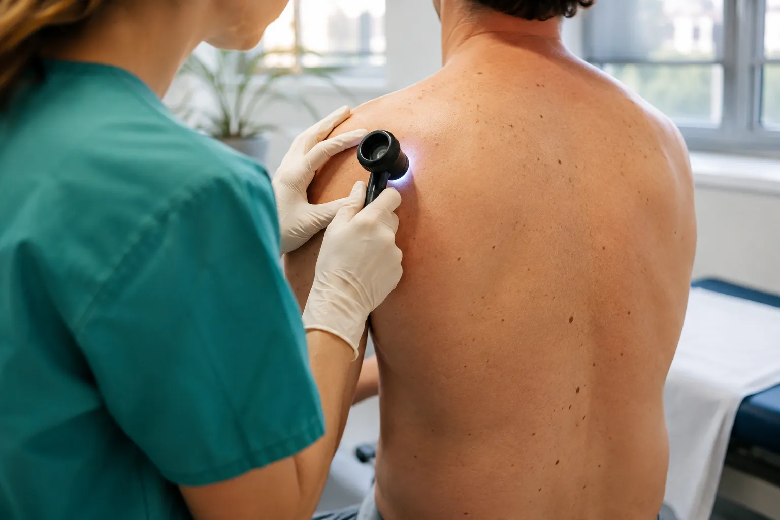
A mole check is a short clinical appointment where a physician takes a brief history, examines your skin with the naked eye, and then looks more closely with a dermatoscope, a handheld device using magnification and polarised light to see structures below the surface. Its purpose is to decide one of three things: the spot can be left alone, it should be tracked with photographs and reviewed later, or it needs testing. The Minor Surgery Center's York Mills clinic at 85 Scarsdale Rd, Unit 101, North York sees patients for skin lesion assessment without a referral, weekdays 9 a.m. to 4 p.m. If a spot is actively changing, have it looked at rather than waiting for a convenient date.
Key Takeaways
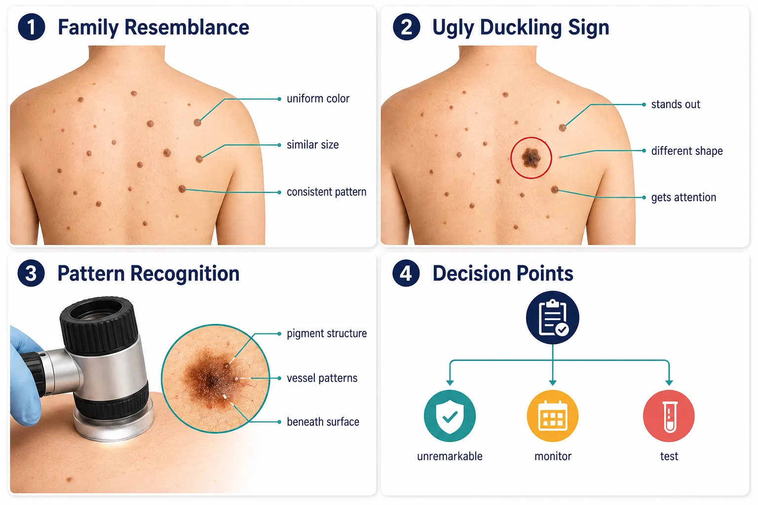
A mole check is an assessment, not a treatment. It answers whether a lesion is unremarkable, worth monitoring, or worth testing.
Clinicians screen by pattern, not by measuring every mole against a checklist. The spot that stands out from its neighbours, the "ugly duckling", gets the attention.
Dermoscopy reveals pigment networks and vessel patterns invisible from across the room. It reduces unnecessary biopsies and flags lesions that look ordinary to the eye.
Change over time is the strongest single signal in skin cancer detection, and change can only be judged against a dated record. That's the entire argument for baseline photography.
Most people at average risk don't need annual whole-body screening; people with many moles, atypical moles, or a personal or family history of melanoma generally do need scheduled surveillance.
A monthly self-exam works when you use good light, a hand mirror, and a second person for your back and scalp, and when it sends you to a clinician rather than reassuring you.
No referral is required at many Ontario private-pay skin lesion clinics. Coverage differs between a suspicious-lesion assessment and a routine cosmetic or screening request, so confirm at booking.
What a Mole Check Is, and What It Is Not
A mole check is a decision-making appointment. A physician examines the skin and answers three questions: is this within the range of normal for you, does it need to be watched, or does it need to be tested?
That's the whole scope. It's short, it's non-invasive, and nothing is cut unless a separate decision is made.
What it isn't matters just as much:
It isn't a removal appointment. Removal is a different procedure with its own consent, technique and aftercare. If you want the full picture on that side, the mole removal service in York Mills page covers methods and recovery properly.
It isn't a cosmetic consult. A check assesses risk. Whether a mole bothers you aesthetically is a valid conversation, but it's a separate one, the difference is laid out in this comparison of a mole check versus a mole removal.
It isn't a diagnosis. This is the part people misunderstand most. Clinical examination and dermoscopy narrow the possibilities and guide the decision. A definitive diagnosis of melanoma or any skin cancer comes from pathology, tissue examined under a microscope. No physician, however experienced, and no imaging device can replace that step.
Sometimes a removal can follow in the same visit, when the finding is clear-cut and the clinical situation allows. That's convenient, not the point. The check is the step where someone competent decides what should happen next.
A mole check doesn't tell you a spot is fine. It tells you what should happen to that spot.
The Ugly Duckling Sign: How Clinicians Actually Scan a Whole Back
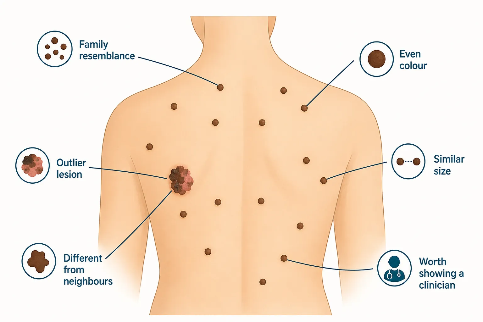
Most people's moles share a family resemblance. When a clinician scans a back with forty moles on it, they aren't measuring each one, they're looking for the one that doesn't belong to the family.
That's the ugly duckling sign, and it's the practical logic behind most whole-body screening. Your moles tend to run to a type: similar size, similar brown, similar edge quality, similar degree of raised-ness. Genetics and sun history give them a shared signature. A lesion that breaks the pattern is worth a closer look, even if, taken on its own, it would pass any checklist you care to apply.
Why pattern beats a checklist
The ABCDE rule (asymmetry, border, colour, diameter over roughly 6 mm, evolution) is a useful complementary tool when you're assessing one single lesion, and it's worth knowing. But it has blind spots, and pattern recognition covers several of them.
Small early melanomas. A lesion 3 mm across can be melanoma. It fails the diameter criterion entirely, and at that size asymmetry and border irregularity are hard to judge by eye. What it may do is look nothing like the other moles nearby.
Amelanotic and hypomelanotic lesions. Some melanomas carry little or no pigment. They can read as pink, skin-coloured, or faintly red, and a rule built around dark irregular pigment misses them. In a field of brown moles, a persistent pink papule is the outlier.
Uniform-looking melanomas. Early lesions can be evenly coloured and neatly bordered. They still stand apart from their neighbours in tone or texture.
People with many atypical moles. If your baseline is dozens of moles that all look somewhat irregular, ABCDE flags nearly everything and stops being informative. Pattern-based screening resets the question: irregular compared to your other moles, not compared to a textbook.
The version you can use at home
Stand in front of a mirror and let your eye pass over a region, one shoulder blade, one calf, one forearm, without stopping on anything in particular. Then ask a simple question: did anything pull your eye?
If one spot keeps drawing your attention while the rest blend together, that's the one to have examined. Also worth noting:
A mole that's the only one of its kind anywhere on your body.
A mole that's clearly darker, lighter, pinker or rougher than every other mole in the same area.
A mole your partner or hairdresser mentions before you noticed it.
Any spot that bleeds, itches persistently, crusts or won't heal, regardless of how it compares to anything else.
Something standing out doesn't mean it's cancer. Most outliers turn out to be ordinary. The point is that "different from the rest" is a better trigger for booking an appointment than any measurement you can take at home, and it's one you're actually capable of using. If you're not sure how to work through a spot you've noticed, the guide on when you should get a mole checked sets out the thresholds.
What Dermoscopy Shows That the Naked Eye Cannot
A dermatoscope is a handheld device combining magnification with a polarised light source, pressed lightly against the skin. Polarised light cuts through surface glare, so the clinician sees pigment and vessel structures within the upper layers of skin rather than just the outline and colour visible from a metre away.
That difference is not cosmetic. Under a dermatoscope, features appear that simply have no naked-eye equivalent.
What the clinician is looking for
Pigment network. Ordinary moles often show a regular, honeycomb-like mesh of pigment. Disruption, thickening or abrupt ending of that network is meaningful.
Vascular patterns. The arrangement of tiny blood vessels differs between lesion types, which is one of the few ways to get useful information out of a lesion with little pigment.
Structural clues to depth and distribution, how pigment sits, whether it's uniform or clumped, whether structures are symmetrically arranged.
Features that point away from melanoma entirely, such as the patterns typical of seborrhoeic keratoses, haemangiomas or basal cell lesions.
What it changes in practice
Two things, both useful. First, it reduces unnecessary cutting. A fair number of lesions that look alarming to the naked eye show a reassuring, well-organised dermoscopic pattern, and can reasonably be photographed and reviewed rather than excised. Published dermatology literature consistently reports that trained use of dermoscopy improves diagnostic accuracy for pigmented lesions compared with unaided visual examination alone, that's the mainstream position, not a marketing claim.
Second, it works in the other direction. A lesion that reads as unremarkable across a desk can show disorganised structures under magnification and get biopsied on that basis. This is the harder half to explain to patients: sometimes the flat, small, boring-looking spot is the one that gets tested while the big dark one you were worried about does not.
Two honest limits. Dermoscopy is a skill, not a scanner, it doesn't produce an answer, it produces information that a trained clinician interprets. And it does not replace pathology. If dermoscopic features are uncertain or concerning, tissue still goes to a lab. There's a fuller account of the sequence in this explanation of how doctors check a mole for cancer.
Focused Check, Full-Body Skin Exam, or Mole Mapping: Which One You Need
Three different appointments, three different purposes. Most people arriving with one spot in mind need the first one.
Type of appointment / What it covers / Best suited for / How long it takes / What you leave with
Focused single-lesion check
What it covers: History for that spot, naked-eye exam, dermoscopy of the lesion and a look at nearby skin
Best suited for: One mole you keep noticing, a spot that has changed, a lesion someone else pointed out
How long it takes: Roughly 10-20 minutes
What you leave with: A plan for that lesion: leave, monitor with a dated photo, or biopsy
Full-body skin examination
What it covers: Systematic review of the skin surface including scalp, ears, back, feet, between toes and nails, with dermoscopy of anything of interest
Best suited for: Higher-risk patients, anyone with many moles, first baseline visit, significant sun history
How long it takes: Roughly 20-40 minutes
What you leave with: An overall risk picture, a list of lesions to track, and a recommended review interval
Baseline photography and mole mapping
What it covers: Full-body exam plus dated regional photographs and close-ups of selected lesions, stored for comparison
Best suited for: Many or atypical moles, personal or family melanoma history, patients on long-term immunosuppression
How long it takes: Typically 30-60 minutes at the baseline visit; shorter at follow-ups
What you leave with: A dated photographic record and a scheduled comparison visit
How the choice gets made: the clinician weighs why you came, how many moles you have, your skin type and burn history, and whether melanoma runs in your family. Someone in their thirties with fair skin, one spot on the shoulder and no family history usually gets a focused check and a conversation. Someone with sixty moles, several atypical, and a sibling treated for melanoma is a mapping candidate.
Decision rule: if you can point at the thing that's bothering you, start with a focused check. If you couldn't count your moles without help, ask about a full-body skin examination or mole mapping instead.
Mole Mapping and Baseline Photography: How Change Is Tracked Over Time
Change over time is the most useful signal in skin cancer detection, and change can only be judged against a record. That's the entire case for baseline photography. Without a dated image, "has it grown?" is a memory question, and memory is unreliable about a 5 mm brown spot on your own back.
What a baseline session involves
You undress to your underwear in stages and a series of standardised regional photographs is taken, front, back, each side, limbs, and the scalp if hair allows. Individual lesions of interest get close-up images, often with a dermoscopic photograph alongside the clinical one. Everything is dated and stored as part of your medical record.
The photographs are not the assessment. They're the reference point the next assessment gets measured against.
How comparison works at follow-up
At the review visit the clinician examines you first, then pulls the prior images and compares region by region. Three findings matter:
A known lesion has changed in size, colour, border or elevation.
A lesion is present now that wasn't present before, a genuinely new mole in an adult, particularly after about age 40, is worth attention.
Nothing has changed, which is itself information, and often means the interval to the next visit can stay where it is.
Comparing images beats comparing impressions. It's also the mechanism that makes conservative management defensible: a lesion can be watched rather than cut precisely because there's a way to prove whether it stayed put.
Who benefits most
People with a high total mole count, screening by memory stops working past a certain number.
People with clinically atypical moles, where nearly everything looks somewhat irregular and the only meaningful question is whether a particular lesion is changing.
Anyone with a personal history of melanoma or a first-degree relative who's had one.
Organ transplant recipients and others on long-term immunosuppressive medication, who carry higher skin cancer risk overall.
People with heavy childhood sun exposure or blistering sunburns, or a history of tanning bed use.
The honest limits
Photographs record surface appearance. They don't record depth, and depth is what matters most in melanoma prognosis. A lesion can change beneath the surface while its photograph barely shifts, which is why mapping supports clinical examination rather than replacing it, you still get examined, every time.
Image quality also varies with lighting, posture and hair. And mapping is not a substitute for reporting a change you notice yourself; it's there to make your reports easier to evaluate, not to catch things on your behalf between visits. Whether photographic mapping meaningfully improves melanoma detection is discussed in more depth in this look at whether 3D mole mapping detects melanoma.
What you actually leave with: a dated image set in your file, a short list of lesions being tracked by location, a recommended interval for the next comparison, and clear instructions on what would justify coming back sooner.
How Often Should You Have Your Moles Checked?
There's no single correct interval. Frequency follows risk, and the physician who examines you sets the interval for you specifically. The bands below are general guidance used in dermatologic practice, not rules.
Risk band / Who typically falls into it / General check interval
Average risk
Who typically falls into it: Few moles, no atypical moles, no family history of melanoma, no significant burn history, skin that tans more readily than it burns
General check interval: Monthly self-exam; a clinical baseline once as an adult, then as advised or when something changes
Moderate risk
Who typically falls into it: Fair skin that burns easily, red or blond hair, many moles, several blistering childhood sunburns, past tanning bed use, outdoor work or year-round outdoor sport, prior non-melanoma skin cancer
General check interval: Clinical skin exam roughly every 12 months, adjusted by the examining physician
Higher risk
Who typically falls into it: Personal history of melanoma, first-degree relative with melanoma, multiple clinically atypical moles, very high mole count, organ transplant recipients and others on long-term immunosuppressive medication
General check interval: Every 3 to 6 months initially, often with baseline photography, then spaced out on physician advice
Two things override the table entirely.
First: a changing spot is seen promptly, at any risk level. Someone in the average-risk band with a mole that has darkened over six weeks does not wait for a scheduled appointment. The interval governs routine surveillance, not new findings.
Second: risk bands move. A first melanoma, a transplant, a new immunosuppressive medication, or the arrival of several atypical moles all shift you upward. Mention any of these at your next visit so the interval gets revised.
A common mistake worth naming: people who had a clear check three years ago often assume they're covered. A clear result describes the skin on the day it was examined. It says nothing about the mole that appeared last spring.
What to Do Between Visits: A Self-Exam That Actually Works
A monthly self-exam is a reasonable cadence for most adults. It takes about ten minutes and its job is narrow, to notice something new or different and get it examined. It's not there to let you decide a spot is fine.
What you need
Strong, even light. Bathroom overheads throw shadows; a bright lamp or daylight through a window works better.
A full-length mirror and a hand mirror, used together for the back of the legs and buttocks.
A second person for your back, the back of your neck and your scalp. This is the single biggest improvement you can make to a home check. Melanoma on the back is a classic late presentation for exactly this reason.
A phone to record a dated photo of anything you want to re-check, ideally with a coin or ruler beside it for scale.
A working order
Use the same route every month so you don't skip regions:
Face, ears, lips. Include the rim and behind each ear, and the front and back of the earlobes.
Scalp. Part the hair in sections with a comb, or have someone else do it. Slow and boring, and worth it.
Neck, chest, torso. Lift the breasts and check underneath.
Arms. Front and back, then armpits with the arm raised, then hands, palms, backs, between the fingers, and the nail beds.
Back and buttocks. Full-length mirror plus hand mirror, or a partner.
Legs. Front, back and sides, sitting down if that helps.
Feet. Tops, soles, heels, between every toe, and toenails.
Genital and perianal area. Uncomfortable to check, genuinely a site where melanoma occurs.
Where lesions are actually missed
The list is short and consistent: scalp, behind the ears, soles of the feet, between the toes, under the fingernails and toenails, and the buttocks and genital area. These are missed because they're hard to see, not because they're low-risk sites.
Nail findings deserve a specific mention. A new dark streak running lengthwise down a nail, or pigment spreading onto the surrounding skin, should be assessed by a physician rather than watched. Remove nail polish before your appointment if nails are your concern. There's step-by-step guidance for checking hard-to-see areas like the scalp, back, ears, nails and soles if you want to work through it properly.
What to do with what you find
Write it down: date, body location, size, and what caught your eye. Photograph it. Then book. A self-exam that ends in "I'll keep an eye on it for a few more months" has failed at the only task it had. The purpose of noticing is to get the thing looked at, a broader framing of that is set out in this guide to skin self-exams for early detection.
On phone apps and AI mole scanners
Fair assessment: they're useful for one thing and unreliable for another.
Useful for keeping a dated, organised photo record with body-map locations. That genuinely helps, both for your own tracking and for showing a clinician a lesion three months ago versus today.
Not reliable for telling you whether a lesion is dangerous. Consumer skin-analysis apps have shown inconsistent accuracy in independent evaluations, they generally work only on pigmented lesions, and they cannot see the sub-surface structures that a dermatoscope reveals. A reassuring app result on a lesion that's changing is a reason to book, not a reason to relax. The realistic uses and limits are covered in this review of mole check apps.
What Happens If Something Looks Suspicious
The next step is proportionate, not dramatic. The clinician examines the lesion dermoscopically, and if the features are uncertain or concerning, arranges a biopsy so tissue can be examined by a pathologist.
Timing is usually planned within days to a few weeks rather than treated as an emergency, and the pathology report determines what, if anything, follows. If you want the full pathway, lesion types, biopsy approach, what a report says, read the detailed suspicious mole assessment page rather than expecting it here. This page is about the check.
Do You Need a Referral, and Is a Mole Check Covered by OHIP?
Many Ontario clinics that assess skin lesions accept patients directly, with no referral from a family doctor. Some dermatology practices still require one, particularly for new patients or anyone not seen in over a year, so confirm before you travel.
Coverage depends on why you're being seen. Assessment of a specific lesion that a physician considers clinically suspicious is treated differently under OHIP from a routine whole-body screening requested without a clinical indication, and differently again from anything undertaken for cosmetic reasons. Baseline photography and mole mapping are commonly offered as private-pay services.
For private-pay skin assessments in the Toronto area, a focused single-lesion consultation typically falls in the low-to-mid hundreds of Canadian dollars, with full-body examinations and baseline photographic mapping costing more. Treat those as broad, general ranges, not a quote.
Three factors move the number:
Scope. A focused check on one lesion costs less than a systematic whole-body examination.
Photography and mapping. Baseline imaging with stored comparison images adds time and cost, and is usually priced separately.
Whether anything is biopsied on the day. Any procedure and the associated pathology are billed separately from the assessment.
Ask for the exact figure at booking, along with whether your visit may be OHIP-eligible. Mole removal is priced differently again, and that's covered on the York Mills mole removal page rather than duplicated here.
Getting Checked in York Mills: Location, Access and Booking
The Minor Surgery Center's York Mills clinic is at 85 Scarsdale Rd, Unit 101, North York, ON M3B 2R2, open Monday to Friday, 9 a.m. to 4 p.m., reachable at 647-614-1611. It opened in July 2026 and no referral is required.
Scarsdale Road sits just south of York Mills Road and east of Leslie, minutes from the Don Valley Parkway at the York Mills Road exit, with on-site parking. By TTC, it's reachable from York Mills station on Line 1 via an eastbound bus along York Mills Road.
Why the staffing structure matters
Assessment and diagnosis are handled by GP dermatologists, physicians whose practice focuses on skin. Excisions and closures are performed by plastic surgeons. If a check leads to a decision to remove something, the surgical side is done by someone whose training is in cutting and closing skin, which matters most on the face, neck and other cosmetically sensitive areas. Practically, it means you're not referred out and re-queued.
Other locations in the group cover North York on Dufferin Street, plus Vaughan, Mississauga and Oakville, useful if a different site is closer or has an earlier opening. The York Mills clinic page has current details and booking.
How to prepare
No makeup, concealer or self-tanner on the area being examined.
Remove nail polish from all fingers and toes if a nail is your concern.
Skip heavy body lotion on the day, a dermatoscope works better on clean skin.
Come ready to point at the spot. Bring your dated photos if you have them, and be able to say roughly when you first noticed the change.
Wear something you can undress from easily if a full-body exam is planned.
Same-day and next-day appointments are sometimes available for urgent lesion assessment, but availability varies day to day. Phone rather than emailing if your spot is changing quickly, and say so when you call, a rapidly changing lesion is triaged differently, as explained in this guidance on how to handle a rapidly changing mole.
Areas Served Around York Mills
The Scarsdale Road location is a short drive for residents of Hoggs Hollow, the Bridle Path and Lawrence Park, and sits close to Banbury, Don Mills and Graydon Hall on the east side of the DVP. Patients also come from Bayview Village, Leaside, Parkwoods, Willowdale and across the wider North York area, helped by the York Mills Road interchange and the Leslie corridor. If you're travelling from further west or north, the group's North York clinic on Dufferin Street or the Vaughan location may be a shorter trip.
Frequently Asked Questions
How long does a mole check take? A focused check on one or two lesions usually takes 10 to 20 minutes. A full-body skin examination generally runs 20 to 40 minutes, and a baseline photography session with mole mapping can take 30 to 60 minutes.
Does a mole check hurt? No. Examination and dermoscopy are non-invasive, the dermatoscope rests lightly on the skin, sometimes with a drop of gel or alcohol. Discomfort only arises if a biopsy is performed, and that's done under local anaesthetic.
Do I need to undress fully? For a focused single-lesion check, only the relevant area is exposed. For a full-body examination you'll usually undress to your underwear, in stages, with a gown and draping. Tell the clinic in advance if you'd prefer a physician of a particular gender or a chaperone present.
Do I need a referral for a mole check in Ontario? Not at every clinic. Many Ontario skin lesion and minor surgery clinics, including the York Mills location, accept patients directly. Some dermatology practices do require a referral from a family doctor or walk-in clinic, so confirm when you book.
How soon can I be seen? Same-day or next-day assessment is sometimes possible for a lesion that's changing, but it depends on the day's schedule. Phone and describe the change rather than booking the next routine slot online.
Can a mole be removed the same day it's checked? Sometimes. If the assessment supports removal and there's surgical time available, it can happen in one visit. It isn't guaranteed, and it isn't always the right decision, some lesions are better photographed and reviewed than removed immediately.
How often should I have my moles checked? It depends on your risk. Average-risk adults with few moles and no family history often need only a baseline plus monthly self-exams. Fair skin, many moles or heavy sun history generally means about annually, and a personal or family history of melanoma often means every 3 to 6 months at first. Your physician sets your interval.
Are mole check apps accurate? Not accurate enough to rely on for a decision. Consumer apps have shown inconsistent performance in independent evaluations and can't see the sub-surface structures a dermatoscope reveals. Use them to keep dated photos, never to decide against getting a spot examined.
Should children's moles be checked? Yes, if a mole is new and growing quickly, bleeding, itching, much larger than the child's other moles, or looks clearly different from them. Moles do change during childhood and adolescence, and most changes are normal growth, but a lesion that stands out from the child's own pattern should be examined rather than watched at home.
I only care about one spot. Do I need a full-body exam? Usually not. If you can point at the lesion and you don't have many moles, a focused check is appropriate. A full-body examination is worth adding if you have a high mole count, atypical moles, a family history of melanoma, or you've never had a baseline done.
This article is general information about skin surveillance and is not a substitute for individual medical advice. Any lesion that concerns you should be examined by a physician.
The Bottom Line
A mole check is a decision, not a treatment. Its value comes from being repeated at a sensible interval and recorded well enough that change can actually be measured, one check in isolation tells you about one day.
Three practical steps: do a self-exam this month using good light and a second person for your back, photograph anything that stands out from your other moles, and book an assessment rather than waiting to see what happens.
For a skin lesion assessment in the area, The Minor Surgery Center's York Mills clinic is at 85 Scarsdale Rd, Unit 101, North York, ON M3B 2R2, weekdays 9 a.m. to 4 p.m., 647-614-1611. No referral needed.
References
Canadian Dermatology Association, patient information on sun safety, skin cancer and skin examination.
Canadian Cancer Society, melanoma risk factors and skin self-examination guidance.
Public Health Agency of Canada / Canadian Task Force on Preventive Health Care, statements on skin cancer screening in adults.
Ontario Ministry of Health, OHIP Schedule of Benefits for Physician Services (insured versus uninsured dermatologic services).
International Dermoscopy Society, educational resources on dermoscopic criteria for pigmented lesions.
American Academy of Dermatology, the ugly duckling sign and the ABCDE criteria in melanoma screening.
The Minor Surgery Center’s York Mills clinic, located at 85 Scarsdale Rd, Unit 101 in North York, offers convenient access to specialized surgical care for a range of skin and hand conditions. Our Plastic Surgeons provide treatment for skin cancer, suspicious moles, cysts, lipomas, carpal tunnel syndrome and trigger finger, with a focus on timely care and personalized treatment. In many cases, patients may be able to have their consultation and treatment completed during the same visit when medically appropriate. For expert care in a welcoming clinical setting, contact our York Mills team at (647) 614-1611.
We are the experts in the field of Minor Surgery. All our Surgeons have completed 5 years of post Medical School Surgical Residencies. They are all Board Certified by the Royal College of Physicians and Surgeons of Ontario.They have been trained at Institutions including McGill, McMaster, Queen's, University of British Columbia, University of Toronto, University of Western Ontario, The Mayo Clinic, University of Pennsylvania, University of Southern California, Yale University, and Oxford.
In many clinics, you could wait months—sometimes over a year—for a procedure like cyst or lipoma removal, mole removal, or even carpal tunnel release. But at TMSC, we prioritize fast access to care. In many cases, we can schedule and complete your minor surgery on the same day as your initial consultation—no extra runaround required.
Our clinics are designed with your comfort, safety, and efficiency in mind. We use strict sterilization protocols to minimize any infection risk, and our advanced surgical equipment meets hospital-grade standards. Yet, you’ll never feel like you’ve stepped into a stark, clinical space—our friendly staff ensures you’re welcome and supported the moment you walk through the door.
Cosmetic results matter—even for the smallest procedures. Skin lesions can be excised by non surgeons in Ontario. Furthermore, any MD with 2 years of post graduate training even outside of a surgical field can perform removal. At TMSC we only have Surgeons under our roof. They are trained well above the level of the procedures we offer to ensure the surgery goes smoothly with minimal scaring and preserving your natural contours. Our Surgeons are trained to ensure both function and aesthetics are always front and center.
At TMSC, we specialize in surgical treatment of both benign and malignant skin lesions, as well as minor hand conditions. We offer significantly reduced wait times often allowing you to schedule your surgery on the same day as your consultation.
You can book your free consultation with The Minor Surgery Center in three easy ways: online through Cortico, by phone, or by requesting a callback from our team.
The initial consultation is free. During this consultation, one of our board certified surgeons will examine your condition and recommend a suitable solution.
Directly Online
Request a Callback
By Phone
By Text
Hours
Sunday
Closed
Monday
9 am — 4 pm
Tuesday
9 am — 4 pm
Wednesday
9 am — 4 pm
Thursday
9 am — 4 pm
Friday
9 am — 4 pm
Saturday
Toronto & Oakville Open Vaughan & Mississauga Closed



.avif)
%20(600%20x%20600%20px)%20(2).avif)
.avif)
%20(8).avif)
%20(7).avif)

.png)