
Last updated: July 7, 2026
Does 3D mole mapping detect melanoma earlier? Not consistently, based on the best current evidence. A 2025 randomized clinical trial published in JAMA Dermatology found that adding 3D total-body photography and sequential digital dermoscopy to usual care in high-risk patients increased biopsies and excisions but did not improve melanoma detection per person. For very high-risk patients (many atypical moles, prior melanoma, strong family history), 3D mapping can still be a useful surveillance tool, but it isn't a magic early-warning system for average-risk people.
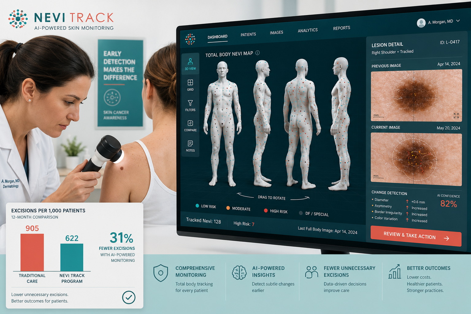
3D mole mapping is a skin surveillance technique that uses multiple synchronized cameras to capture the entire visible skin surface in a single moment, then reconstructs a 3D digital avatar where individual moles can be tagged, measured, and compared across future visits.
The most widely referenced clinical system, VECTRA WB360, uses 92 calibrated digital cameras that fire simultaneously to build a high-resolution rotatable 3D model of the body. [2] Software then flags new lesions, missing lesions, and changes in existing ones between scans. Individual moles of interest can be photographed at higher magnification with a dermatoscope for detailed follow-up, a process called sequential digital dermoscopy.
How a typical session runs:
The core idea is simple: melanoma often shows itself through change. A mole that looks unremarkable today but shifts in shape, colour, or size six months later is more suspicious than the same mole seen in isolation. 3D mapping gives clinicians a reliable reference point for spotting that change.
In theory yes, in practice sometimes. 3D mole mapping is designed to catch melanoma while it's still thin and localized, before it invades deeper tissue or spreads to lymph nodes, and observational studies in specialized clinics have reported that most melanomas found through structured photographic surveillance are 1 mm thick or less.
A 2021 cohort study of 593 very-high-risk patients found that 96% of the 171 new melanomas detected were thin (≤1 mm), and about two-thirds were found with help from total-body photography or sequential digital dermoscopy. That's exactly the stage where surgical removal has excellent outcomes.
The catch: the 2025 randomized trial couldn't confirm that this early-detection benefit translated into more melanomas found per patient once you controlled for the fact that the control group was also getting good routine care. So the honest answer is that 3D mapping can help catch melanoma at a treatable stage, especially in high-risk people, but it doesn't automatically outperform an attentive dermatologist doing regular exams.
Choose 3D mapping if: you have many moles, atypical nevi, or a personal or family history of melanoma, and you want a reliable reference point for tracking change.
3D mole mapping is a surveillance tool, not a diagnostic test, so "accuracy" depends on what you're measuring. It's very accurate at documenting what's on your skin at a point in time and detecting change, much more so than memory or a paper chart. It's less clear that it improves the accuracy of who ultimately gets diagnosed with melanoma versus a thorough clinical exam plus dermoscopy alone.
Here's how the pieces compare:
MethodWhat It DetectsStrengthsLimitsNaked-eye skin examSurface features (ABCDE signs)Fast, free, no tech neededMisses subsurface features; relies on memoryDermoscopy (handheld)Subsurface pigment patternsImproves accuracy over eye aloneSingle lesion at a time; needs training2D total-body photographyFull-body snapshotChange detection over timeFlat images, angles inconsistent3D mole mapping (VECTRA-type)Full-body 3D avatar with tagged lesionsBest change detection, standardized anglesCost, access, doesn't replace examBiopsy + pathologyDefinitive diagnosisGold standardInvasive, only for specific lesions
The 2025 trial's most striking finding was that despite the imaging group having more procedures done, the number needed to excise (how many moles you cut out to find one melanoma) was essentially the same as in the control group. In plain English: adding 3D imaging didn't make excision decisions more efficient.
Not reliably, based on the randomized evidence, but the comparison isn't really fair, because 3D mapping is meant to support dermatologists, not replace them. A skilled dermatologist doing a full-body exam with dermoscopy remains the backbone of melanoma detection. 3D mapping adds a memory aid and a change-detection layer on top of that expertise.
Where 3D mapping tends to help most:
Where it adds less:
If you're deciding between "3D mapping and no dermatologist" versus "dermatologist and no 3D mapping," the dermatologist wins every time. If you're deciding between "dermatologist alone" versus "dermatologist plus 3D mapping," the answer depends on your risk level. For guidance on how doctors actually evaluate moles, see how do doctors check a mole for cancer.
3D mole mapping captures the whole body to track lesions across time; dermoscopy examines one lesion in detail to assess whether it looks benign or suspicious. They answer different questions and are usually used together.
3D mole mapping answers: What's new or changed on this person's skin since last visit?
Dermoscopy answers: What does this specific mole look like below the surface, and is it worrying?
Dermoscopy uses a handheld device with magnification and specialized lighting to reveal subsurface pigment patterns, vessels, and structures invisible to the naked eye. It's typically done during an in-person exam on any lesion of interest.
Sequential digital dermoscopy is a middle layer: it uses dermoscopy repeatedly on the same selected lesion over time. This is different from total-body 3D imaging, which captures everything at lower magnification. Modern high-risk surveillance protocols combine all three: 3D total-body imaging for change detection, dermoscopy for close-up assessment, and sequential digital dermoscopy for lesions worth monitoring rather than immediately removing.
3D mole mapping typically costs between CAD $200 and $600 per session in Canada, and it's generally not covered by provincial health plans (OHIP) or most private insurers because it's classified as a screening or surveillance service rather than a medically necessary diagnostic procedure. Costs vary by clinic, technology used, and whether dermoscopy is included.
Typical pricing tiers:
If a specific lesion is clinically suspicious and gets biopsied or excised for pathology, that portion is typically covered when medically indicated. The imaging itself usually isn't. For a broader look at what mole-related procedures cost in Ontario, our mole removal cost calculator breaks down surgical fees by lesion type and location. You can also read our detailed guide on mole removal cost in Toronto.
Some extended health benefits plans reimburse dermatology screening under wellness allowances, so it's worth checking your specific plan.
3D mole mapping makes the most sense for patients at genuinely elevated risk of melanoma, not average-risk adults looking for one-off reassurance. Expert guidance and observational research consistently point to a specific high-risk profile.
Consider 3D mole mapping if you have any of these:
Probably skip formal 3D mapping if:
For a broader risk-assessment framework, our guide on when you should get a mole checked walks through the specific warning signs that warrant a clinical evaluation regardless of imaging.
Yes, 3D mole mapping can miss melanoma, and this is one of the most important things patients need to understand before relying on it. The technology has real blind spots, and treating a "clear" scan as absolute reassurance is dangerous.
Common ways 3D mapping can miss melanoma:
The takeaway: 3D mapping is a helpful layer, not an insurance policy. Between scans, self-checks matter. See how to check moles at home and how to check hard-to-see areas for practical technique.
Most high-risk surveillance protocols recommend 3D mole mapping every 6 to 12 months, with the exact interval based on individual risk. Very high-risk patients (recent melanoma, atypical mole syndrome plus family history, or genetic risk) often get imaging every 3-6 months during the first two years, then every 6-12 months thereafter.
Typical intervals by risk tier:
Between imaging visits, monthly self-exams remain important, along with clinical skin checks by a dermatologist or trained surgeon. The imaging isn't a replacement for hands-on examination, it's a longitudinal record that makes the exam more precise.
If 3D mole mapping flags a change, the next step is a clinical evaluation of that specific lesion, usually with dermoscopy, followed by biopsy if the mole meets criteria for suspicion. The imaging system doesn't diagnose melanoma; it identifies candidates for closer investigation.
Typical workflow after a flagged lesion:
If the pathology comes back as stage 0 melanoma or stage 1 melanoma, five-year survival is excellent, often above 95% for stage 0.
Probably not. If you have fewer than 20-30 moles, no atypical nevi, no personal or family history of melanoma, and average sun exposure, formal 3D mole mapping usually isn't cost-effective. A thorough clinical skin exam with dermoscopy, repeated annually or biannually, covers most of what you'd get from imaging at a fraction of the price.
When few-mole patients should still consider it:
When it's likely overkill:
For most average-risk patients, the smarter investment is learning to do a good self-exam, seeing a clinician for anything that changes, and having any concerning lesion evaluated promptly rather than photographed and watched.
Yes, but selectively. Most dermatologists and skin cancer societies recommend 3D mole mapping (or the broader category of total-body photography plus sequential digital dermoscopy) specifically for high-risk patients, not as a universal screening tool. Institutional adopters like the University of Chicago Medicine describe mole mapping as an early-detection service targeted at patients with many moles, atypical nevi, or personal or family history of melanoma. [1]
Major society positions can be summarized as:
What most experts don't recommend: 3D mole mapping as a routine annual test for average-risk adults, or as a substitute for hands-on clinical examination.
AI-assisted analysis is one of the most promising developments in 3D mole mapping. Recent research has shown that machine learning models trained on 3D total-body photography can help triage lesions, distinguish melanoma from benign nevi, and track changes more consistently than manual review. [3][5]
A 2024 dataset called SLICE-3D released over 400,000 lesions extracted from 3D total-body photographs to accelerate AI training, and 2025 model competitions demonstrated that using intra-patient context (comparing a lesion against the patient's other moles) improved detection performance. Consumer apps like NeviScan are also bringing simplified versions of change-tracking technology to smartphones, letting users photograph moles and share images with clinicians. [4]
The reality check: AI models remain adjuncts, not diagnostic tools. They can help flag suspicious lesions for expert review, but the current evidence doesn't support skipping the clinician. Automated lesion tracking research is also still working through basic challenges like accurately pairing the same lesion across multiple scans. [5]
Treating a clean scan as a green light to ignore new symptoms. If a mole itches, bleeds, or changes between scans, get it evaluated immediately, don't wait for the next imaging appointment.
Photographing a suspicious mole instead of biopsying it. Any lesion that already looks clinically suspicious (asymmetry, irregular border, multiple colors, diameter over 6mm, evolution) should go directly to biopsy, not into a monitoring queue.
Using consumer apps as a replacement for professional care. Smartphone apps can help with self-tracking, but they aren't validated diagnostic tools.
Choosing 3D mapping over dermatologist access. If you have to pick one, pick the expert clinician.
Assuming coverage. Confirm cost before booking, most Canadian provincial plans don't cover imaging.
Skipping self-exams between visits. Nodular melanomas can appear and grow between 6-month scans. Monthly self-checks catch what interval imaging misses.
3D mole mapping isn't a breakthrough that will catch every melanoma earlier. The best 2025 randomized evidence shows that adding it to routine care in high-risk patients increased biopsies and costs without clearly improving per-person melanoma detection. That's a genuinely important finding.
But nuance matters. Observational studies in specialized clinics continue to report that structured photographic surveillance helps catch thin, curable melanomas in patients who need it most. The difference likely lies in how the technology is delivered: expert clinicians using imaging as an adjunct to careful examination get better results than workflows where less-experienced staff rely on the images alone.
If you're at high risk, ask about 3D mapping as part of a broader surveillance plan, not as a substitute for expert care. If you're at average risk, focus on regular self-exams, sun protection, and getting any changing lesion evaluated promptly. If you have a specific worrying mole right now, don't add it to a monitoring list, get it looked at. Our Toronto mole and cyst clinic and locations in Mississauga, Markham, and Oakville offer surgeon-led assessment, biopsy, and excision when needed.
Is 3D mole mapping painful?
No. It's completely non-invasive, you just stand in a booth or in front of a camera array while photos are taken. No touching, no discomfort.
How long does a 3D mole mapping session take?
The imaging itself takes seconds. The full appointment, including clinical review and any close-up dermoscopy, usually runs 30-60 minutes.
Can 3D mole mapping detect all types of skin cancer?
It's optimized for melanoma and pigmented lesions. It can incidentally capture basal cell and squamous cell carcinomas, but it's less sensitive to non-pigmented cancers, especially amelanotic melanoma.
Do I need a doctor's referral for 3D mole mapping?
Not usually, most private clinics accept direct bookings. Some dermatology practices require referral if imaging is bundled with a specialist consult.
How is 3D mole mapping different from a regular skin check?
A regular skin check is a clinician looking at your skin in person, often with a dermatoscope. 3D mapping adds a photographic record for change detection across visits.
Will 3D mole mapping replace my dermatologist visits?
No. It's an adjunct. You still need clinical exams; the imaging just gives your clinician a reliable reference point.
Can I use smartphone apps instead of clinical 3D mapping?
Apps can help with self-tracking of specific moles, but they don't replace clinical imaging or professional interpretation. Treat them as a supplement.
Is 3D mole mapping safe during pregnancy?
Yes, it's just photography, with no radiation or contrast. If you're pregnant and concerned about a specific mole, see our guide on mole removal during pregnancy.
How accurate is AI-based mole analysis?
Current AI models can perform comparably to dermatologists on curated image sets but are less reliable in real-world use. They should support, not replace, clinical judgment.
What if a mole changes between scheduled scans?
Don't wait for the next scan. See a clinician promptly for any mole that changes in size, shape, color, symptoms, or bleeding.
Is 3D mole mapping worth it if I've never had melanoma?
Only if you have significant risk factors (many moles, atypical nevi, strong family history, genetic risk). For low-risk people, routine clinical exams are usually sufficient.
Where can I get 3D mole mapping in the Greater Toronto Area?
Several private dermatology and skin cancer clinics in the GTA offer imaging services. For a curated list, see our roundup of the 15 best mole mapping clinics in Toronto.
Does 3D mole mapping detect melanoma earlier? Sometimes, for the right patients, in the right hands. It's a legitimate surveillance tool that helps clinicians spot changes in high-risk skin, but it isn't a shortcut around good clinical care, and the strongest 2025 evidence shows it can also drive more procedures without clearly improving outcomes when bolted onto existing high-risk workflows.
Your practical next steps:
If you're in the Greater Toronto Area and want a surgeon-led assessment, biopsy, or excision for any mole you're worried about, The Minor Surgery Center offers same-week appointments across multiple locations. When a lesion needs to come off, it should come off, imaging is for tracking, surgery is for treating.
[1] Mole Mapping - https://www.uchicagomedicine.org/conditions-services/dermatology/treatments-and-services/mole-mapping
[2] Vectra 3D - https://theskindoctor.melbourne/treatments/vectra-3d/
[3] AI in Melanoma Detection Research - https://pubmed.ncbi.nlm.nih.gov/36708077/
[4] NeviScan - https://neviscan.com/
[5] Lesion Tracking in 3D Total Body Photography (arXiv) - https://arxiv.org/abs/2412.07132
[6] Mole Mapping (Aim at Melanoma Foundation) - https://www.aimatmelanoma.org/melanoma-101/early-detection-of-melanoma/mole-mapping/