Skin Cancer Clinic for Etobicoke and West Toronto: Assessment, Biopsy and Removal Close to Home
Last updated: September 3, 2026
Quick Answer
A skin cancer clinic examines a suspicious spot, uses dermoscopy to look below the surface, takes a biopsy when pathology is needed, and removes confirmed or high-risk lesions surgically with a planned closure. For patients across Etobicoke and west Toronto, the nearest location of The Minor Surgery Center is at 1224 Dundas Street West, Unit 101 in Mississauga, a short drive from Highway 427 and the QEW, with on-site parking and no referral required. Assessment and removal are often completed in one visit when that's clinically appropriate. If a spot bleeds, scabs over, appears to heal and then breaks down again, it needs to be looked at rather than watched.
Key Takeaways
A sore that will not stay healed is one of the most reliable warning signs of a basal cell or squamous cell carcinoma, and it's routinely mistaken for a stubborn pimple or a shaving nick.
No lesion is diagnosed by sight alone. A diagnosis comes from a pathologist reading tissue under a microscope.
Most basal cell carcinomas appear on the head and neck, where removal is only half the operation, the closure or reconstruction is the other half.
Freezing or lasering a suspicious lesion destroys the tissue and leaves nothing to test, which is why suspected skin cancers are excised instead.
Mohs micrographic surgery removes tumour in stages with margins checked during the operation. It's a specialised service, and good care includes knowing when to refer a patient to a Mohs surgeon.
Skin cancer is less common in people with darker skin but is often found later, frequently on the palms, soles, between the toes or under a nail.
A first skin cancer raises the odds of a second, so scheduled follow-up checks matter as much as the original removal.
No referral is needed to book at 1224 Dundas Street West, Unit 101, Mississauga, call 647-614-1611, weekdays 9 a.m. to 4 p.m.
What a Skin Cancer Clinic Does, and Where Its Limits Are
A skin cancer clinic handles the front end of skin cancer care: examination, dermoscopy, risk assessment, biopsy with pathology, and surgical removal with margins and a planned closure. It also handles the referral onward when a case belongs in a cancer centre.
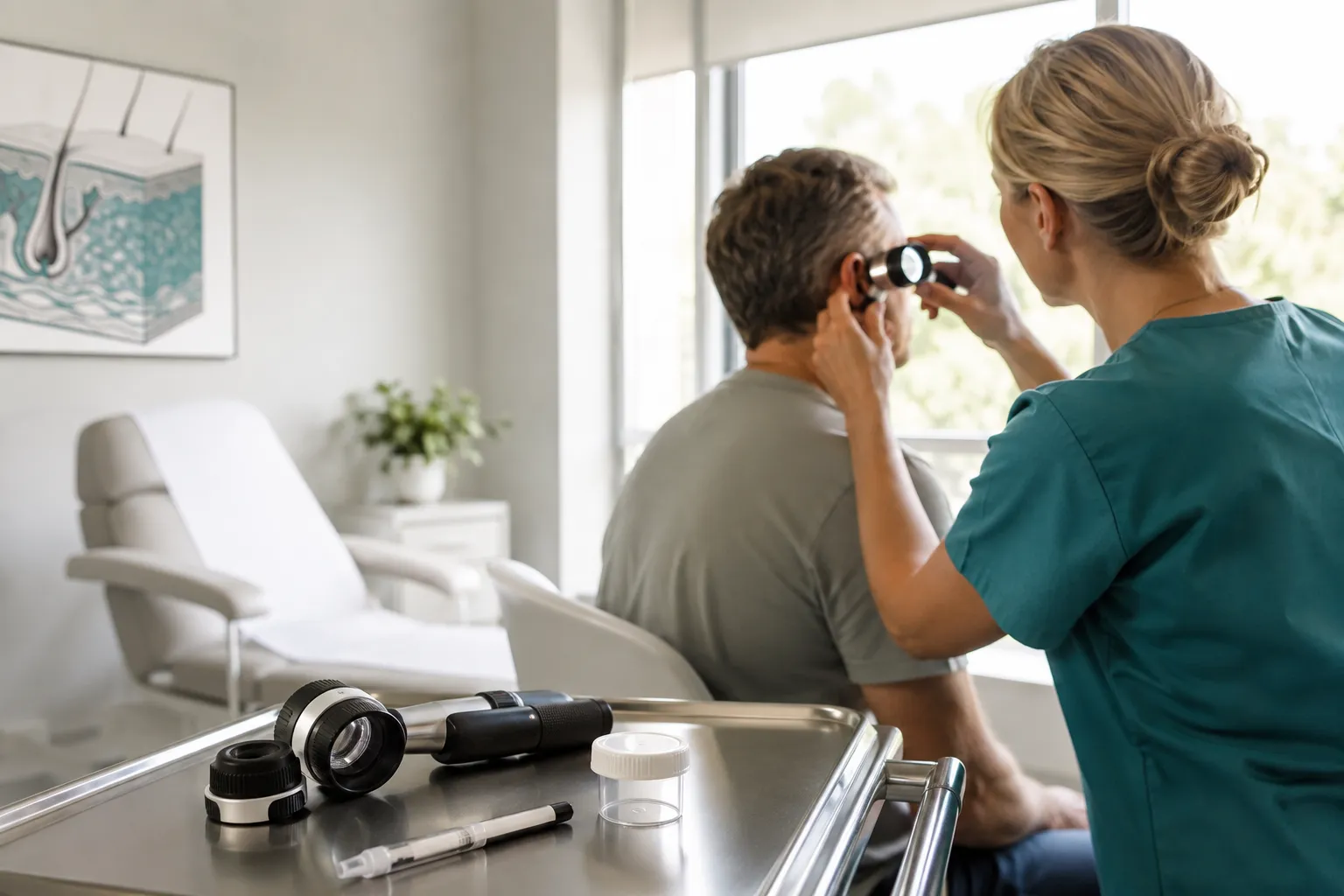
In practice, a visit covers five things. The clinician takes a history, how long the spot has been there, what's changed, past sun exposure, prior skin cancers, family history. The lesion is examined with a dermatoscope, a handheld magnifier with polarised light that shows pigment and vessel patterns the naked eye cannot see. The lesion is then risk-stratified: reassure and monitor, biopsy, or excise. If tissue is taken, it goes to a laboratory. If cancer is confirmed, the clinic either completes the definitive excision or refers.
A clear line is worth drawing here. Laser and cosmetic clinics can change how a lesion looks. They cannot diagnose it. A lesion treated with a laser or liquid nitrogen without pathology is a lesion whose identity is now permanently unknown, and if it was a carcinoma, the surface may heal while tumour continues underneath. If you're weighing where to go, our overview of how to find a skin cancer clinic in Toronto walks through what to ask.
What this kind of clinic does not do: chemotherapy, immunotherapy, radiation, sentinel lymph node biopsy, or the management of advanced or metastatic disease. Those belong to a cancer centre such as Princess Margaret or Sunnybrook's Odette Cancer Centre, and part of a clinic's job is recognising the cases that need to go there.
Choose a diagnostic clinic if the spot is new, changing, bleeding, non-healing or simply different from your other marks. Choose a cosmetic provider only after a lesion has been confirmed benign by a clinician who examined it.
The Spot That Bleeds, Scabs and Comes Back
The single most common presentation that turns out to be skin cancer is a small area that scabs, seems to heal, then breaks down again weeks later. A wound that will not stay healed is a diagnostic question, not a nuisance.
People rarely arrive worried about it. They arrive because they're tired of it. It catches on a towel. It bleeds when they shave. They've been calling it a pimple that never came to a head, a scratch that keeps reopening, or a patch of dry skin that won't respond to moisturiser. Basal cell and squamous cell carcinomas behave exactly like that, and this pattern is what our guide to skin cancer that looks like a pimple describes in more detail.
Other patterns worth an in-person look:
A pearly, shiny or translucent bump, sometimes with tiny visible blood vessels crossing it, most often on the nose, cheek or ear.
A rough, scaly or crusted patch that persists for months, feels like sandpaper, and may sting or itch.
A firm nodule that's growing, tender or not, especially on the ear, lip, scalp or back of the hand.
A dark line or band under a fingernail or toenail without a remembered injury.
A spot that simply doesn't match your other moles and marks in colour, shape, border or behaviour.
Change over time is the most useful signal you have. A mark that has looked identical for thirty years is far less concerning than one that has altered over three months. If you're unsure what "changed" means in practice, the walkthrough on how to check your own skin for early cancer detection gives a practical method.
One caution: no list, including this one, can clear a spot. Benign things bleed and scab too. Seborrheic keratoses, angiomas and irritated moles all mimic cancer, and cancers all mimic them back. That's what the examination and, where indicated, the biopsy are for.
Skin Cancer on the Face, Nose, Ear, Eyelid and Lip
The majority of basal cell carcinomas occur on the head and neck, because that's where cumulative sun exposure is highest across a lifetime. These sites are treated differently from a lesion on the back or shoulder, because tissue there is limited, functionally important and unforgiving.
On the trunk, skin is loose and there's slack to borrow. On the face, there isn't. Every millimetre removed has to be accounted for, and a closure that pulls in the wrong direction can shift a feature permanently. That's why site drives the plan as much as the diagnosis does.
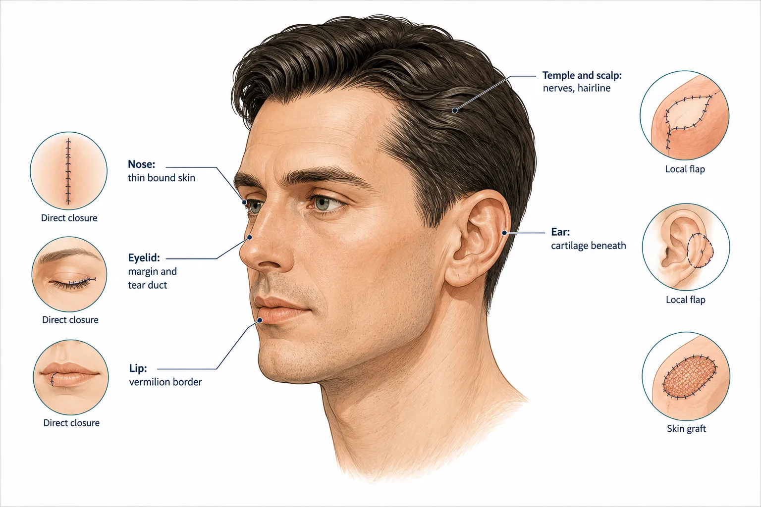
The nose. Skin over the nasal tip and ala is thin, tightly bound to underlying cartilage, and has almost no slack. Pull too hard on a closure and the nostril rim lifts or notches. Small defects here often close directly, but a defect over the tip or rim frequently needs a local flap that borrows tissue from the cheek or nasal sidewall, or a graft. Lesions here also have a habit of extending further than they look, which is covered in our detailed page on skin cancer on the nose and how it's diagnosed and treated.
The ear. Cartilage sits directly beneath thin skin with very little fat in between. Grafts don't take reliably on bare cartilage, so the closure plan has to be decided before the first cut, not after. Squamous cell carcinomas on the ear and its helical rim are also considered higher-risk than the same tumour on a limb, which affects margin planning and follow-up.
The eyelid. The lid margin, lash line and tear drainage system are all within a few millimetres of each other. A closure that shortens the lid vertically can turn the lid outward or inward, and either causes chronic irritation or watering. Lesions at the inner corner sit close to the tear duct. Eyelid margin and canthal lesions are commonly referred for specialist oculoplastic or Mohs care rather than excised in a general setting.
The lip. The vermilion border, the crisp line where lip meets skin, is visible from across a room, and a closure that misaligns it by a millimetre reads as an obvious asymmetry. The border is marked before local anaesthetic goes in, because the injection distorts the tissue. Lower-lip lesions also carry more squamous cell carcinoma risk than upper, and lip cancer behaves more assertively than a comparable tumour on the cheek.
The temple and scalp. Branches of the facial nerve run superficially at the temple, so depth matters. On the scalp, skin is thick and stiff with little stretch, and the hairline determines how visible a scar will be. Scalp lesions hide under hair for years and are often found by a barber or hairdresser before the patient notices, which is why the material on skin cancer on the scalp emphasises deliberate checking rather than waiting for symptoms.
On the face, the removal is half the operation. The reconstruction is the other half, and it should be planned before the lesion comes out.
The reconstruction options are straightforward to describe and less straightforward to choose between. Direct closure brings the edges together and works when there's enough local slack and the scar can be hidden in a natural crease or wrinkle line. A local flap moves adjacent skin into the defect on its own blood supply, which matches colour and texture well and is often the best answer on the nose or cheek. A skin graft takes skin from elsewhere, behind the ear, the neck, the collarbone, and is used when there simply isn't tissue to borrow, accepting that colour and texture match are less exact.
The practical point: at these sites, having a plastic surgeon involved from the outset changes what's possible. At the Dundas Street West clinic, GP dermatologists handle assessment and diagnosis, and plastic surgeons perform excisions and closures. That matters when a lesion on the nasal rim turns out to need removing and the closure has to be designed, not improvised.
Mohs Surgery: What It Is, When It Is Used, and When You Are Referred Out
Mohs micrographic surgery removes a skin cancer in stages, checking the margins under a microscope during the operation, so the surgeon only takes more tissue where tumour is still present. It spares the maximum amount of healthy skin, which is why it's favoured for tumours on the nose, eyelid, ear and lip.
Here's how it works in plain terms. The visible tumour is removed with a narrow margin. That tissue is processed and mapped on site while you wait, usually with a bandage on and often over several hours. The surgeon reads the edges. If tumour reaches an edge, another thin layer is taken from that specific direction only. The cycle repeats until the margins are clear. Then the defect is repaired.
When Mohs is typically preferred:
Tumours on the central face, nose, eyelids, lips, ears, and around the eyes
Recurrent tumours, or those that were previously treated and came back
Aggressive histological subtypes, infiltrative, morpheaform or micronodular basal cell carcinoma, for example
Lesions with poorly defined clinical borders
Large tumours, or those in patients who are immunosuppressed
Sites where sparing every millimetre of tissue changes the functional result
When standard surgical excision is entirely reasonable: a well-defined, low-risk basal cell carcinoma on the trunk, arm or leg, where there's tissue to spare and a wider margin costs nothing functionally. Most non-melanoma skin cancers fall into this group, and standard excision with margins is the accepted treatment.
Now the honest part. Mohs requires a fellowship-trained Mohs surgeon plus an on-site histology lab, and not every clinic, including this one, provides it. Recognising which cases should be referred to a Mohs surgeon rather than excised locally is part of competent care, not a failure of it. In Ontario, Mohs is available through a limited number of hospital and academic centres, and access is through referral.
What to ask at your consultation:
Given the site and the biopsy result, is standard excision appropriate here, or should this go to a Mohs surgeon?
Who will do the closure, and what type of closure is planned?
What happens if the pathology comes back with a positive margin?
If a re-excision is needed, will it be done here or elsewhere?
A clinician who answers those four questions directly is giving you what you need. If you've already been given a plan you're uncertain about, our discussion of when to seek a second opinion on skin cancer covers reasonable grounds for asking.
Skin Cancer in Skin of Colour: The Assumption That Delays Diagnosis
Skin cancer is less common in people with brown and Black skin, but it's more often found at a later stage, and later diagnosis, not different biology alone, is a major reason outcomes can be worse. Melanin provides meaningful but partial protection. It doesn't provide immunity.
This matters across Etobicoke and west Toronto, where many residents have South Asian, Caribbean, African, Eastern European or Filipino backgrounds and may have been told, sometimes by a clinician, that skin cancer isn't something they need to think about. That advice is out of date, and the practical consequence is that people wait.
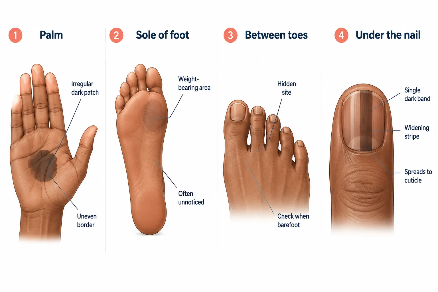
The pattern of where it appears is different, and that's the useful thing to know. In darker skin, melanoma more often shows up on sites with little or no sun exposure:
The palms of the hands
The soles of the feet, including weight-bearing areas
Between the toes
Under fingernails and toenails
Inside the mouth, and on mucosal surfaces
These are places people don't associate with sun and rarely examine. A dark patch on the sole gets attributed to a callus or a stain. A stripe under a nail gets attributed to a knock from months ago.
Acral lentiginous melanoma, in plain terms, is a melanoma that arises on the palms, soles or nail units. It often begins as a flat brown or black patch with an uneven border and uneven colour, sometimes with a raised area developing later. It's not caused by sun exposure, and it occurs across all skin tones, but it makes up a larger share of melanomas diagnosed in people with darker skin, largely because the sun-driven types are less frequent.
A dark band running lengthwise under a single nail deserves assessment rather than an assumption. Features that raise concern include a band that involves one nail rather than several, one that is widening or becoming more irregular over time, one with varying shades of brown and black rather than a uniform colour, and one where pigment extends onto the cuticle or surrounding skin. Nail changes with splitting, thinning or distortion of the nail plate also warrant a look. A true bruise under a nail grows out with the nail over weeks to months; pigment that stays put in the same position doesn't behave that way. None of this means a dark band is cancer, most aren't, but it does mean the difference is decided in a clinic, not at home.
Basal cell and squamous cell carcinomas also occur in darker skin, and they can look different from the pictures in textbooks and on websites. Basal cell carcinomas may appear pigmented, brown, blue-grey or black, rather than the classic pearly pink, and can be mistaken for a benign mole or seborrheic keratosis. Squamous cell carcinomas in Black patients more often arise in areas of chronic scarring, chronic inflammation or long-standing wounds rather than on the most sun-exposed skin. The comparison in our article on skin cancer in fair versus dark skin sets these differences out side by side.
Two practical points. First, sun protection is worthwhile at every skin tone, sunscreen, shade, hats and long sleeves reduce cumulative damage regardless of how easily you burn, and any suggestion otherwise is wrong. Second, add the soles, the spaces between your toes, and your nails to whatever skin check you already do. Those are the sites most often skipped.
What Happens at an Assessment, Step by Step
An assessment usually runs 20 to 40 minutes and ends with a clear recommendation: reassure and monitor, biopsy, or remove. Nothing is diagnosed as cancer on the day without tissue.
The sequence is consistent:
History. How long the spot has been present, what has changed and over what period, whether it bleeds or itches, previous skin cancers, family history of melanoma, sunburns and tanning history, outdoor work, immunosuppressant medication or a transplant, and any prior radiation to the area.
Examination. Focused on the lesion of concern, with a look at surrounding skin. A full-body skin examination is appropriate if you have many atypical moles, a personal or family history of melanoma, or heavily sun-damaged skin.
Dermoscopy. The dermatoscope shows pigment networks, vessel patterns and structures beneath the surface. It changes management often, it identifies lesions that look alarming but are clearly benign, and flags ones that look bland but aren't.
Photography. Lesions are photographed with a measurement reference so the next visit compares like with like. Memory is unreliable about size; photographs aren't.
Risk stratification and plan. The clinician explains what the lesion most likely is, how confident that assessment is, and what happens next. If a biopsy or excision is indicated and the schedule allows, it can often be done in the same visit.
Common mistake: applying makeup, concealer or self-tanner to the area before the appointment, which obscures colour and vessel detail. Come with the area clean.
Biopsy and Pathology: How the Diagnosis Is Made
A skin cancer diagnosis is made by a pathologist examining tissue, not by a clinician looking at skin. That's true no matter how experienced the examiner or how classic the appearance.
Three approaches are used, chosen by lesion type and site. A shave biopsy takes a thin horizontal slice and suits raised, clearly surface-level lesions. A punch biopsy uses a small circular blade to take a full-thickness core, useful for flat or thick lesions and for rashes. An excisional biopsy removes the whole lesion with a narrow margin and is generally preferred for suspected melanoma, because depth of invasion determines staging and a partial sample can underestimate it. Our overview of how a mole biopsy and skin cancer screening work goes into more detail on each.
All of these are done under local anaesthetic. The injection stings for a few seconds; the procedure itself shouldn't hurt. Most sites need a stitch or two, or none at all.
The pathology report names the lesion, and for a malignancy typically describes the subtype, how deep it goes, whether margins are clear, and features that affect risk such as perineural invasion. In Ontario, routine dermatopathology turnaround is commonly around one to three weeks, sometimes longer if additional stains or a second opinion are needed.
The point that matters most: freezing or lasering a lesion destroys it and leaves nothing to test. A suspected skin cancer is excised, not burned off, because the tissue is the evidence. If a lesion was treated cosmetically and has come back, tell the clinician, that history changes the plan.
After a Diagnosis: Margins, Re-Excision and Follow-Up
A diagnostic biopsy is deliberately narrow, so a wider excision often follows once the tumour type and subtype are known. Margins are the ring of normal-looking tissue taken around the tumour, and how wide they need to be depends on what the pathology says.
The logic is simple. A biopsy answers "what is this?" with as little tissue as possible. Definitive treatment answers "is it all out?" and needs a margin. For basal and squamous cell carcinomas the required margin depends on subtype, size and site; for melanoma, margins are set by the measured depth of invasion. Once the report is in, the second procedure is planned properly, with the right margin and the right closure, rather than guessed at.
Some cases move to a cancer centre. Referral is usual for melanoma beyond a thin lesion, where staging investigations or a sentinel lymph node biopsy may be discussed; for squamous cell carcinoma with high-risk features such as perineural invasion or nodal involvement; for tumours too large or too deeply invasive for office-based surgery; and where radiation or systemic therapy is on the table. In the Toronto region that generally means Princess Margaret Cancer Centre or Sunnybrook's Odette Cancer Centre.
Now the part most pages skip.
A first skin cancer is a marker, not an endpoint. Having had one basal or squamous cell carcinoma substantially raises the likelihood of developing another, and reported studies consistently show a meaningful proportion of patients develop a second non-melanoma skin cancer within a few years. The same applies after a melanoma. The reason is straightforward: the field of skin that produced one tumour has the same accumulated damage across the rest of it.
What surveillance actually looks like. Follow-up should be scheduled rather than left for you to remember. In practice that often means a check at around three to six months after treatment, then every six to twelve months, with the interval set by tumour type, site, whether margins were clear, and how much sun damage is present overall. Melanoma follow-up is usually more frequent and longer-running, and is directed by the treating specialist.
What's being looked for at each visit. Three things: recurrence at the original scar, new primary lesions elsewhere, and, for higher-risk squamous cell carcinoma and melanoma, the regional lymph nodes. Scars are palpated as well as inspected, since recurrence can be felt as firmness beneath a scar that looks fine.
What you're being asked to do between visits. Watch the treated area and the surrounding skin. A firm nodule at or beside a scar, a scar that starts to bleed or ulcerate, or new numbness or tingling in the area are all reasons to come back before the scheduled appointment rather than after. Actinic keratoses, the rough, scaly patches that often accompany sun damage, are also monitored, since a small proportion progress; our explainer on actinic keratosis and how it relates to skin cancer describes what to look for.
Edge case worth naming: patients on immunosuppressive medication and organ transplant recipients develop skin cancers more often, faster, and with more aggressive behaviour. Surveillance intervals for that group are shorter, and any new lesion should be assessed promptly rather than watched.
Do You Need a Referral, and Is Any of This Covered by OHIP?
No referral is needed to book at The Minor Surgery Center. Some dermatology practices in Etobicoke and Toronto require a family physician referral, and some walk-in dermatology clinics take patients directly on a first-come basis, so it's worth confirming before you drive anywhere.
On coverage: in Ontario, assessment and treatment of a lesion that is clinically suspicious for skin cancer is handled differently from a cosmetic request. A medically necessary consultation and the treatment of a suspected malignancy are generally insured services when performed by a physician who bills OHIP. Removal of a lesion for appearance alone is not insured, and the patient pays privately.
Routine whole-body screening requested without any clinical indication sits in a grey area and may not be insured. Pathology on tissue sent to a licensed Ontario laboratory is typically covered when the biopsy is medically indicated. Coverage also depends on the individual clinic's billing arrangement, not every practice bills OHIP for every service, so confirm your own situation when you book rather than assuming. If you don't have OHIP coverage, ask about the self-pay consultation fee up front.
This article is general information and not a substitute for assessment by a physician who can examine your skin.
Costs and What Drives Them
If your visit is diagnostic and the clinic bills OHIP, there's often no out-of-pocket cost for the consultation, the biopsy or the removal. Private-pay pricing applies to cosmetic removals and to patients without provincial coverage.
Where fees do apply in Ontario, broad typical ranges look roughly like this: a private consultation commonly falls in the range of about $50 to $200, and a straightforward private lesion removal often runs from roughly $200 to $600 per lesion, with more if pathology is added or the closure is complex. Treat those as general market ranges rather than a quote.
What moves the number:
Diagnostic or cosmetic. This is the single biggest factor, because it determines whether OHIP is involved at all.
Number and size of lesions. Several lesions in one sitting usually cost less per lesion than several separate visits.
Body site. Face, eyelid, ear, nose and lip take longer and need a planned closure.
Pathology. Sending tissue to a laboratory adds a lab charge when the case isn't insured.
Closure complexity. A direct closure, a local flap and a skin graft are three different operations in time and cost.
An exact figure comes from the consultation, once someone has seen the lesion. Any clinic quoting a firm price for facial surgery over the phone is guessing.
Getting Seen From Etobicoke and West Toronto
The closest location to Etobicoke is 1224 Dundas Street West, Unit 101, Mississauga, on the Dundas Street West corridor, a short drive from both Highway 427 and the QEW, with on-site parking. Phone 647-614-1611. Hours are Monday to Friday, 9 a.m. to 4 p.m. No referral is required.
From the Kingsway, Islington or Markland Wood it's a straightforward run west on Dundas. From Mimico, Humber Bay or Alderwood, the QEW to Highway 427 north connects to Dundas within minutes. From Bloor West Village, the Junction or Roncesvalles, Dundas Street West runs the whole way. The same group also has clinics in North York at 2920 Dufferin Street, in York Mills at 85 Scarsdale Road, and in Vaughan and Oakville, if one of those is an easier drive on a given day.
On the team structure. GP dermatologists carry out the assessment, dermoscopy and diagnosis. Plastic surgeons perform the excisions and closures. That combination is the reason a lesion on the nasal rim or near the lid margin can be planned properly rather than handed off, the person deciding the margin and the person designing the closure are in the same building. Lesion removal in this catchment is also covered on our page about skin lesion removal for Etobicoke patients.
Urgent care or dermatology? For a spot that is bleeding, scabbing, scaling or not healing but is otherwise stable, a dermatology or minor surgery clinic is the right destination, urgent care and emergency departments don't perform diagnostic skin biopsies or planned excisions, and you'll be redirected. Go to urgent care or the emergency department for a rapidly enlarging painful lump with fever or spreading redness, bleeding that won't stop with fifteen minutes of firm pressure, or a wound with signs of significant infection.
On wait times. Direct-access clinics that don't require a referral generally see patients within days to a couple of weeks, while referral-based dermatology in the GTA can run considerably longer, often weeks to several months for a non-urgent lesion. Call and describe the lesion; a spot that's bleeding or growing is triaged differently from a mole you'd simply like checked.
How to prepare. Three small things make the appointment better:
Leave the area clean, no makeup, concealer, foundation or self-tanner on the lesion.
Remove nail polish from all nails if a nail is your concern, not just the affected one.
Be ready to say how long it's been there and what specifically has changed. A photo from a few months ago on your phone is genuinely useful.
Wear something that gives easy access to the area, and bring your list of medications, particularly blood thinners.
Areas Served Across Etobicoke and West Toronto
Patients travel to the Dundas Street West clinic from across Etobicoke, the Kingsway, Islington-City Centre, Mimico, New Toronto, Long Branch, Alderwood, Humber Bay Shores, Markland Wood, the Royal York corridor, Rexdale and Thistletown, and from the west end of Toronto proper, including Bloor West Village, the Junction, Swansea, High Park, Roncesvalles and Parkdale. For patients further west along the lake, the same services are described on our skin cancer clinic page for Oakville and the Halton region.
Frequently Asked Questions
Do I need a referral for a skin cancer clinic in Etobicoke? Not at The Minor Surgery Center, patients can book directly by calling 647-614-1611. Some Etobicoke dermatology practices do require a family physician referral, and some walk-in dermatology clinics see patients same-day with no appointment, so confirm the requirement before travelling.
How soon can I be seen? Direct-access clinics that don't need a referral typically offer appointments within days to about two weeks. Referral-based specialist dermatology in the GTA is often longer. When you call, describe the lesion, bleeding, growing or non-healing spots are prioritised over routine mole checks.
How do I know if a spot needs checking? Get it looked at if it bleeds, scabs and reopens, has lasted more than four to six weeks without healing, is growing or changing, is a new dark or irregular mark, or is a dark band under a single nail. When you're unsure, that uncertainty is itself the reason to have it examined.
Does a biopsy hurt? The local anaesthetic stings for a few seconds as it goes in. After that, the biopsy itself shouldn't be painful. Most people describe pressure or tugging rather than pain, and the site is usually mildly sore for a day or two afterwards.
How long do results take? Routine skin pathology in Ontario commonly comes back within about one to three weeks. Cases needing extra stains or a second pathologist's review can take longer. You should be told how results will reach you and roughly when, before you leave the appointment.
Can the lesion be removed the same day? Often, yes. When a lesion is clearly suitable for removal and the schedule allows, consultation and excision are completed in one visit. Larger lesions, lesions on the eyelid, nose or lip, and cases where the pathology result should guide the margin are usually booked as a second appointment.
Will it leave a scar on my face? Any excision leaves a scar. The realistic aim is a scar that's fine, flat and placed along a natural crease or feature line so it becomes hard to notice over months. Facial closures are planned specifically for this, and no clinician can promise a particular cosmetic result in advance.
What is Mohs surgery, and will I need it? Mohs micrographic surgery removes a skin cancer in stages while margins are examined under a microscope during the procedure, sparing the most healthy tissue. It's typically used for recurrent tumours, aggressive subtypes, and lesions on the nose, eyelid, ear and lip. Most non-melanoma skin cancers don't require it, and it's provided at a limited number of Ontario centres by referral.
Can people with darker skin get skin cancer? Yes. It's less common in brown and Black skin, but it's more often diagnosed at a later stage. It appears more frequently on the palms, soles, between the toes, under nails and inside the mouth, and any persistent new dark patch or changing nail stripe at those sites should be assessed.
What happens if it turns out to be cancer? For most basal and squamous cell carcinomas, treatment is a surgical excision with an appropriate margin and a planned closure, completed in an office setting, followed by scheduled follow-up checks. Melanoma and high-risk squamous cell carcinoma are referred to a cancer centre for staging and any further treatment. You'll be told the diagnosis, the plan and who's doing what.
The Bottom Line
A spot that will not heal is a diagnostic question, and the answer comes from pathology, not from looking at it, and not from reading about it. That applies whether the mark is on your nose, your scalp, the sole of your foot, or under a fingernail, and it applies at every skin tone.
The next step is a single phone call. The Minor Surgery Center location nearest Etobicoke and west Toronto is at 1224 Dundas Street West, Unit 101, Mississauga, reachable at 647-614-1611, Monday to Friday from 9 a.m. to 4 p.m. No referral needed. Come with the area clean, know roughly how long it's been there, and let someone examine it properly.
Meta title: Skin Cancer Clinic Etobicoke: Assessment, Biopsy & Removal
Meta description: Skin cancer clinic serving Etobicoke and west Toronto. Dermoscopy, biopsy and surgical removal with closure, no referral needed. Call 647-614-1611 to book.
Tags: skin cancer clinic Etobicoke, skin cancer screening Toronto, mole biopsy Ontario, basal cell carcinoma, squamous cell carcinoma, melanoma, Mohs surgery, skin cancer in skin of colour, acral lentiginous melanoma, facial skin cancer reconstruction, skin lesion removal, OHIP skin cancer coverage
The Minor Surgery Center’s York Mills clinic, located at 85 Scarsdale Rd, Unit 101 in North York, offers convenient access to specialized surgical care for a range of skin and hand conditions. Our Plastic Surgeons provide treatment for skin cancer, suspicious moles, cysts, lipomas, carpal tunnel syndrome and trigger finger, with a focus on timely care and personalized treatment. In many cases, patients may be able to have their consultation and treatment completed during the same visit when medically appropriate. For expert care in a welcoming clinical setting, contact our York Mills team at (647) 614-1611.
We are the experts in the field of Minor Surgery. All our Surgeons have completed 5 years of post Medical School Surgical Residencies. They are all Board Certified by the Royal College of Physicians and Surgeons of Ontario.They have been trained at Institutions including McGill, McMaster, Queen's, University of British Columbia, University of Toronto, University of Western Ontario, The Mayo Clinic, University of Pennsylvania, University of Southern California, Yale University, and Oxford.
In many clinics, you could wait months—sometimes over a year—for a procedure like cyst or lipoma removal, mole removal, or even carpal tunnel release. But at TMSC, we prioritize fast access to care. In many cases, we can schedule and complete your minor surgery on the same day as your initial consultation—no extra runaround required.
Our clinics are designed with your comfort, safety, and efficiency in mind. We use strict sterilization protocols to minimize any infection risk, and our advanced surgical equipment meets hospital-grade standards. Yet, you’ll never feel like you’ve stepped into a stark, clinical space—our friendly staff ensures you’re welcome and supported the moment you walk through the door.
Cosmetic results matter—even for the smallest procedures. Skin lesions can be excised by non surgeons in Ontario. Furthermore, any MD with 2 years of post graduate training even outside of a surgical field can perform removal. At TMSC we only have Surgeons under our roof. They are trained well above the level of the procedures we offer to ensure the surgery goes smoothly with minimal scaring and preserving your natural contours. Our Surgeons are trained to ensure both function and aesthetics are always front and center.
At TMSC, we specialize in surgical treatment of both benign and malignant skin lesions, as well as minor hand conditions. We offer significantly reduced wait times often allowing you to schedule your surgery on the same day as your consultation.
You can book your free consultation with The Minor Surgery Center in three easy ways: online through Cortico, by phone, or by requesting a callback from our team.
The initial consultation is free. During this consultation, one of our board certified surgeons will examine your condition and recommend a suitable solution.
Directly Online
Request a Callback
By Phone
By Text
Hours
Sunday
Closed
Monday
9 am — 4 pm
Tuesday
9 am — 4 pm
Wednesday
9 am — 4 pm
Thursday
9 am — 4 pm
Friday
9 am — 4 pm
Saturday
Toronto & Oakville Open Vaughan & Mississauga Closed



.avif)
%20(600%20x%20600%20px)%20(2).avif)
.avif)
%20(8).avif)
%20(7).avif)

.png)