

Last updated: September 15, 2026
Mole mapping is a whole-body photography system that tracks dozens or hundreds of moles over months and years, built for people with many moles or a personal or family history of melanoma. A mole check is a focused, same-day assessment of one or a few specific spots that look new, changed, or unusual. The right choice depends on how many moles you have, your risk history, and whether you already have a concerning lesion right now, and in many cases, the smartest path is a GP dermatologist skin check that decides which pathway you actually need.
The core difference is scope. Mole mapping documents your entire skin surface using standardized photography so a clinician can track many moles over time, while a mole check is a direct, hands-on look at one or a few specific spots you're worried about right now. One is a surveillance system; the other is a diagnostic response to a specific concern.
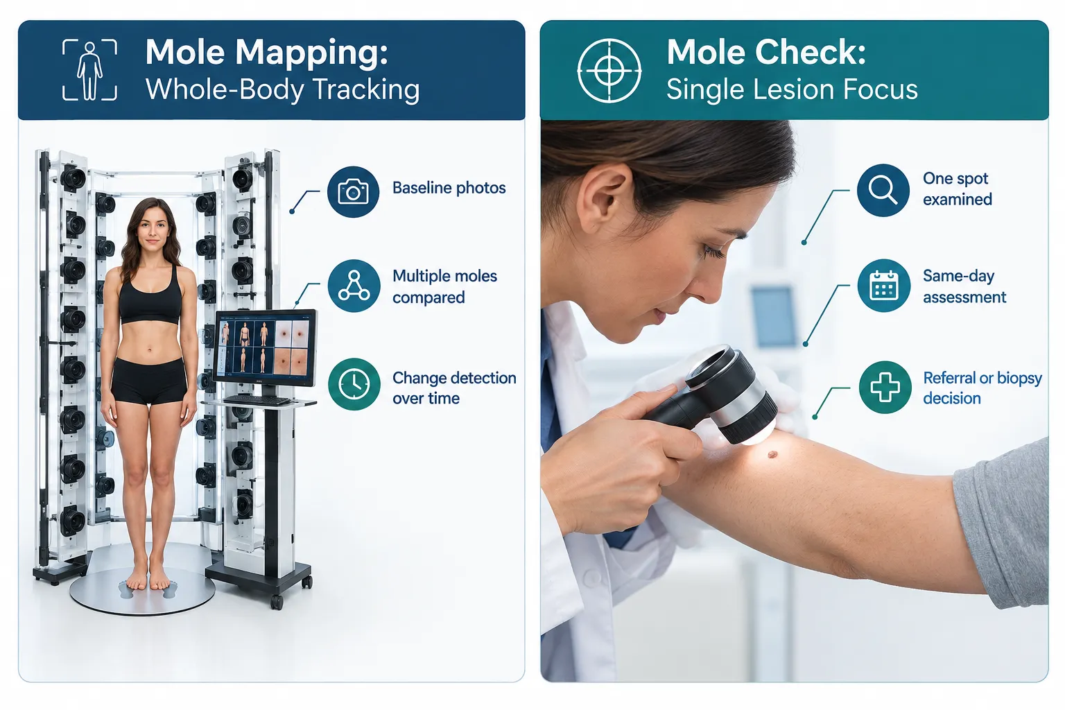
Think of it this way: mole mapping answers the question "has anything on my body changed since last year?" A mole check answers "what is this particular spot, and does it need to come off?"
Mole mapping typically involves:
A mole check typically involves:
Clinics increasingly frame this choice for patients directly as "mole mapping or skin check?", because the answer changes based on how many moles a patient has and whether there's already a specific lesion of concern [9]. If you have one new dark spot on your arm, you don't need a 90-minute full-body photography session. If you have 120 moles and a father who had melanoma, a single-spot check misses the point entirely: you need a system that remembers what your skin looked like last year.
For a deeper side-by-side look at how these two services stack up against a full-body option, see this related comparison of mole mapping versus full body mole check.
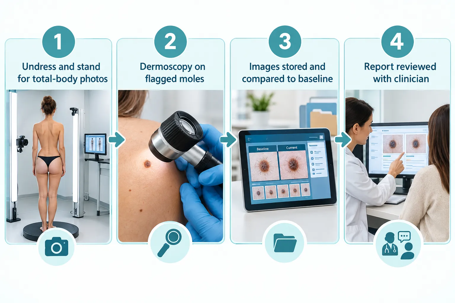
A mole mapping appointment usually takes 30 to 90 minutes and involves standing for a series of full-body photographs, followed by dermoscopy on any lesions that look different from the rest. The exact time depends on the technology used and how many moles need closer dermoscopic review.
A typical mole mapping session follows this pattern:
Follow-up mole mapping visits are usually shorter because the system only needs to re-photograph and compare against the existing baseline, not build one from scratch.
Common mistake: Patients sometimes assume mole mapping replaces the need to mention a specific worrying spot. It doesn't. If you have one lesion that's already bleeding, itching, or growing fast, say so at booking, that lesion may need dermoscopy and possible biopsy the same day, independent of the mapping schedule.
For patients whose main concern is a single changing, bleeding, or irregular spot rather than full-body tracking, a suspicious mole assessment is usually the faster route.
Most clinicians recommend an initial mole mapping baseline followed by repeat imaging every 6 to 12 months, though the exact interval depends on personal risk factors. People with a personal history of melanoma or highly atypical moles sometimes need shorter intervals, while lower-risk patients with many benign-appearing moles may space sessions out further.
Frequency generally depends on:
Decision rule: if your clinician builds a mole mapping baseline and doesn't set a specific follow-up date before you leave, ask for one. Mole mapping only works as a comparison tool, a single session with no follow-up provides a snapshot, not a change-detection system.
A mole check, by contrast, doesn't run on a fixed calendar. You book one when a specific spot changes, and the follow-up (if any) is based on that lesion's behavior, not a standing schedule.
Mole mapping is most valuable for people with more than 50 moles, atypical mole syndrome, a personal or family history of melanoma, or significant childhood sun exposure and blistering sunburns. People with few moles and no elevated risk factors generally don't need full mole mapping and are better served by an annual skin check or a targeted assessment when something changes [4][9].
Strong candidates for mole mapping:
Who typically doesn't need full mole mapping:
Is mole mapping necessary if you don't have many moles? Generally, no. If you have a handful of moles and no elevated risk factors, a standard skin check with dermoscopy on any spot of concern covers the same ground more efficiently. Mole mapping earns its value when there are too many moles to reliably track by memory or photographs on your phone. With only a few moles, a clinician can mentally track them year to year without needing a digital baseline system.
Edge case: someone with only 15 moles but one atypical-looking lesion and a parent who died of melanoma should still get a same-day mole check on that lesion, mapping the other 14 moles can come later, but the concerning one shouldn't wait for a scheduling slot.
Mole mapping can help detect melanoma earlier in high-risk patients by making subtle changes between visits easier to spot, but recent trial evidence on 3D total-body photography shows mixed results compared to usual dermatologic care. Real-world data from high-risk cohorts followed with structured mole mapping programs has shown earlier detection patterns, but mole mapping is a surveillance tool, not a diagnostic test on its own [2][3].
What the evidence actually shows:
Why this matters practically: mole mapping is a change-detection system. It works by comparing this year's images to last year's. The earlier the abnormal change is caught, the sooner a biopsy can confirm or rule out melanoma. But mole mapping itself never diagnoses melanoma, it flags candidates for closer dermoscopic review and, when warranted, biopsy.
"Mole mapping is a surveillance tool that makes change easier to catch. It is not a substitute for tissue diagnosis when a lesion looks suspicious."
If a mapped mole changes between visits, the next step is the same as it would be for a mole found any other way: dermoscopy, and if needed, biopsy. Learn more about what happens after imaging flags a concern by reviewing this guide on mole biopsy, suspicious mole testing, pathology, and removal.
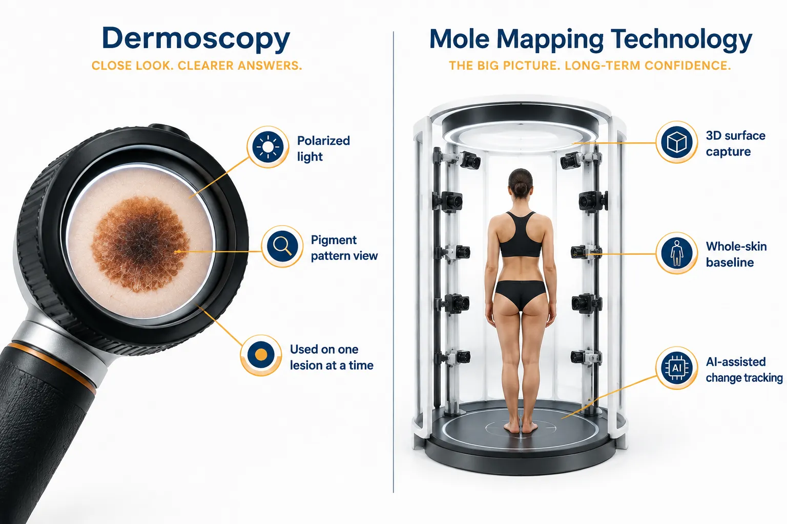
Mole mapping and dermoscopy solve different problems: mole mapping tracks change across your entire skin surface over time, while dermoscopy is a magnification tool used to examine the internal structure of a single lesion at a single point in time. Most modern mole mapping programs use both, total-body photography to find outlier moles, then dermoscopy to examine those outliers closely [6][8].
FeatureMole MappingDermoscopyMole CheckScopeEntire body, many molesOne lesion at a timeOne or a few lesionsTechnology2D/3D total-body camerasHandheld magnifying dermatoscopeVisual exam, sometimes with dermatoscopeTime needed30-90 minutesA few minutes per lesion10-20 minutes typicallyBest forTracking change over months/yearsExamining internal pigment pattern of a lesionAssessing a specific new or changing spotOutputStored baseline image archiveReal-time clinical impressionSame-day clinical decisionReplaces biopsy?NoNoNo
Decision rule: choose dermoscopy alone (as part of a mole check) if you have one lesion of concern and no large mole burden to track. Choose mole mapping if you need a system that remembers all your moles well enough to notice when one of them starts behaving differently than the rest.
A common misunderstanding is that dermoscopy and mole mapping are competing options. In practice, they're complementary, dermoscopy is often the second step within a mole mapping session, applied to any mole the photography system or clinician flags as an outlier [8].
The most advanced mole mapping systems now use 3D total-body photography combined with AI-assisted lesion comparison, which can automatically flag moles that look statistically different from a patient's other moles. Adoption of these 3D platforms has grown in major dermatology centers, and newer AI-driven systems introduced in 2025 and 2026 aim to reduce the manual comparison workload for clinicians [3][7].
Categories of mole mapping technology:
Practical trade-off: more advanced technology generally means more consistent image capture and easier year-over-year comparison, but it doesn't replace the need for a clinician to interpret the results and decide whether dermoscopy or biopsy is warranted. A high-tech camera system with no clinical follow-through is just a photo archive.
If you're evaluating clinics on their imaging technology specifically, this roundup of 15 mole mapping clinics in Toronto breaks down what different providers offer in terms of camera systems and follow-up protocols.
Home mole-mapping apps and personal photography can help you track visible changes between professional visits, but they cannot replace a clinician's dermoscopy, clinical judgment, or biopsy decision-making. Apps are a monitoring aid, not a diagnostic tool, and image quality from phone cameras varies too much to reliably detect the subtle changes that matter [8][10].
What home tracking can reasonably do:
What home tracking cannot do:
Recent independent reviews of consumer 3D mole-mapping apps have found meaningful limitations in image consistency and lesion-matching accuracy compared to clinical systems, which matters because inconsistent images make year-over-year comparison unreliable [10]. For a closer look at how these consumer tools measure up, see this analysis of whether 3D mole mapping apps are reliable.
Why a dermatologist recommends professional mole mapping over just monitoring moles at home: self-monitoring relies on memory and casual observation, which works poorly once someone has more than a few dozen moles. A dermatologist-run mole mapping program standardizes lighting, angle, and distance for every photo, uses dermoscopy to examine anything unusual, and keeps a permanent digital record that doesn't degrade the way memory does. For patients with high mole counts or risk factors, that structural consistency is the entire point of the service.
Decision rule: use home photo-tracking as a supplement between professional visits, never as a substitute for them. If a mole changes noticeably between clinic visits, don't wait for the next scheduled mapping session, book a mole check.
Mole mapping generally costs more than a single-lesion mole check because it involves more equipment, more time, and a stored digital archive, while a basic mole check is often faster and cheaper since it targets one or a few spots. Insurance and public health coverage for either service varies significantly by region and by whether the visit is framed as medically necessary versus elective screening.
Cost factors to expect:
Is mole mapping covered by insurance? Coverage depends heavily on jurisdiction and clinical justification. In many public health systems, a medically necessary assessment of a specific suspicious lesion is more likely to be covered than routine full-body mapping done purely for peace of mind, though patients with documented high-risk status (melanoma history, atypical mole syndrome) sometimes qualify for covered surveillance imaging. Always confirm coverage specifics with your clinic and insurer before booking, since policies change and vary by provider network.
Practical trade-off: if cost is the deciding factor and you don't have significant risk factors, a mole check on your specific concern is usually the more economical starting point. If you have dozens of moles and real risk factors, the higher upfront cost of mole mapping is often justified by the long-term value of a comparison baseline, a single mole check appointment can't give you that.
For patients weighing options without a referral, see this overview of rapid-access mole check clinics in Toronto that don't require a GP referral to book.
If mole mapping flags a mole as changed, new, or statistically different from your other moles, the next step is dermoscopy on that specific lesion, followed by a biopsy if the clinician still has concerns after magnified examination. Mole mapping identifies candidates for closer review; it does not itself confirm or rule out melanoma.
Typical pathway after a flagged finding:
Common mistake: assuming a flagged mole automatically means cancer. Most flagged lesions in mole mapping programs turn out to be benign on closer review, the flagging system is designed to be sensitive (catch anything unusual) rather than specific (only flag true cancers), which is intentional, since missing a melanoma is far worse than over-flagging a benign mole [2].
If your mole mapping report flags something, don't wait for your next scheduled mapping session to act on it. Book a focused follow-up. Understanding what a pathology report actually says can also help you interpret the outcome; this breakdown of demystifying your pathology report after mole removal walks through common terminology.
The Minor Surgery Center (TMSC) addresses both sides of this decision by offering GP dermatologist skin checks for assessment and a surgical pathway for biopsy or removal once a lesion needs tissue sampling. This structure avoids sending patients through unnecessary extra steps between screening and treatment.
How the pathway typically works at TMSC:
This model is designed around the reality that most patients don't walk in needing full-body mole mapping, they walk in with a specific spot they're worried about. For those patients, a same-day, GP dermatologist-led mole check assessment with a clear route to biopsy if needed is often more useful than a longer mapping session built for tracking many moles.
Who this fits best: patients with one or a few concerning lesions who want assessment and, if necessary, removal without bouncing between multiple providers. Patients whose primary need is full-body tracking of many moles over years may still benefit from a dedicated mole mapping program elsewhere or in combination with periodic TMSC skin checks on any specific lesion that changes.
Choosing between the two comes down to how many moles you have, your risk history, and whether you already have a specific lesion of concern right now. Use the checklist below as a starting point, then confirm with a clinician.
Choose mole mapping if:
Choose a mole check if:
Choose both, in sequence, if:
Quick example: A 45-year-old with 80 moles and no family history books mole mapping for a baseline, then notices a new dark spot on their calf six months before their scheduled follow-up. The correct move isn't to wait for the mapping appointment, it's to book a targeted mole check on that spot right away, since a fast-growing lesion shouldn't sit on a calendar built for routine surveillance.
Is mole mapping the same as a skin cancer screening?
No. Mole mapping is a tracking and comparison tool used within a broader skin cancer screening approach. A full skin cancer screening can include a mole check, dermoscopy, and mole mapping depending on your risk profile.
Do I need a referral to get a mole check?
Not always. Some clinics, including certain rapid-access providers, offer mole checks without requiring a GP referral first, which can shorten the wait for patients with an urgent concern.
How many moles is "too many" to track without mapping?
There's no strict number, but once you have more than roughly 50 moles or moles that look similar to each other, tracking them by memory becomes unreliable, and mole mapping starts to add real value [4].
Can a mole check turn into a mole mapping session on the same visit?
Typically no, since mole mapping requires dedicated equipment and time for full-body photography. Most clinics book these as separate appointment types, though a clinician may recommend mole mapping as a next step after a mole check.
Does a normal mole mapping result mean I'm in the clear until next year?
It means no concerning changes were found at that visit. New moles or changes can still appear between sessions, so any new or changing spot should still be checked promptly rather than waiting for the next scheduled mapping visit.
Are 3D mole mapping apps as good as clinic-based systems?
Independent reviews suggest consumer 3D mole-mapping apps have real limitations in image consistency and lesion-matching accuracy compared to professional systems, making them useful as a supplement but not a substitute for clinical mole mapping [10].
What's the difference between a mole check and a biopsy?
A mole check is the assessment step, a clinician looks at the lesion and decides what to do next. A biopsy is a procedure where tissue is removed and sent to pathology for a definitive diagnosis. A mole check may or may not lead to a biopsy.
If I've had melanoma before, do I need mole mapping instead of regular checks?
Most clinicians recommend structured mole mapping in addition to, not instead of, regular clinical checks for melanoma survivors, since combining photographic tracking with hands-on exams offers more complete surveillance than either alone.
Mole mapping and a mole check solve different problems, and confusing the two leads either to unnecessary appointments or missed opportunities to catch something early. Mole mapping is a surveillance system built for patients with many moles or elevated risk who need year-over-year comparison. A mole check is the right tool when one or a few specific lesions need a clinical opinion now.
Next steps:
Patients who want a GP dermatologist opinion on a specific mole, with a direct path to biopsy or removal if needed, can start with a full-body skin cancer screening and mole assessment to determine the right next step.
[1] Total Body Photography - https://spotdoc.com/clinical-guides/total-body-photography
[2] jamanetwork - https://jamanetwork.com/journals/jamadermatology/fullarticle/2831500
[3] Total Body Photography - https://oncodaily.com/oncolibrary/total-body-photography
[4] Mole Mapping - https://www.uchicagomedicine.org/conditions-services/dermatology/treatments-and-services/mole-mapping
[6] Difference Between Mole Mapping Vs Mole Check - https://dermasurge.co.uk/difference-between-mole-mapping-vs-mole-check/
[7] Optimascan Is Here - https://spotcheck.clinic/optimascan-is-here/
[8] Mole Mapping Total Body Photography - https://www.dermadex.ca/blog/mole-mapping-total-body-photography
[9] Mole Mapping Or Skin Check A Guide To Smarter Screening - https://cliffordclinic.com/mole-mapping-or-skin-check-a-guide-to-smarter-screening/
[10] pubmed.ncbi.nlm.nih.gov - https://pubmed.ncbi.nlm.nih.gov/41167046/