Last updated: July 15, 2026
Skin Cancer Removal in Burlington, Milton & Halton Region
Skin cancer removal in Burlington, Milton and Halton Region typically involves an initial lesion assessment, a biopsy to confirm diagnosis, and a surgical excision once pathology results are back. Treatment depends on the type of skin cancer, its size and location, and physician assessment, and most straightforward basal cell and squamous cell carcinoma excisions are covered by OHIP with a valid referral. Melanoma or complex cases are often referred to specialized surgical or oncology teams.
Key Takeaways
- Skin cancer removal is a staged process: assessment, biopsy, pathology, then surgical excision when appropriate.
- Basal cell carcinoma (BCC) and squamous cell carcinoma (SCC) are the two most common types treated through standard excision in the Halton area.
- Suspicious moles or possible melanoma need biopsy and pathology first; treatment pathways depend entirely on the diagnosis.
- OHIP generally covers medically necessary skin cancer removal when a physician confirms the lesion is not purely cosmetic.
- Mohs micrographic surgery is now available locally in Burlington for certain high-risk or recurrent skin cancers [1][2].
- Recovery from a standard excision usually takes one to three weeks for the surface wound to heal, though full scar maturation takes longer.
- Skin cancer can return after removal, especially with incomplete margins, chronic sun exposure, or a history of multiple skin cancers.
- Residents of Oakville, Burlington, Milton, Georgetown, Hamilton and the wider Halton Region can access assessment and surgical removal without traveling into downtown Toronto for every step of care.
Skin Cancer Removal in Burlington, Milton and Halton Region: What to Expect
Skin cancer removal in Burlington, Milton and Halton Region generally follows a defined clinical pathway: a physician assesses the lesion, arranges or performs a biopsy if the diagnosis is unclear, reviews the pathology report, and then plans surgical removal based on what the biopsy shows. This is different from a routine mole check because the starting point is usually a specific concern, a spot that has changed, bled, or already come back with an abnormal biopsy result.
Patients typically reach this stage in one of three ways:
- A family doctor or walk-in clinic noticed a suspicious lesion and referred the patient for assessment.
- A biopsy has already been done elsewhere, and the pathology report confirms basal cell carcinoma, squamous cell carcinoma, or another skin cancer.
- The patient has noticed a mole or patch of skin that is changing shape, color, or size and wants it checked before it becomes a bigger problem.
Unlike a general skin cancer clinic visit focused purely on screening, this stage of care is about action: confirming the diagnosis and moving toward removal when removal is the right next step. GP dermatologists are typically the ones who assess suspicious moles and lesions where applicable, while plastic surgeons perform the biopsies, excisions, and surgical removals once a treatment plan is agreed upon.
For patients across Oakville, Burlington, Milton, Georgetown, Hamilton and the broader Halton Region, having a local pathway for this, rather than a long wait for a hospital dermatology referral, can shorten the time between "something looks wrong" and "it's been removed and checked."
When Is Skin Cancer Removal Recommended?
Skin cancer removal is recommended when a biopsy confirms a malignant or pre-malignant lesion, or when a lesion has features that are highly suspicious for skin cancer even before biopsy results return. The decision to remove a lesion is never made on appearance alone, it's based on clinical exam findings, dermoscopy, and, in most cases, tissue pathology.
There are a few situations where removal is typically discussed:
- Confirmed diagnosis. A biopsy has already shown basal cell carcinoma, squamous cell carcinoma, or another cancerous or pre-cancerous change (such as actinic keratosis progressing toward SCC).
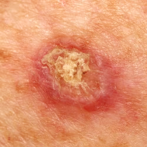
- High clinical suspicion. The lesion has classic warning signs, irregular borders, uneven color, rapid growth, bleeding, or crusting that doesn't heal, even before a formal biopsy is done.
- Prior skin cancer history. Patients who have had one skin cancer removed have a meaningfully higher chance of developing another, so new or changing spots get closer attention.
- Recurrence at a prior excision site. A scar or area that was previously treated and now shows new changes needs prompt reassessment.
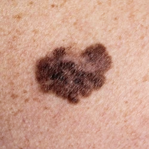
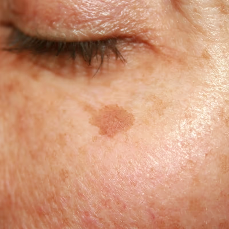
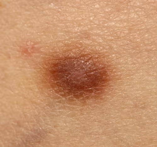
When should you get a mole checked for cancer? A mole should be checked promptly if it shows any of the "ABCDE" warning signs: Asymmetry, Border irregularity, Color variation, Diameter growth beyond 6mm, or Evolution in size, shape, or texture over weeks to months. Bleeding, itching, or a sore that won't heal after several weeks are also reasons to get it looked at rather than waiting for a routine annual check.
Decision rule: if a spot has changed noticeably in the last one to three months, or it bleeds without an obvious injury, book an assessment rather than monitoring it at home. Waiting is reasonable for stable, long-standing moles that haven't changed; it is not reasonable for anything new or evolving.
Common mistake: assuming a mole is "probably fine" because it doesn't hurt. Most early skin cancers, including melanoma, are painless. Pain is a poor indicator of risk, visual and textural changes matter far more.
Types of Skin Cancer We Commonly Assess
The three skin cancers most commonly assessed and treated in this region are basal cell carcinoma, squamous cell carcinoma, and suspicious moles that may represent melanoma. Each behaves differently, grows at a different pace, and carries a different level of urgency, which is why diagnosis has to come before any treatment plan is discussed.
Basal Cell Carcinoma Removal
Basal cell carcinoma removal usually involves surgical excision of the lesion along with a small margin of healthy-looking skin, based on the size, depth, and location confirmed by biopsy. BCC is the most common skin cancer overall, and it rarely spreads to other organs, but it can grow locally into deeper tissue if left untreated for a long time.
Typical factors that affect the removal plan:
- Size and depth of the lesion on imaging or clinical exam.
- Location, areas like the nose, ears, and eyelids often require more careful planning to preserve function and appearance.
- Subtype confirmed on biopsy, since some BCC subtypes are more aggressive than others.
- Prior treatment history, including any earlier excisions in the same area.
For a deeper look at what recovery looks like after this specific procedure, see this guide on basal cell carcinoma recovery and what to expect after removal. More background on the condition itself is available on the basal cell carcinoma removal and treatment page.
Squamous Cell Carcinoma Removal
Squamous cell carcinoma removal follows a similar surgical logic to BCC removal but often carries slightly more urgency, since SCC grows faster and carries a higher, though still relatively low, risk of spreading if untreated. Diagnosis is confirmed by biopsy before any excision is planned.
Key differences worth knowing:
- SCC often develops from long-standing sun damage or actinic keratosis, so patients with a history of these changes are monitored more closely.
- Larger or deeper SCC lesions, or those near lymph node-rich areas, may need more thorough surgical margins and follow-up.
- Immunosuppressed patients (organ transplant recipients, for example) are treated with extra caution because SCC can behave more aggressively in this group.
Suspicious Mole or Melanoma Assessment
A suspicious mole needs a biopsy before any treatment decision is made, melanoma cannot be diagnosed by appearance alone, and treatment plans differ enormously depending on the pathology result. If a biopsy confirms melanoma, especially at a deeper or more advanced stage, care is typically coordinated with a specialized surgical oncology or dermatology team, since melanoma management can involve wider excision margins, sentinel lymph node evaluation, or referral to a cancer center.
This page focuses on assessment, biopsy guidance, and appropriate surgical removal, it does not replace hospital-based oncology care when a melanoma diagnosis requires that level of treatment. For background on how melanoma staging affects prognosis and next steps, see this discussion of whether stage 3 melanoma is a death sentence and this overview of finding the right melanoma surgeon in the Toronto area.
"Treatment for any suspicious lesion depends on diagnosis, location, pathology results, and individual physician assessment, no removal plan is made on appearance alone."
Biopsy, Pathology and Next Steps
A biopsy is the step that turns "suspicious lesion" into a confirmed diagnosis, and it is almost always required before a definitive skin cancer removal plan is finalized. There are a few biopsy techniques used depending on the lesion's size, depth, and location.
Common biopsy types:
- Shave biopsy, removes a thin top layer of skin, useful for raised or superficial lesions.
- Punch biopsy, removes a small cylindrical sample that includes deeper skin layers, useful when depth matters.
- Excisional biopsy, removes the entire visible lesion with a margin, sometimes doubling as treatment if margins come back clear.
Once tissue is sent to pathology, results generally take one to two weeks to come back, though this can vary by lab and complexity of the case. The pathology report tells the physician:
- What type of skin cancer (if any) is present.
- How deep or aggressive the cells appear under the microscope.
- Whether the margins from an excisional biopsy were clear or involved.
If margins are involved (meaning cancer cells extend to the edge of the removed tissue), a second, wider excision is usually recommended. This is a normal part of the process, not a sign that something went wrong, margins are checked precisely because leaving cancer cells behind increases the chance of recurrence. For a closer look at how biopsy and pathology work together for suspicious lesions, see this mole biopsy guide covering testing, pathology and removal.
What Happens During Surgical Skin Cancer Removal?
Surgical skin cancer removal is typically an outpatient procedure done under local anesthetic, where the confirmed lesion is excised along with a margin of surrounding tissue, and the wound is closed with stitches or, occasionally, left to heal by secondary intention. The exact technique depends on the lesion's size, depth, location, and the pathology diagnosis.
A typical excision follows these steps:
- Local anesthetic is injected around the lesion so the area is numb during the procedure.
- Marking the margin, the surgeon outlines the area to be removed, including a safety margin of normal-looking skin around the visible lesion.
- Excision, the marked tissue is surgically removed and sent to pathology for confirmation of clear margins.
- Closure, the wound is closed with sutures, using techniques chosen to minimize tension and support cosmetic healing.
- Dressing and instructions, a dressing is applied, and the patient receives specific aftercare guidance before leaving.
Does skin cancer removal hurt? The procedure itself is generally not painful because local anesthetic numbs the area before any cutting begins. Patients may feel a brief sting during the anesthetic injection, and some pressure or tugging sensation during the excision, but not sharp pain. Mild soreness for a day or two afterward, similar to a minor cut, is common and usually manageable with over-the-counter pain relief.
How long does skin cancer removal take? A straightforward excision for a single BCC or SCC lesion usually takes 30 to 60 minutes from anesthetic to closure, though this varies with lesion size, location, and the complexity of the repair. Lesions on the face or near sensitive structures like the eyelid or nose may take longer due to the care needed for both function and appearance.
Edge case: larger lesions, lesions requiring flap or graft reconstruction, or cases needing Mohs surgery involve longer appointments and sometimes multiple visits, since tissue is checked under the microscope in stages before the wound is closed.
Mohs Surgery vs Excision for Skin Cancer
Mohs micrographic surgery and standard excision are both effective treatments for skin cancer, but they differ in how tissue margins are checked, and the right choice depends on the tumor's size, location, and risk profile. Standard excision removes the lesion with a set margin and sends it to an outside lab for pathology review afterward. Mohs surgery removes the lesion in thin layers, examining each layer under a microscope on-site before deciding whether more tissue needs to be removed.
FactorStandard excisionMohs surgeryBest suited forLower-risk, well-defined BCC/SCC on trunk or limbsHigh-risk, recurrent, or poorly defined tumors, especially on faceMargin checkingSent to outside lab, results in daysChecked on-site, layer by layer, same visitTissue sparingStandard margin, more predictable timingSpares more healthy tissue, useful for cosmetic areasTime commitmentSingle visit, quicker overallCan take several hours across multiple layers same day
Halton Dermatology & Skin Surgery in Burlington became the first clinic in the city, and the westernmost in Ontario, to offer Mohs micrographic surgery, a gold-standard treatment reserved for higher-risk and recurrent skin cancers [1]. Treatment plans there are tailored based on tumor risk, location, and size, with Mohs surgery specifically recommended for high-risk or recurrent basal cell carcinoma, squamous cell carcinoma, Bowen's disease, melanoma, and lentigo maligna cases [2].
Choose Mohs surgery if the lesion is on the face, ears, or other cosmetically or functionally sensitive area, has recurred after prior treatment, or has poorly defined borders on exam. Choose standard excision if the lesion is well-defined, lower risk, and located somewhere tissue sparing is less critical, such as the trunk or upper arms. The final decision always rests with the treating physician based on biopsy results and tumor characteristics.
Skin Cancer Removal Cost in Burlington Ontario and OHIP Coverage
Medically necessary skin cancer removal is generally covered by OHIP in Ontario, including Burlington, when a physician confirms the lesion is not purely cosmetic and a referral or direct assessment supports treatment. Costs to the patient, when they arise, usually relate to non-covered add-ons rather than the core removal itself.
What's typically covered under OHIP:
- Physician assessment of a suspicious or symptomatic lesion.
- Biopsy to confirm diagnosis.
- Surgical excision of confirmed basal cell carcinoma, squamous cell carcinoma, or melanoma.
- Follow-up visits related to the cancer diagnosis and treatment.
What may not be covered:
- Removal of lesions confirmed as benign and removed purely for cosmetic reasons.
- Certain elective scar-revision procedures done after the medically necessary excision is complete.
- Some private clinic add-ons or expedited services outside standard OHIP-funded pathways.
Common mistake: assuming any mole or skin lesion removal is automatically billed to OHIP. Coverage hinges on medical necessity, not the patient's personal preference to have something removed. For more detail on how this distinction works in practice, see is mole removal covered by OHIP. Costs and coverage should always be confirmed directly with the treating clinic, since billing codes and referral requirements can vary by case.
Recovery After Skin Cancer Removal: Scarring, Aftercare and Timeline
Recovery after skin cancer removal generally involves one to two weeks for the surface wound to close, though full scar maturation and softening can take several months to a year. Recovery time depends heavily on the size of the excision, its location, and whether stitches or more complex reconstruction were needed.
Skin cancer removal surgery recovery time, broken down:
Aftercare basics for a smoother recovery:
- Keep the wound clean and dry as instructed, changing dressings on schedule.
- Avoid strenuous activity or stretching the surgical site until cleared by the physician.
- Protect the healing scar from direct sun exposure, since UV can darken and prolong scar discoloration.
- Watch for signs of infection, increasing redness, warmth, swelling, or discharge, and report these promptly.
- Follow up as scheduled so the physician can confirm proper healing and discuss final pathology if not already reviewed.
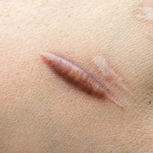
Skin cancer removal scarring and aftercare depend on lesion size, location, skin type, and how well aftercare instructions are followed. Scars on the face tend to heal more favorably due to good blood supply, while scars on the chest or back may be more prone to raised or thickened healing. For guidance on scar care products, see this comparison of silicone, vitamin E, and petroleum jelly for scar care after removal.
Can Skin Cancer Come Back After Removal?
Yes, skin cancer can come back after removal, particularly if surgical margins were not fully clear, the original lesion was a more aggressive subtype, or the patient continues significant sun exposure without adequate protection. Recurrence rates vary widely depending on the type of skin cancer, its original size and depth, and the treatment method used.
Factors that raise recurrence risk:
- Incomplete margins on the original pathology report.
- Larger or deeper lesions at the time of initial treatment.
- Aggressive histologic subtypes identified on pathology.
- Location in high-risk zones such as the nose, ears, or areas with prior radiation.
- Ongoing sun exposure without consistent sunscreen use or protective clothing.
This is why follow-up matters as much as the removal itself. Physicians typically recommend periodic skin checks after a skin cancer diagnosis, since having one skin cancer increases the likelihood of developing another, either at the same site or elsewhere on the body. Patients with a history of skin cancer are generally advised to continue regular self-checks between formal visits and report any new or changing spots promptly rather than waiting for the next scheduled appointment.
Best Time to Get Skin Cancer Removed
The best time to get skin cancer removed is as soon as a diagnosis is confirmed, since most skin cancers grow slowly but steadily, and earlier treatment generally means a smaller excision, simpler closure, and better cosmetic outcome. There is no clinical benefit to delaying treatment once a biopsy confirms a malignant or pre-malignant lesion.
That said, a few practical timing considerations come up in real scheduling:
- Season isn't a medical factor, but many patients prefer scheduling excisions in cooler months to make sun avoidance during healing easier, especially for facial procedures.
- Upcoming travel or major events should be flagged early, since sutures and visible healing can affect appearance for one to three weeks.
- Waiting for pathology results before a definitive excision is normal and expected, this is different from delaying care after a diagnosis is already confirmed.
Quick example: a patient with a biopsy-confirmed BCC on the nose who waits eight months "to see if it changes" risks the lesion growing larger and requiring a more complex reconstruction than if it had been excised soon after diagnosis. Early, prompt treatment after confirmed diagnosis consistently produces simpler surgical outcomes.
Dermatologists in Burlington, Milton and Halton Region: Finding the Right Care Pathway
Several dermatology, plastic surgery, and skin clinics across Burlington, Milton, and the wider Halton Region offer parts of the skin cancer assessment and removal pathway, and choosing the right one depends on the stage of care needed. Some clinics focus on cosmetic dermatology, others on surgical excision, and a smaller number offer advanced techniques like Mohs surgery.
Examples of the local landscape include:
- Halton Dermatology & Skin Surgery in Burlington, which offers surgical treatment for basal cell carcinoma, squamous cell carcinoma, Bowen's disease, melanoma, and lentigo maligna, with Mohs surgery available for high-risk or recurrent cases [1][2].
- AroRegen in Milton, where physician-led procedures include removal of cysts, lipomas, moles, and skin tags, with dermoscopy used to support accurate diagnosis before removal [3].
- Milton-based plastic surgery services, including biopsy-proven excision and reconstruction for basal cell and squamous cell carcinomas, operating out of Milton District Hospital and a private surgical center in Burlington [5].
- Dermetics in Burlington, offering mole, skin tag, and cyst removal consultations that assess size, depth, and location before treatment to help minimize scarring [4].
- Laser and cosmetic clinics in Milton and Burlington, which primarily handle benign lesion removal, such as seborrheic keratoses and cherry hemangiomas, rather than confirmed skin cancer excision [6][9].
Skin cancer removal clinic near me Halton searches often turn up a mix of dermatology practices, plastic surgery offices, and medical spas, it's worth confirming which stage of care a clinic actually provides (assessment only, biopsy, or full surgical excision) before booking, since not every clinic handles every step. Patients in Oakville and nearby communities can also access assessment and surgical removal locally rather than relying solely on Burlington or Milton-based providers, keeping travel time shorter for follow-up visits.
Why Choose TMSC Oakville for Skin Cancer Removal Near Burlington, Milton & Halton Region
Choosing a clinic for skin cancer removal near Burlington, Milton, or Halton Region should come down to whether the team can manage the full pathway, assessment, biopsy coordination, and surgical excision, under one roof, with clear communication about pathology results and next steps. The Minor Surgery Center's Oakville clinic supports this pathway for patients across the Halton area.
What this looks like in practice:
- GP dermatologists assess suspicious moles and lesions where applicable, helping determine whether biopsy is needed and what type is most appropriate.
- Plastic surgeons perform biopsies, excisions, and surgical removals when a treatment plan calls for it, with attention to both clearing the lesion and supporting good cosmetic healing.
- Clear referral guidance for patients whose pathology results point toward a diagnosis, such as melanoma, that requires hospital or oncology-level care beyond standard local excision.
- Coordinated follow-up, so patients understand pathology results, scar care expectations, and when to return for further monitoring.
Care always depends on diagnosis, lesion location, pathology findings, and individual physician assessment, no clinic can promise a specific outcome before biopsy results are known. Patients considering care in the area can also review this breakdown of top skin cancer screening and treatment clinics in the wider region for additional context on what to look for when comparing providers. Learn more about the physician background supporting these procedures on the Dr. Mark Shafarenko profile page.
Areas Served Near Oakville and Halton Region
Skin cancer removal support through TMSC Oakville is designed for patients across Oakville and the surrounding Halton Region, including communities where travel to a downtown hospital dermatology department can mean long wait times for a straightforward excision. Local access matters most for patients who already have a confirmed or suspected diagnosis and want to move to treatment without unnecessary delay.
Communities and neighborhoods commonly served include:
- Oakville, including Bronte, Glen Abbey, West Oak Trails, and Palermo
- Burlington
- Milton
- Georgetown
- Hamilton
- Halton Region more broadly
For patients specifically in Milton looking for related mole and cyst removal services alongside skin cancer assessment, see the Milton Mole & Cyst Removal page. Oakville residents can also review the Downtown Oakville Mole & Cyst Removal page, and Burlington patients comparing local options may find this guide to the best mole removal clinic in Burlington useful for understanding what to expect before booking an assessment.
FAQs
What is the skin cancer removal procedure? Skin cancer removal is a surgical excision performed under local anesthetic, where the confirmed lesion and a margin of surrounding tissue are removed and the wound is closed with sutures. The exact approach depends on the biopsy-confirmed diagnosis, lesion size, and location.
How much does skin cancer removal cost in Burlington, Ontario? Medically necessary skin cancer removal is generally covered by OHIP when confirmed by physician assessment, so most patients do not pay out of pocket for the core assessment, biopsy, and excision. Costs may apply for elective cosmetic add-ons or services outside standard OHIP-funded care, and specifics should be confirmed with the treating clinic.
How long does skin cancer removal take? A standard excision for a single lesion usually takes 30 to 60 minutes, though larger lesions, facial locations, or Mohs surgery cases can take longer due to more detailed tissue checking and reconstruction.
What is the recovery time after skin cancer removal surgery? Surface healing typically takes one to two weeks, with sutures removed within that window depending on location. Full scar softening and fading can continue for up to a year.
Does skin cancer removal hurt? The area is numbed with local anesthetic before the procedure, so significant pain during the excision is uncommon. Mild soreness for a day or two afterward is normal and usually managed with over-the-counter pain relief.
Can skin cancer come back after it's been removed? Yes, recurrence is possible, especially with incomplete margins, aggressive subtypes, or continued sun exposure. This is why follow-up monitoring after treatment is an important part of long-term care.
Is Mohs surgery better than standard excision? Neither is universally "better", Mohs surgery is generally preferred for high-risk, recurrent, or cosmetically sensitive lesions because it spares more healthy tissue, while standard excision works well for lower-risk, well-defined lesions. The right choice depends on tumor characteristics and physician assessment.
What types of skin cancer can be removed surgically? Basal cell carcinoma and squamous cell carcinoma are commonly removed through standard excision or Mohs surgery. Melanoma treatment depends heavily on staging and may require wider excision margins, sentinel lymph node evaluation, or referral to a specialized oncology team.
Is skin cancer removal covered by OHIP? Yes, in most cases, when a physician confirms the lesion is medically necessary to remove rather than purely cosmetic. Coverage details can vary, so it's worth confirming with the specific clinic providing care.
When should I get a mole checked for possible cancer? Get a mole checked if it shows asymmetry, irregular borders, uneven color, growth beyond about 6mm, or any noticeable change in shape, size, or texture over recent weeks or months. Bleeding, itching, or a sore that won't heal are also reasons to seek assessment promptly.
Are there dermatologists and skin cancer clinics in Burlington, Milton, and Halton Region? Yes, the area has a mix of dermatology practices, plastic surgery clinics, and skin surgery centers offering different stages of care, from assessment and biopsy through to advanced Mohs surgery for eligible cases [1][2][3][5].
Does removing a mole or lesion cause cancer to spread? No, appropriately performed biopsy and excision do not cause cancer to spread. This is a common misconception worth clarifying with a physician if it's a concern; see does removing a mole cause cancer for more detail.
Book a Consultation
Concerned about a skin cancer or suspicious lesion near Burlington, Milton or Halton Region? The Minor Surgery Center's Oakville clinic can assess skin lesions and discuss biopsy or surgical removal options when appropriate. Bring any prior biopsy or pathology reports to the appointment, along with a list of when the lesion was first noticed and any changes observed since then, this speeds up the assessment and helps guide next steps quickly.
Conclusion
Skin cancer removal in Burlington, Milton and Halton Region follows a clear sequence: assessment, biopsy when needed, pathology review, and surgical excision matched to the confirmed diagnosis. Basal cell carcinoma and squamous cell carcinoma are commonly managed through standard excision or, for higher-risk cases, Mohs surgery, while suspicious moles and possible melanoma require biopsy-confirmed diagnosis before any treatment plan is set. OHIP typically covers medically necessary removal, recovery generally spans one to two weeks for initial healing with longer-term scar maturation, and recurrence risk means follow-up care matters as much as the initial procedure.
Next steps for patients in the region: if a lesion has changed recently, bled, or simply looks different from surrounding moles, book an assessment rather than waiting for the next annual check. Bring any existing pathology reports, note when changes were first noticed, and ask directly about whether biopsy, excision, or referral to a specialist pathway is the right next step for that specific lesion.
References
[1] haltonderm.ca - https://haltonderm.ca/?utm_source=openai [2] Haltondermatologyskinsurgery - https://www.medifinder.com/en/clinics/canada/burlington/haltondermatologyskinsurgery/?utm_source=openai [3] aroregen.ca - https://aroregen.ca/?utm_source=openai [4] Mole Skin Tag And Cyst Removal - https://dermetics.ca/treatments/mole-skin-tag-and-cyst-removal?utm_source=openai [5] miltonplasticsurgery.ca - https://miltonplasticsurgery.ca/?utm_source=openai [6] Milton - https://laserit.ca/services/mole-removal/milton.aspx?utm_source=openai [7] Medical Spa - https://www.nhchalton.com/milton/medical-spa/?utm_source=openai [8] Cosmetic Dermatologists Milton - https://dermetics.ca/locations/cosmetic-dermatologists-milton?utm_source=openai [9] radiancelaser.ca - https://www.radiancelaser.ca/?utm_source=openai



%20Treatment.avif)