Lipoma Removal in Etobicoke and West Toronto: What a Fatty Lump Is and When It Should Come Out
Last updated: September 3, 2026
Quick Answer
A lipoma is a benign growth of mature fat cells that sits in a thin capsule, usually just under the skin, and typically feels soft, doughy and easy to slide under your fingers. Removal is a short in-office excision: the area is frozen with local anaesthetic, a small incision is made, the fatty lump is lifted out with its capsule intact, and the skin is closed with fine sutures. The tissue is then sent to a pathology lab, because no clinician can confirm what a lump is by feel or from a photograph. Patients across Etobicoke and west Toronto can arrange lipoma removal without a referral at The Minor Surgery Center's clinic at 1224 Dundas Street West in Mississauga, minutes from Highway 427.
Key Takeaways
A lipoma is a benign fatty tumour under the skin. Most are soft, mobile, slow-growing and painless, and many people have more than one.
A small, soft, stable, painless lipoma can reasonably be left alone and watched. Removal is a choice, not a requirement, unless there are worrying features.
Removal is done under local anaesthetic in a treatment room, not under general anaesthetic, and a straightforward case takes about 20 to 40 minutes from freezing to dressing.
Every excised specimen goes to pathology. A lipoma is confirmed by a pathologist, never by appearance alone.
Features that prompt imaging or a specialist opinion include size over roughly five centimetres, a lump sitting deep to the muscle fascia, firmness or fixation, rapid growth, new pain, or recurrence at a previously operated site.
Liposarcoma, a malignant tumour of fat tissue, is rare. The overwhelming majority of soft, mobile, slow-growing lumps under the skin are benign, and the reason lumps get investigated is that the two cannot be told apart by touch.
No cream, oil, supplement or diet has been shown to dissolve a lipoma. Removing the capsule completely is what keeps it from coming back at that spot.
Removal for appearance alone is not an OHIP-insured service in Ontario; private-pay excision in the GTA commonly falls somewhere in a broad range from a few hundred dollars to a few thousand, depending on size, depth and complexity.
What a Lipoma Actually Is
A lipoma is a benign tumour made of mature fat cells wrapped in a thin fibrous capsule, sitting most often in the layer of fat between the skin and the muscle. It's not a cancer, it doesn't spread, and having one doesn't mean something is wrong with you.
The classic feel is distinctive once you know it. Press on a lipoma and it gives slightly, soft, a bit doughy, like a small water balloon under a towel. Push it sideways and it usually slides, because it isn't attached to the skin above or the muscle below. Most are painless. Most grow so slowly that people notice them by accident and then can't say how long they've been there.
The common sites are the shoulders, upper back, back of the neck, upper arms, thighs, chest and abdomen. They can appear almost anywhere there's fat, including the forehead and the forearm. Size ranges widely: many stay at a centimetre or two for years, some reach the size of a golf ball or larger, and a smaller number sit deeper than the usual subcutaneous layer. Our overview of what a lipoma is and how these fatty lumps behave under the skin covers the deeper variants in more detail.
One more plain fact: multiple lipomas are common. Finding a second or third one on your arm or back a few years after the first isn't a sign that something has changed for the worse.
Lipoma or Something Else? Telling a Fatty Lump From Its Look-Alikes
Several ordinary lumps get mistaken for lipomas, and the differences are mostly in how they feel and where they sit. What follows is how a clinician sorts through them during an examination, useful context, not a way to diagnose yourself.
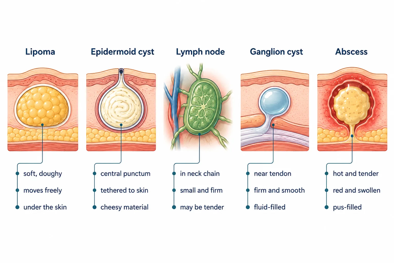
Epidermoid or sebaceous cyst. These sit in the skin rather than under it, so they feel firmer and more tethered, move the skin and the lump moves with it. Many have a tiny central dot, a punctum, where the follicle opens to the surface, and some occasionally discharge thick, foul-smelling material. If you want the full side-by-side, we've written a detailed comparison of how to tell a lipoma from a cyst.
Enlarged lymph node. Nodes live in predictable chains, the sides of the neck, under the jaw, the armpit, the groin. They tend to feel firmer and more rubbery than a lipoma, are often slightly tender, and frequently follow a sore throat, a dental problem or a skin infection nearby. A node that appeared quickly and is shrinking behaves very differently from a lipoma that has been sitting quietly for three years.
Ganglion cyst. These form next to a joint or along a tendon sheath, most often on the back of the wrist or on a finger. They feel tense and rubbery rather than doughy, are anchored to the deeper structures, and often change size with activity. Location is the biggest clue: a lump directly over the wrist joint is much more likely to be a ganglion than a lipoma.
Dermatofibroma. A small, firm, button-like nodule fixed within the skin, usually less than a centimetre, often brownish or pink, and commonly on the lower legs. Pinch the skin around it and the centre tends to dimple inward. Nothing like the soft mobility of a lipoma.
Abscess. A collection of pus, so it is hot, red, tense and genuinely painful, and it usually appeared over days rather than years. Fever or spreading redness makes this urgent and it needs to be seen promptly rather than booked as an elective procedure.
These distinctions guide a clinician's thinking. They do not replace an examination, and they can't be settled from across a room or over a photo. A lump that nobody can confidently identify gets removed and tested, that's the whole logic of a proper skin lesion assessment before any removal in Etobicoke.
Lipoma or Liposarcoma? The Question Worth Taking Seriously
Lipomas are common and benign. Liposarcoma is a rare malignant tumour arising from fat tissue, and while it is uncommon, it can look and feel similar to a lipoma from the outside, particularly in the early stages. That resemblance is exactly why soft-tissue lumps get investigated rather than assumed.
Start with the reassuring part, because it is the accurate part. A small, soft, mobile, slow-growing lump sitting just under the skin is benign in the overwhelming majority of cases. Liposarcomas are reported to be a small fraction of soft-tissue tumours overall, and they tend to arise in the deeper tissues, inside muscle compartments of the thigh, the retroperitoneum, the shoulder girdle, rather than in the thin fat layer under the skin of the forearm.
What changes a clinician's index of suspicion is a specific set of features. Any one of these is a reason to image, biopsy or refer rather than simply excise and reassure:
Size above roughly five centimetres. Larger lesions are more likely to warrant imaging before anything is cut, and size is one of the most consistently cited thresholds in surgical guidance.
Depth below the muscle fascia. A lump you can't slide because it's anchored under or within muscle is a different problem from one floating in subcutaneous fat.
Firm or fixed rather than soft and mobile. A hard lump that doesn't move deserves an explanation.
Rapid growth. A lump that has visibly changed over weeks or a few months, rather than years.
New pain, especially pain that isn't clearly from pressure on a nerve or from rubbing against clothing.
Recurrence at a site where a "lipoma" was removed before. This is an important one. A fatty lump reappearing where one was previously taken out is a reason to look more carefully, not a reason to repeat the same operation.
A lipoma and a low-grade liposarcoma can feel identical through skin. Neither feel nor a photograph gives a diagnosis. Only tissue examined under a microscope does.
That last point matters more than any list. Imaging can describe a lesion and measure it. A surgeon can describe how it looked and behaved during the operation. But the definitive answer comes from a pathologist, which is why every specimen is sent. If you want the longer discussion of the differences and how the assessment runs, our piece on when a fatty lump needs more than reassurance goes further into the clinical reasoning.
None of this is a reason to panic about a soft lump on your shoulder. It's a reason to have it looked at by someone who examines these regularly, and to have it tested if it comes out.
Size, Depth and Location: Why No Two Lipoma Operations Are the Same
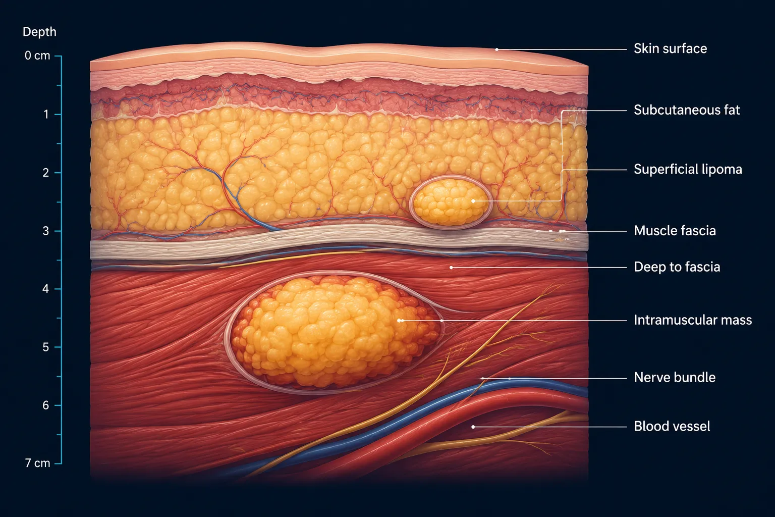
Depth relative to the muscle fascia matters more than width. A three-centimetre lipoma sitting in subcutaneous fat on the forearm is a short procedure in a treatment room. A three-centimetre lipoma sitting inside the deltoid muscle is a genuinely different operation.
Superficial subcutaneous lipomas, the common kind, sit above the fascia in a plane that separates cleanly. The incision is usually shorter than the lump's diameter, because the fat is compressible and can be delivered through a smaller opening once the capsule is freed. Closure is often a single layer, sometimes two.
Larger lipomas need proportionally longer incisions and more attention to what's left behind. A ten-centimetre lipoma on the back leaves a real cavity, and that cavity can fill with fluid if it isn't managed, closed in layers, sometimes with a compressive dressing. Our discussion of the surgical challenges specific to large lipomas covers the planning involved.
Intramuscular and subfascial lipomas are a separate category. They interdigitate with muscle fibres rather than sitting in a neat capsule, which makes complete removal harder and makes imaging beforehand more or less mandatory. These are the cases most likely to need a different setting than a standard treatment room, and the ones where intramuscular lipoma symptoms and surgical considerations are worth reading before booking anything.
Location changes the operation for two separate reasons: tension and anatomy.
The shoulder, upper back and posterior neck are the most common lipoma sites, and they are also among the highest-tension skin areas on the body. Skin there is thick and constantly pulled by movement, which means a closure has to hold against that tension and the resulting scar tends to widen more than the same scar would on a forearm. This is why lipoma removal on the back is planned differently from removal on a limb, often with deeper buried sutures to take load off the skin edges.
Anatomy is the other constraint. In the neck, important nerves and vessels run close to the surface, and a lump in the posterior triangle sits near structures worth respecting, which is one reason removing a lipoma on the neck is approached carefully. The armpit contains the brachial plexus and axillary vessels. The groin has femoral structures. The volar forearm and wrist have superficial sensory nerves that produce a patch of numbness if they're bruised. And on the face, where fat layers are thin and every millimetre of scar is visible, the priorities shift toward incision placement in natural lines, the reasoning behind our notes on facial lipomas and minimizing scarring.
Decision rule worth remembering: if you can slide the lump easily in every direction and it feels soft, it's almost certainly superficial and the operation is likely to be simple. If it won't move, or it feels firmly anchored when the underlying muscle contracts, expect imaging first.
When Imaging Is Done Before Removal, and When It Is Not
Most small, soft, mobile, superficial lumps don't need a scan before removal. Imaging is ordered when the clinical picture is ambiguous or when the lump has a feature that makes depth and extent important to know before an incision is made.
Ultrasound is the usual first step. It's quick, involves no radiation, and answers the practical questions: is this actually fat, how big is it in all three dimensions, does it sit above or below the fascia, is there abnormal blood flow inside it, and does it have internal features that don't fit a simple lipoma. In Ontario, ultrasound for a soft-tissue lump is generally accessible when it's clinically indicated.
MRI comes into play when a lesion appears to sit within or beneath muscle, when the ultrasound isn't reassuring, or when a lipomatous mass is large enough that a surgeon needs a map before operating. MRI shows the relationship of the mass to nerves, vessels and muscle compartments better than anything else, and it characterises fat with high reliability.
Here's the important limit. Imaging characterises and measures. It can strongly suggest a benign lipoma, and it can raise concern about something more, but it does not deliver a definitive tissue diagnosis. A radiologist's report saying "consistent with a lipoma" is a probability statement, not a verdict. That's why imaging and pathology serve different jobs, and why the presence of a reassuring ultrasound doesn't remove the need to send an excised specimen to the lab. Our explanation of what lipoma imaging can and cannot tell you breaks this down further.
The practical reality: nobody scans every soft lump. A 1.5 cm doughy, mobile bump on the upper arm that's been unchanged for four years does not need an MRI. A firm 7 cm mass on the thigh that grew over five months needs imaging before anyone touches it. Most cases sit clearly at one end or the other, and the examination is what decides.
Common mistake to avoid: assuming a scan is the more thorough choice and therefore always better. Ordering imaging for a clearly benign superficial lump mostly adds weeks of waiting and a small chance of an incidental finding that leads to more tests. The judgement call belongs at the examination.
Do You Actually Need It Removed?
A lipoma that's small, soft, painless and not growing can reasonably be left alone and monitored. There's no medical obligation to remove a stable benign fatty lump.
People go ahead with removal for reasons that are all legitimate:
It's getting bigger, or looks like it is.
It hurts, or it presses on a nerve and produces aching or tingling. Whether a lipoma can be painful depends largely on where it sits and what it's pushing against.
It catches, on a bra strap, a waistband, a seatbelt, a backpack strap, a razor.
It's somewhere visible: the forehead, the jawline, the forearm, the back of the neck above a collar.
It's changing in any way they can describe.
The uncertainty itself is the problem. Knowing what it is, definitively, is a reasonable thing to want.
A clinician may recommend removal even if you're inclined to leave it, and the reasons are the ones from the liposarcoma section: size, depth, firmness, fixation, rapid growth, new pain, or recurrence at a previously excised site. In those situations the removal isn't cosmetic, it's diagnostic, and it isn't really optional.
If you choose to watch instead, watch actively. Note the size against something concrete (a coin, a measurement) and note the date. If it's the same size and feel a year from now, that's genuinely useful information. If it isn't, you'll know quickly.
What the Procedure Involves, Step by Step
A straightforward lipoma excision is done in a treatment room under local anaesthetic, and most patients are in and out inside an hour, walking out with a dressing on.
Assessment and marking. The lump is examined, its borders felt out and marked, and the incision is planned along a natural skin crease or along the direction of relaxed skin tension where the anatomy allows. Consent, allergies, blood thinners and previous scarring get covered here.
Cleaning and draping. The skin is prepared with antiseptic and a sterile drape is placed.
Local anaesthetic. A fine needle delivers freezing around and under the lipoma. This is the only genuinely uncomfortable part, a brief sting and a stinging spread that lasts several seconds, similar to dental freezing. Within a minute or two the area is numb. You'll feel pressure and movement during the procedure, and you shouldn't feel sharpness. If you do, more anaesthetic is added.
Incision. Usually shorter than the width of the lipoma itself, because fat compresses and delivers.
Enucleation. The capsule is identified and the lipoma is separated from the surrounding tissue in its own plane and lifted out whole. This is the part that determines whether it comes back. Scooping out fat leaves capsule behind; taking the capsule with the lesion is what makes recurrence at that site uncommon.
Checking the cavity. Bleeding points are controlled and the cavity is inspected for any remaining lobule of fat, lipomas can be multilobulated and a stray lobe left behind explains many apparent "recurrences."
Closure. Deeper absorbable sutures close the dead space and take tension off the skin, then the skin is closed with fine sutures. Layered closure matters most on the back, shoulder and anywhere a cavity was left.
Dressing. A simple dry dressing, occasionally with a compressive layer over a larger cavity.
Specimen to pathology. The lipoma goes into a labelled container and is sent to the lab.
Timing: a small superficial lipoma commonly takes 20 to 30 minutes of procedure time, and a larger or deeper one can take 45 minutes to well over an hour. If you're curious about the real-world clock, we've written specifically about how long lipoma removal surgery actually takes.
Most patients drive themselves home after a local-anaesthetic excision, though it depends on the site and how you feel; the practical considerations are covered in our note on whether you can drive home after lipoma removal.
Why Every Lipoma Goes to the Lab
A lump that looks and feels exactly like a lipoma is confirmed as a lipoma by a pathologist examining the tissue under a microscope. Not by the surgeon's impression, not by the ultrasound, and not by how cleanly it came out.
The pathology report answers three things. First, what the tissue actually is, mature adipose tissue with a capsule, or something else. Second, whether there are atypical features, which is the distinction between a benign lipoma and an atypical lipomatous tumour or liposarcoma. Third, whether the excision appears complete at the margins, which matters if the diagnosis turns out to be anything other than a plain lipoma.
Results usually take about one to two weeks in Ontario, sometimes a little longer depending on the lab. Reports are reviewed by the clinician and communicated to the patient, a benign result is straightforward information, and anything unexpected triggers a conversation and, where appropriate, a referral into the sarcoma or surgical oncology pathway.
This is also the practical argument against any method that destroys the lesion rather than removing it. Dissolving, burning or draining a lump leaves nothing to send. If the identity of that lump was never established, and it wasn't, because appearance can't establish it, you've traded a definitive answer for a cosmetic result. The same reasoning applies to why a cyst sac has to come out whole rather than being squeezed empty.
Recovery, Activity and Scarring
Most people go back to desk work the same day or the next, and the wound is closed and dressed before you leave. The main restriction is on stretching or loading the closure for the first couple of weeks.
Day of. Expect the freezing to wear off over two to four hours, followed by soreness that plain acetaminophen usually handles. Keep the dressing dry and in place. Some bruising around the site is normal, and on the back or shoulder it can spread downward with gravity over the next few days.
First week. Most protocols allow showering the day after surgery, patting the area dry and re-dressing. Avoid soaking, swimming and hot tubs until the wound is sealed. Keep lifting and reaching to a minimum if the lump was on the trunk, shoulder or neck.
Sutures. Non-absorbable sutures generally come out around six to seven days on the face and roughly ten to fourteen days on the body and limbs. Absorbable sutures dissolve on their own over a few weeks. Areas under tension keep sutures in longer.
Weeks two to six. Light activity resumes early. For lifting, gym work and anything that pulls across the wound, two to four weeks is typical for a body site, and longer for a large back or shoulder excision. The general timeline is covered in more detail in our guide to how long recovery from lipoma removal surgery takes.
Reasons to call. Increasing redness spreading outward, increasing pain after day three, warmth, discharge, fever, or a growing soft swelling under the wound. That last one is often a seroma, a fluid collection after lipoma removal is more common after larger excisions and usually settles, but it should be assessed rather than guessed at. Persistent numbness or shooting pain near the scar can reflect a bruised sensory nerve, which is discussed in our piece on nerve pain after lipoma removal.
Scars, honestly. A lipoma excision leaves a linear scar roughly the length of the incision. It will be pink or red for the first several weeks, often firm and slightly raised at six to eight weeks, and then it flattens and fades over six to twelve months. A forearm scar typically settles fine. A shoulder or upper back scar tends to widen more, because of skin tension there, and that's a limitation of anatomy rather than technique. Keeping the scar out of the sun, or covered with sunscreen, over the first year makes a measurable difference to how dark it stays.
Can a Lipoma Come Back?
A lipoma removed completely with its capsule rarely comes back at that same site. When one does reappear, it usually means a lobule of the original was left behind, or that the lesion was more extensive than it appeared.
New lipomas can still appear elsewhere on the body, because removing one doesn't change whatever tendency led to it forming. That's not a failure of the surgery, it's a different lump.
There's one important qualifier. A fatty mass regrowing at a site where one was previously excised is one of the features that prompts a closer look rather than a simple repeat removal. In that situation, imaging beforehand and careful pathology afterward are the appropriate response.
Multiple Lipomas and What Changes
Some people have several lipomas, and a smaller number have many, a dozen or more across the arms, trunk and thighs. Having multiple lipomas doesn't change what each individual one is, but it does change how a plan is built.
They're assessed together and prioritised, not removed all at once. Priority goes to the ones that are symptomatic, growing, sitting somewhere that catches or gets bumped, or showing any feature that warrants investigation. A cosmetically annoying but stable one-centimetre lump on the thigh sits lower on the list than a four-centimetre one on the shoulder that aches under a backpack.
Two or three small superficial lipomas in the same region can often be removed in one sitting, since they share the same anaesthetic and the same visit. Spreading a larger number across separate appointments keeps the local anaesthetic dose sensible and keeps recovery manageable, since each site has its own wound to look after.
If you have a strong family pattern, a parent and siblings with the same thing, or a very high number of them, mention it. That's relevant history worth documenting, and it may affect how the clinician approaches assessment. It isn't a reason for alarm.
Non-Surgical Options and Home Remedies: What Actually Works
There's no cream, oil, supplement, herbal paste, diet or heat treatment shown to dissolve a lipoma. If a product promises to shrink a fatty lump through the skin, the evidence isn't there.
Two clinical alternatives are worth describing accurately. Steroid injection has been used to shrink some small lipomas; results are inconsistent, the lump generally doesn't disappear, and nothing is obtained for pathology. Liposuction can debulk a large lipoma through a very small opening, which is genuinely useful in specific situations where scar length is the dominant concern. The trade-off is real: suction removes fat but tends to leave the capsule behind, so recurrence is more likely, and there's no intact specimen for the lab. Our review of non-surgical lipoma removal options and what they can realistically achieve covers the details.
Squeezing, lancing or cutting a lipoma at home is a bad idea for practical reasons rather than moral ones. It causes bleeding into a fatty cavity, introduces bacteria into a space that drains poorly, produces a worse scar than a planned incision, and removes any chance of having the tissue examined. The specific risks are set out in our article on the real dangers of DIY lipoma extraction.
Is Lipoma Removal Covered by OHIP in Ontario?
The distinction OHIP draws is medical necessity. Removal of a lump that is symptomatic, enlarging, or clinically suspicious is handled differently from removal requested purely because of how it looks, and cosmetic removal of a stable benign lesion is not an insured service in Ontario.
In practice, most people who come in specifically because a lipoma bothers them cosmetically pay privately. When a lump has features that require investigation, the pathway and the billing can look different, and that's a determination made at the assessment rather than in advance.
Private extended health plans and health spending accounts sometimes cover minor surgical procedures, and coverage varies substantially between employers and insurers. Confirm your own situation at the consultation rather than assuming either way. Our detailed breakdown of whether lipoma removal is covered by OHIP or private insurance goes through the specifics.
What Lipoma Removal Costs Around Etobicoke and West Toronto
Private-pay lipoma excision in the Greater Toronto Area commonly falls somewhere in a broad range, roughly a few hundred dollars for a small, simple, superficial lump, up to a few thousand for a large or deep one near important structures. Reported Canadian ranges for private excision typically span from around $300 to $3,000 per lipoma.
What moves the number:
Size and depth. A 1 cm subcutaneous lump and a 9 cm subfascial mass are not the same procedure.
Location on the body. High-tension or anatomically sensitive sites take longer and require more careful closure.
Closure complexity. Single-layer versus layered closure with buried sutures.
Number removed in one sitting. Additional lesions in the same visit are usually priced incrementally rather than at full rate.
Pathology processing, which is a standard part of the procedure rather than an optional add-on.
An exact figure comes from the consultation, once someone has examined the lump and knows what the operation involves. For broader context on GTA pricing, see our 2026 price guide for lipoma removal in Toronto.
Getting It Done Near Etobicoke and West Toronto
The nearest clinic serving Etobicoke and west Toronto is at 1224 Dundas Street West, Unit 101, in Mississauga, straight along the Dundas Street West corridor, a short drive from Highway 427 and the QEW, with on-site parking. The phone number is 647-614-1611 and hours are weekdays, 9 a.m. to 4 p.m. No referral is required.
For patients who find another location easier, The Minor Surgery Center also has clinics in North York at 2920 Dufferin Street, in York Mills at 85 Scarsdale Road, and in Vaughan and Oakville. Anyone coming down the 427 from Rexdale or across from the Kingsway generally finds Dundas Street West the simplest run.
The staffing matters for a lump like this. GP dermatologists handle skin assessment, examining the lesion, sorting a lipoma from its look-alikes, and deciding whether imaging is warranted before anything is removed. Plastic surgeons perform the excisions and closures. For a lipoma on the shoulder, the back of the neck, the jawline or anywhere else that shows, the closure is the part that determines how the scar looks a year later, and having that done by someone whose training centres on soft-tissue closure is the practical reason for the split. If you're weighing the options generally, we compare which doctor to see for lipoma removal and the roles of a dermatologist versus a plastic surgeon for moles, cysts and lipomas in Toronto.
On the question of urgent care versus a specialist clinic: a walk-in or urgent care centre is the right place for a lump that's hot, red, spreading and painful, because that's likely an abscess and needs prompt drainage or antibiotics. It isn't the right place for a stable fatty lump you want properly assessed and excised, because urgent care doesn't typically do planned excisions with layered closure and pathology. A minor surgery clinic or a plastic surgeon's office is the appropriate setting for that.
One more practical point: consultation and removal can often be completed in the same visit when the assessment supports it. That's not universal, a lump needing imaging first won't be removed that day, but for a straightforward superficial lipoma it usually is. Patients also frequently ask about cyst removal in Etobicoke and west Toronto and mole removal across Etobicoke and west Toronto, which are handled at the same locations.
Areas Served Across Etobicoke and West Toronto
Patients travel to the Dundas Street West clinic from across Etobicoke, the Kingsway, Islington-City Centre, Mimico, New Toronto, Long Branch, Alderwood, Humber Bay Shores, Markland Wood, the Royal York corridor, and Rexdale and Thistletown up near the airport, as well as from west Toronto neighbourhoods including Bloor West Village, the Junction, Swansea, High Park, Roncesvalles and Parkdale. For most of those addresses it's a straight run along Dundas or a short hop off the 427 or the QEW, and the on-site parking means you're not circling for a spot before an appointment.
Frequently Asked Questions
Does lipoma removal hurt? The only part most people describe as uncomfortable is the local anaesthetic going in, a brief sting for a few seconds. After that the area is numb, and you feel pressure and movement rather than sharpness. Afterward, expect soreness for a few days that plain acetaminophen usually manages.
How long does lipoma removal take? A small superficial lipoma usually takes about 20 to 30 minutes of procedure time, and a larger or deeper one can run 45 minutes to over an hour. Allow roughly an hour in the clinic overall for a straightforward case, including assessment, consent and dressing.
Do I need a referral for lipoma removal in Etobicoke? No. Patients can book directly without a referral from a family doctor, which is why the no-referral model is common at minor surgery clinics across the GTA.
Can I drive myself home afterward? Usually yes, because the procedure is done under local anaesthetic with no sedation. It depends on the site and how you feel, a large excision on the right shoulder may make steering uncomfortable, so arranging a ride is sensible for bigger cases.
Will lipoma removal leave a scar? Yes. Any excision leaves a linear scar roughly the length of the incision. It fades over six to twelve months, and how well it settles depends heavily on location, forearm scars generally do well, while shoulder and upper back scars tend to widen because of skin tension there. Sun protection over the first year helps.
How big does a lipoma have to be before it should come out? There's no universal size threshold for a benign lipoma; a small stable one can be left alone indefinitely. That said, a lump larger than about five centimetres is generally investigated more thoroughly before removal, because size is one of the features that prompts imaging.
Can a lipoma turn into cancer? A benign lipoma is not generally considered to transform into a malignant tumour. The clinical concern is different: a lesion that was assumed to be a lipoma may turn out on pathology to be an atypical lipomatous tumour or a liposarcoma, which is why excised tissue is always sent to the lab.
Can more than one lipoma be removed in the same visit? Often yes, particularly for two or three small superficial lipomas in the same region. Larger numbers are usually staged across visits to keep the anaesthetic dose reasonable and recovery manageable, since each site has its own wound.
How long until I can lift weights or go back to the gym? Light activity can resume within days. For lifting and anything that pulls across the wound, two to four weeks is typical for a limb or trunk site, and longer after a large back or shoulder excision. The closure needs time to gain strength before it's loaded.
Is lipoma removal covered by OHIP? Removal purely for appearance is not an insured service in Ontario. When a lump is symptomatic, enlarging or clinically suspicious, the situation is assessed differently, and that determination is made at the consultation. Some private extended health plans and health spending accounts cover minor surgery.
The Bottom Line
Most soft, mobile, slow-growing lumps under the skin are exactly what they appear to be. The honest caveat is that nobody, not a surgeon, not a radiologist, not you with a photograph, can confirm that by looking. What settles it is an examination by someone who assesses these regularly and, if the lump comes out, a pathology report on the tissue.
If a fatty lump is catching on your clothing, growing, aching, or simply sitting in the back of your mind, the next step is a straightforward one. Book an assessment at 1224 Dundas Street West, Unit 101, Mississauga, or call 647-614-1611 during weekday hours. Bring anything useful, a photo from a year ago, a rough sense of when you first noticed it, any previous excision at the same site.
This article is general information about lipomas and their removal, not medical advice for your specific situation. A lump should be assessed in person by a qualified clinician.
Meta title: Lipoma Removal Etobicoke: When a Fatty Lump Must Come Out
Meta description: Lipoma removal Etobicoke explained by clinicians: how excision works, when imaging is needed, why pathology matters, costs, OHIP and recovery. No referral needed.
Tags: lipoma removal Etobicoke, lipoma vs liposarcoma, minor surgery Toronto, lipoma excision, soft tissue lump, lipoma pathology, OHIP coverage, west Toronto clinic, lipoma imaging, lipoma recovery, plastic surgeon Toronto, fatty lump under skin
The Minor Surgery Center’s York Mills clinic, located at 85 Scarsdale Rd, Unit 101 in North York, offers convenient access to specialized surgical care for a range of skin and hand conditions. Our Plastic Surgeons provide treatment for skin cancer, suspicious moles, cysts, lipomas, carpal tunnel syndrome and trigger finger, with a focus on timely care and personalized treatment. In many cases, patients may be able to have their consultation and treatment completed during the same visit when medically appropriate. For expert care in a welcoming clinical setting, contact our York Mills team at (647) 614-1611.
At TMSC, we specialize in surgical treatment of both benign and malignant skin lesions, as well as minor hand conditions. We offer significantly reduced wait times often allowing you to schedule your surgery on the same day as your consultation.
You can book your free consultation with The Minor Surgery Center in three easy ways: online through Cortico, by phone, or by requesting a callback from our team.
The initial consultation is free. During this consultation, one of our board certified surgeons will examine your condition and recommend a suitable solution.
Directly Online
Request a Callback
By Phone
By Text
Hours
Sunday
Closed
Monday
9 am — 4 pm
Tuesday
9 am — 4 pm
Wednesday
9 am — 4 pm
Thursday
9 am — 4 pm
Friday
9 am — 4 pm
Saturday
Toronto & Oakville Open Vaughan & Mississauga Closed




.avif)
%20(600%20x%20600%20px)%20(2).avif)
.avif)
%20(8).avif)
%20(7).avif)

.png)