

Last updated: September 29, 2026
Superficial BCC vs eczema or psoriasis often comes down to one clue: response to treatment. Eczema and psoriasis usually improve with topical steroids or moisturizers within weeks, while a superficial basal cell carcinoma (sBCC) stays put, slowly spreads, and often shows a thin pearly or thread-like border under close inspection. Any single pink, scaly patch that will not clear after four to six weeks of appropriate eczema or psoriasis treatment deserves a dermatologist's look and, often, a biopsy [1][8].
Superficial BCC typically presents as a single, slow-growing, thin pink-to-red plaque with a fine, slightly raised, pearly or thread-like border, while eczema usually shows up as multiple itchy, ill-defined red patches with dryness or oozing. The border is the biggest visual clue: BCC borders have a subtle shine or "rolled" quality, while eczema borders blend into surrounding skin.
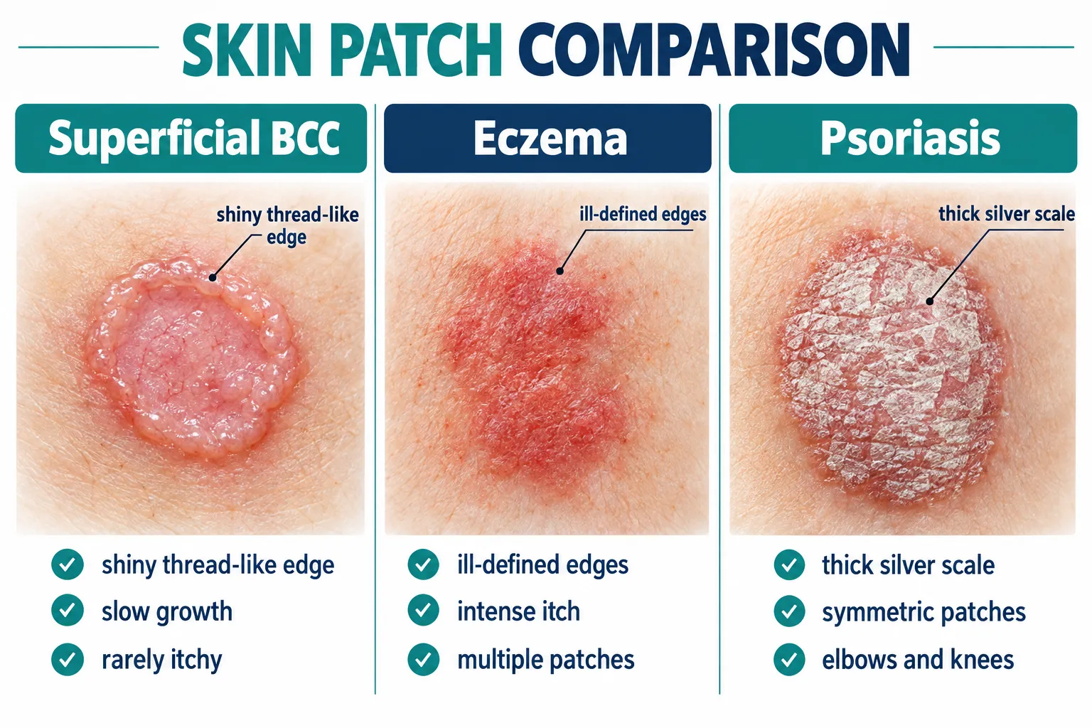
Superficial basal cell carcinoma is the flattest and most deceptive subtype of BCC. Instead of the classic nodular, pearly bump most people associate with skin cancer, sBCC forms a slightly scaly, faintly translucent plaque that can sit quietly on the trunk, shoulders, or back for years [2][5]. Because it lacks a dramatic bump or ulceration in early stages, it is frequently confused with common inflammatory skin conditions.
Key visual differences include:
Choose to suspect sBCC over eczema if the patch is solitary, has been present for more than two to three months, shows a subtle shiny border, and has not responded to a reasonable trial of emollients or mild topical steroids. A common mistake is assuming that any scaly patch on sun-exposed skin is automatically eczema simply because eczema is more familiar to patients and even some clinicians on first glance [3][6].
For a broader look at how this cancer type compares with other forms, see this overview of the four types of skin cancer.
The clearest way to tell BCC apart from psoriasis is scale thickness and symmetry: psoriasis produces thick, silvery-white scale on symmetric body areas like elbows and knees, while superficial BCC produces thin, minimal scale on a single asymmetric patch with a pearly edge. Psoriasis also tends to run in families and flare with stress or illness, patterns rarely seen with BCC.
Psoriasis is an autoimmune condition driven by overactive skin cell turnover. It classically produces well-demarcated, thick plaques covered in silvery scale, most often on the scalp, elbows, knees, and lower back, and it usually appears on both sides of the body in a mirrored pattern [7][10]. Superficial BCC, by contrast, is a localized cancerous growth that stays confined to one area and does not mirror on the opposite side of the body.
Practical distinguishing features:
FeatureSuperficial BCCPsoriasisDistributionSolitary, one-sidedOften symmetric, multiple plaquesScaleThin, fine, sometimes absentThick, silvery, adherentBorderPearly, thread-like, subtle shineSharply demarcated, no shineFamily historyNot typically relevantFrequently positiveNail changesNonePitting, thickening commonResponse to steroidsLittle to no improvementUsually improves
A common mistake in primary care is treating a solitary "psoriasis-like" plaque with a standard topical steroid without checking for classic psoriasis features elsewhere on the body, such as nail pitting or scalp involvement. If those systemic signs are absent and the plaque stands alone, sBCC should move up the list of possibilities [6][10].
Edge case: Psoriasis can occasionally present as a single plaque early in the disease course, which is exactly when it looks most like sBCC. In this scenario, dermoscopy or a short observation period with follow-up is the safest next step rather than assuming either diagnosis.
Yes, superficial BCC is one of the most commonly misdiagnosed skin cancers precisely because it mimics inflammatory dermatoses so convincingly. Clinical studies and dermatology reviews consistently list eczema (nummular dermatitis) and psoriasis among the top differential diagnoses for sBCC [2][3][8].
The overlap happens for a few concrete reasons:
Example: A 45-year-old patient notices a coin-sized pink patch on the upper back. It is treated as nummular eczema with a moderate-strength steroid cream for six weeks. The patch shrinks slightly at first, from reduced surrounding inflammation, but then plateaus and slowly regrows. This pattern, partial response followed by stagnation or regrowth, is a hallmark warning sign that should prompt biopsy rather than a stronger steroid.
Superficial BCC vs eczema or psoriasis confusion is also common because primary care visits are short and biopsy is not always the first step for a seemingly benign, common-looking rash. Recent primary-care, oriented guidance stresses that any solitary, non-resolving plaque, regardless of how "typical" it looks for eczema or psoriasis, should be tracked closely and biopsied if it fails to improve within a defined treatment window [6].
Eczema and psoriasis are both far more common in the general population than superficial BCC, but sBCC is one of the most frequently diagnosed BCC subtypes among skin cancers, and its prevalence rises sharply with age and cumulative sun exposure. In practical terms, a random scaly patch is statistically more likely to be eczema or psoriasis, but sBCC is common enough that it cannot be dismissed on probability alone.
Eczema affects a large share of the population across all age groups, often starting in childhood, while psoriasis affects a smaller but still substantial percentage of adults, often with a genetic component [7][10]. Superficial BCC, on the other hand, is a cancer of accumulated sun damage and is typically diagnosed in adults, though notably it appears in younger adults more often than other BCC subtypes such as nodular or morpheaform BCC [2].
This creates an important clinical nuance:
Decision rule: Weigh age, sun exposure history, and number of lesions together rather than assuming a single scaly patch is "probably eczema because eczema is common." Probability arguments should never replace direct examination or biopsy when a lesion behaves atypically.
Superficial BCC is usually asymptomatic or only mildly itchy, while eczema is defined by intense, often disruptive itching and psoriasis produces moderate itching along with tightness or discomfort from scale buildup. Itch intensity is a genuinely useful clue, though it is not foolproof since a minority of sBCC lesions do itch or feel tender, especially if irritated by clothing or scratching.
Breaking down the symptom pattern:
Common mistake: Assuming that "if it itches, it can't be cancer." While sBCC is less itchy on average, a subset of lesions, particularly those that have been present for a long time or are located in areas prone to friction, can develop secondary irritation and mild itch. Itch absence supports sBCC but itch presence does not rule it out.
Edge case: A lesion that itches mildly but also bleeds slightly after minor trauma, scratching, or shaving is behaving in a way that is unusual for ordinary eczema and should raise suspicion for a cancerous process rather than simply intensifying steroid treatment.
Superficial BCC is caused by cumulative ultraviolet (UV) damage to skin cells, particularly intermittent or long-term sun exposure, while eczema stems from a combination of genetic skin barrier dysfunction and immune system overreaction to irritants, allergens, or environmental triggers. Psoriasis, similarly, is autoimmune-driven rather than sun-driven. Understanding cause helps explain who is most at risk for each condition.
Superficial BCC risk factors:
Eczema (atopic dermatitis) risk factors:
Psoriasis risk factors:
Decision rule: A patient with a strong family history of eczema and multiple itchy, symmetric patches since childhood is very unlikely to have sBCC. A patient with decades of outdoor sun exposure, no personal history of eczema or psoriasis, and a single new patch after age 40 should be evaluated with sBCC on the differential list. For a related look at how sun damage manifests differently from cancer risk, see this comparison of age spots versus cancer spots.
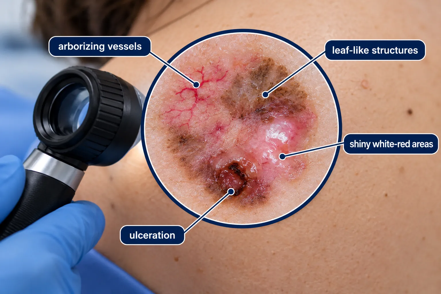
Dermoscopy, a handheld magnification tool used by dermatologists, reveals specific vascular and structural patterns in superficial BCC that are absent in eczema and psoriasis, making it one of the most reliable non-invasive ways to separate the three conditions before biopsy. Trained use of dermoscopy significantly improves diagnostic accuracy compared to visual inspection alone [1][9].
Distinguishing dermoscopic features of superficial BCC include:
Dermoscopy is particularly useful for separating sBCC from Bowen's disease (a form of squamous cell carcinoma in situ) and psoriasis, two conditions that can otherwise look remarkably similar on plain visual exam [1][9]. Clinics offering in-office dermoscopy skin checks in Toronto use this exact technology to shorten the path to an accurate diagnosis without requiring every suspicious patch to go straight to biopsy.
Common mistake: Relying on dermoscopy alone as a final answer. Dermoscopy narrows the differential and guides the decision to biopsy, but it is not a substitute for histologic confirmation when findings are ambiguous or when the lesion still behaves suspiciously despite reassuring dermoscopic features [1][9].
Superficial BCC does not resolve on its own. Left untreated, it typically persists indefinitely and grows slowly outward over months to years, in contrast to eczema or psoriasis flare-ups, which can settle down with treatment or even spontaneously calm between flares.
This is one of the most important practical distinctions for patients trying to decide whether to "wait and see." Eczema and psoriasis are chronic but fluctuating conditions: a flare can worsen for a week and then improve substantially with treatment, moisturizing, or removal of a trigger. Superficial BCC follows a different trajectory entirely.
Decision rule: If a "rash" has been present continuously, without a single period of meaningful clearing, for longer than three months despite reasonable treatment, that continuous, non-fluctuating course is itself a diagnostic clue pointing away from eczema or psoriasis and toward a possible skin cancer.
Common mistake: Waiting indefinitely because the lesion is "not painful" or "not growing quickly." Superficial BCC's slow pace is exactly what makes it dangerous to ignore. It rarely causes urgent symptoms early on, which removes the natural motivation to seek care that a painful or rapidly changing lesion would create.
Yes, it is entirely possible to have both conditions simultaneously, either in different locations or, less commonly, with a BCC developing within or near a long-standing area of chronic eczema. The two conditions are unrelated in cause, so having one does not protect against or predispose to the other, aside from shared risk from chronic skin irritation and inflammation in rare cases.
Practical implications of this overlap:
Edge case: A patient with a decades-long history of eczema on the trunk notices that one patch has stopped responding to their usual steroid routine while the rest of their skin clears normally. This selective non-response in a single spot, distinct from the behavior of the rest of the eczema, is the practical signal to seek a dedicated evaluation of that one patch rather than assuming it is simply a "stubborn eczema flare."
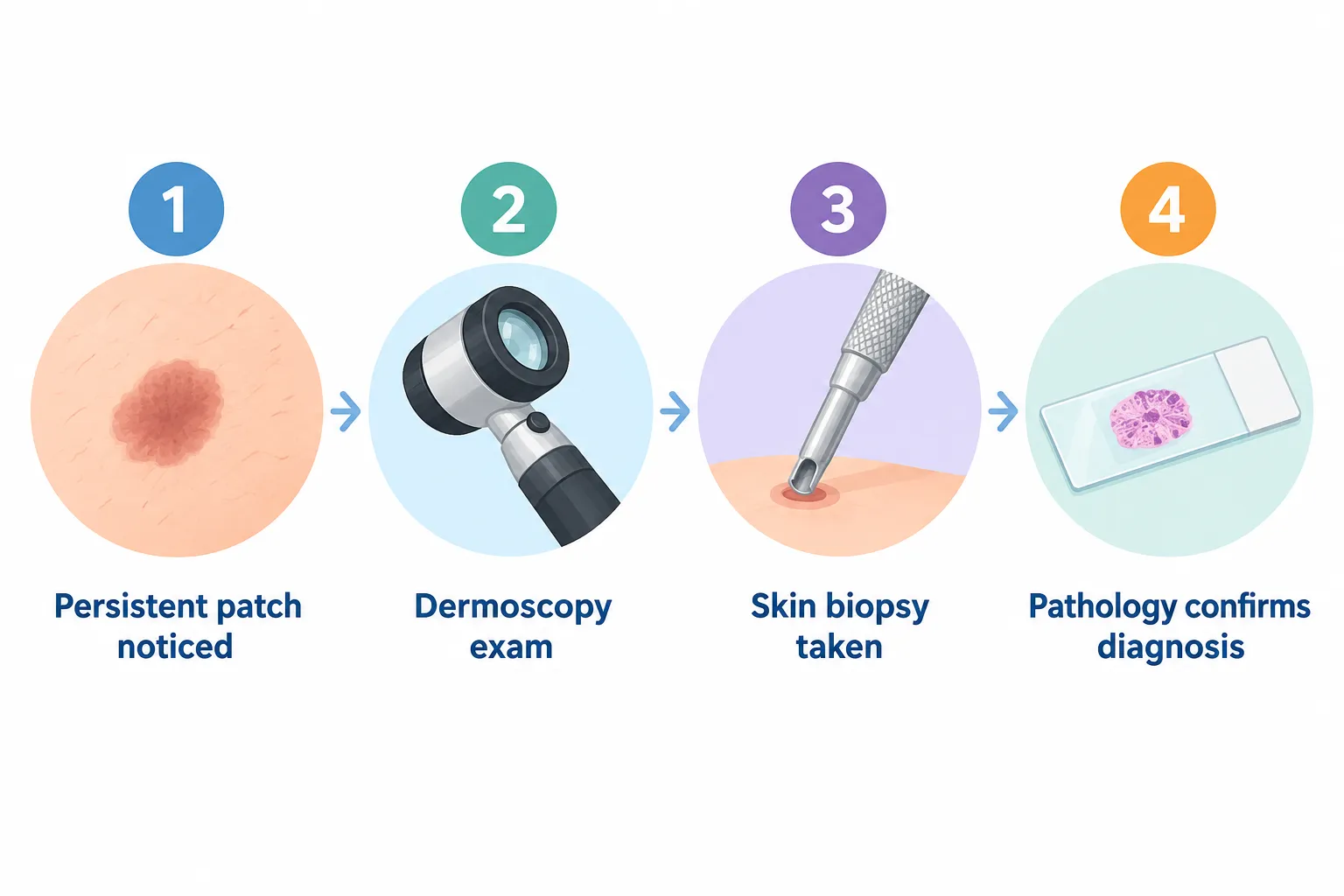
A skin biopsy is the only definitive way to distinguish superficial BCC from eczema or psoriasis. A dermatologist or surgeon removes a small sample of the lesion under local anesthetic, and a pathologist examines the tissue under a microscope for characteristic cancer cell patterns that are absent in inflammatory skin conditions.
The biopsy process typically follows these steps:
Histologically, superficial BCC shows nests of basaloid cells budding from the epidermis with a characteristic peripheral palisading pattern, findings that are completely distinct from the inflammatory cell infiltrates, spongiosis (fluid within the epidermis), and epidermal thickening seen in eczema and psoriasis biopsies [2][8]. This histologic clarity is why biopsy remains the gold standard even when dermoscopy is highly suggestive.
Common mistake: Delaying biopsy because the lesion "looks mild" or because a patient is reluctant to have any procedure. A biopsy for a suspected superficial BCC is a brief, low-risk office procedure, and the alternative, an incorrect assumption of eczema or psoriasis, can allow a cancer to continue growing untreated for months or years [3][6].
For patients being evaluated for a possible skin cancer more broadly, a full body skin exam is often recommended alongside biopsy of the specific lesion of concern, since patients with one suspicious lesion are statistically more likely to have others.
Superficial BCC is highly treatable once correctly diagnosed, with options ranging from prescription topical creams and photodynamic therapy for very thin lesions to minor surgical excision or curettage and electrodesiccation for confirmed cases. Cost and choice of treatment depend primarily on lesion size, location, depth, and whether it is a first occurrence or a recurrence.
Common treatment approaches include:
Choice of treatment depends on lesion size, location, patient health, and cosmetic priorities, and should be made in consultation with a dermatologist or skin surgery specialist after biopsy confirmation [2][6]. Costs vary by province, clinic, and whether the procedure is covered under provincial health insurance versus performed at a private clinic, so patients should confirm coverage details directly with their chosen provider rather than relying on general estimates.
Decision rule: Small, thin, low-risk superficial BCCs on the trunk are often well-suited to simpler in-office procedures, while lesions on the face, recurrent lesions, or larger plaques typically warrant referral to a surgical specialist for excision or Mohs surgery. Clinics that provide minor surgical procedures, such as this mole, cyst, and skin lesion surgery center, can often confirm the diagnosis and complete treatment in a coordinated pathway.
People most likely to develop superficial BCC are fair-skinned adults over 40 with a history of significant sun exposure, though sBCC specifically is diagnosed in younger adults more often than other BCC subtypes, making age under 40 an insufficient reason to dismiss a suspicious plaque [2].
High-risk groups include:
Example: A 34-year-old former competitive swimmer with fair skin and a childhood spent largely outdoors develops a persistent pink patch on the shoulder. Age alone might suggest eczema is far more likely, but the combined sun exposure history and solitary nature of the lesion make sBCC a reasonable concern worth ruling out, illustrating why demographic assumptions should never override direct examination.
No, superficial BCC does not resolve spontaneously the way an eczema or psoriasis flare can settle down between episodes. Once a basal cell carcinoma forms, it requires active treatment; it will not shrink, clear, or disappear through diet changes, moisturizing, avoiding triggers, or simply waiting.
This is a critical safety point because eczema and psoriasis patients are conditioned by their own experience to expect fluctuation: flares that worsen with stress or weather and then improve with treatment or time. Applying that same expectation to a lesion that turns out to be sBCC leads directly to delayed diagnosis.
Common mistake: Interpreting a temporary reduction in redness (often from reduced surface inflammation after steroid use) as evidence the lesion is clearing. A true response to treatment means the lesion disappears entirely and does not return; partial calming followed by plateau or slow regrowth is a warning sign, not a good sign.
A dermatologist evaluation is warranted for any solitary skin patch that has persisted longer than four to six weeks despite appropriate eczema or psoriasis treatment, has a pearly or shiny border, bleeds easily with minor trauma, or is located on chronically sun-exposed skin in a patient over 40. Earlier evaluation is reasonable for anyone uncertain or anxious about a changing spot, regardless of these specific criteria.
Concrete triggers for prompt evaluation:
Recent expert consensus trends from 2024 through 2026 increasingly emphasize routine dermoscopy and earlier biopsy thresholds for solitary plaques rather than extended trial-and-error treatment periods, reflecting growing recognition of how often sBCC hides behind an eczema or psoriasis label in clinical practice. Booking a full body skin check is a practical way to have any concerning spot, along with the rest of the skin, assessed systematically by a trained professional rather than relying on self-diagnosis from photos or general descriptions found online.
Edge case: Patients with darker skin tones sometimes present with less visually obvious pearly borders on sBCC, making the lesion's persistence and lack of response to treatment even more important as diagnostic clues than border appearance alone.
The table below summarizes the practical distinctions covered throughout this guide for quick reference.
FactorSuperficial BCCEczemaPsoriasisNumber of lesionsUsually solitaryOften multipleOften multiple, symmetricTypical ageAdults, including younger adultsAny age, often childhood onsetAdults, genetic linkItch levelMild or noneOften intenseModerateBorderPearly, thread-likeIll-definedSharply defined, no shineScaleThin or absentVariable, can weepThick, silveryCourse over timeSlow, continuous growthFluctuates, flares and clearsFluctuates, responds to treatmentResponse to steroidsLittle to noneUsually improvesUsually improvesResolves without treatmentNoCan improve or clearCan improve significantlyDiagnostic confirmationBiopsy requiredClinical exam, sometimes patch testingClinical exam, sometimes biopsy
For related conditions that can also cause confusing crusty or scaly patches, see this guide on crusty spots on skin and this explanation of contact dermatitis, which can further complicate the picture when a rash develops from an irritant rather than atopic eczema.
A patch that quietly outlasts every cream tried on it is telling you something. Persistence, not pain, is the loudest warning sign superficial BCC gives.
Is superficial BCC dangerous if left alone for a few months?
Superficial BCC grows slowly, so a short delay of a few weeks while pursuing appropriate diagnosis is unlikely to cause serious harm, but it will not resolve on its own and should still be formally diagnosed and treated once identified [2][8].
Can a dermatologist tell BCC from eczema just by looking at it?
Experienced dermatologists can often form a strong suspicion through visual exam and dermoscopy, but a biopsy is still required to confirm superficial BCC vs eczema or psoriasis with certainty [1][6].
Does superficial BCC bleed like a wound?
Superficial BCC can bleed slightly with minor trauma, such as shaving or scratching, and may be slow to fully heal, which is unusual for typical eczema or psoriasis patches [8].
Is superficial BCC the most common type of BCC?
Superficial BCC is one of the more frequently diagnosed BCC subtypes and is notably more common in younger adults compared to other BCC types such as nodular BCC [2].
Can topical steroids make superficial BCC worse?
Topical steroids do not directly worsen sBCC, but they can mask surface inflammation and create a false impression of improvement, which delays correct diagnosis and treatment [3][6].
What is the fastest way to rule out cancer versus eczema?
Dermoscopy combined with a defined trial period of appropriate eczema or psoriasis treatment is the fastest non-invasive approach; if there is no meaningful improvement within four to six weeks, biopsy should follow promptly [1][6].
Does superficial BCC only appear on sun-exposed skin?
Superficial BCC most commonly appears on sun-exposed areas like the trunk, shoulders, and back, though it can occasionally develop on less exposed skin, particularly in patients with significant cumulative UV history or immunosuppression [2][5].
Can eczema turn into skin cancer over time?
Eczema itself does not transform into basal cell carcinoma; the two are caused by different mechanisms, though a separate sBCC can coincidentally develop nearby or within an area affected by long-standing eczema.
How painful is a biopsy for a suspected superficial BCC?
A biopsy is performed under local anesthetic and is generally described as a brief pinching or pressure sensation during numbing, with minimal discomfort afterward.
Should every scaly patch be biopsied just to be safe?
Not every scaly patch needs an immediate biopsy; most respond as expected to standard eczema or psoriasis treatment. Biopsy becomes appropriate specifically when a solitary lesion fails to improve, has atypical features, or persists beyond a reasonable treatment trial [6].
Superficial BCC vs eczema or psoriasis is rarely obvious at a glance, and that overlap is exactly why so many cases get mislabeled early on. The most reliable signals are practical ones: a single patch instead of several, a subtle pearly or thread-like border, minimal itch, and, most tellingly, no real improvement after a proper course of standard eczema or psoriasis treatment. Dermoscopy adds a valuable layer of certainty, but biopsy remains the only way to close the question for good.
Next steps for anyone dealing with a persistent, unexplained patch:
A persistent patch that will not respond to reasonable treatment deserves a direct answer, not another round of guessing.
[1] Basal Cell Carcinoma Dermoscopy - https://dermnetnz.org/topics/basal-cell-carcinoma-dermoscopy
[2] Nbk482439 - https://www.ncbi.nlm.nih.gov/books/NBK482439/
[3] Pmc4445438 - https://pmc.ncbi.nlm.nih.gov/articles/PMC4445438/
[5] Basal Cell Carcinoma - https://www.merckmanuals.com/professional/oncology/cancers-of-the-skin/basal-cell-carcinoma
[6] P161 - https://www.aafp.org/afp/2012/0715/p161
[7] Nbk470301 - https://www.ncbi.nlm.nih.gov/books/NBK470301/?report=printable
[8] Basal Cell Carcinoma - https://dermnetnz.org/topics/basal-cell-carcinoma
[9] Pmc2980434 - https://pmc.ncbi.nlm.nih.gov/articles/PMC2980434/
[10] P339 - https://www.aafp.org/afp/2020/0915/p339
Meta Title: Superficial BCC vs Eczema or Psoriasis: Key Differences
Meta Description: Learn how to tell superficial BCC vs eczema or psoriasis apart using border features, itch patterns, dermoscopy, and treatment response clues.
Tags: superficial BCC, basal cell carcinoma, eczema, psoriasis, skin cancer symptoms, dermoscopy, skin biopsy, skin cancer screening, nummular dermatitis, misdiagnosed skin conditions, skin cancer treatment, dermatology