When people notice unusual changes on their skin, particularly crusty spots, it can cause significant concern and anxiety. These skin irregularities can appear suddenly or develop gradually over time, leaving individuals wondering about their cause and whether they require medical attention. Understanding what crusty spot on skin pictures reveal about various conditions is crucial for proper identification and timely treatment.
Skin changes are among the most visible health indicators, and crusty spots represent a diverse category of dermatological conditions ranging from benign to serious. The appearance, texture, color, and location of these spots can provide valuable clues about their underlying cause. While some crusty spots are harmless and resolve on their own, others may indicate conditions requiring professional medical evaluation and treatment.
The importance of visual identification through crusty spot on skin pictures cannot be overstated. These images serve as valuable educational tools, helping individuals recognize patterns and characteristics that distinguish one condition from another. However, it's essential to understand that visual comparison should never replace professional medical diagnosis, as many skin conditions can appear similar despite having different causes and treatment requirements.
• Crusty spots on skin can range from harmless conditions like seborrheic keratoses to serious concerns like skin cancer
• Visual characteristics including color, texture, size, and location provide important diagnostic clues • Professional medical evaluation is essential for accurate diagnosis and appropriate treatment planning • Early detection and intervention often lead to better treatment outcomes and reduced complications • Regular skin self-examinations combined with professional screenings help identify changes promptly
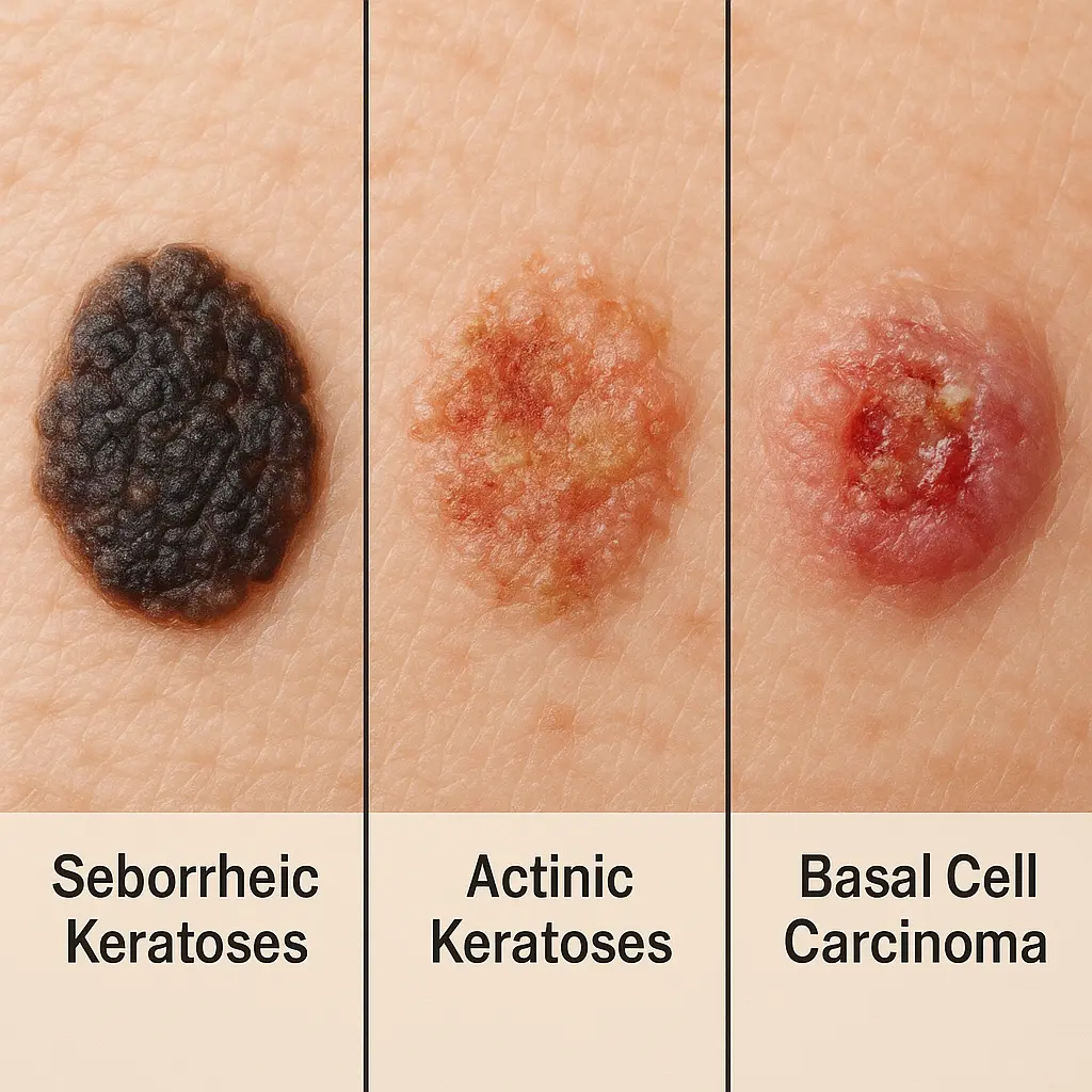
Seborrheic keratoses represent one of the most frequently encountered types of crusty spots on the skin. These benign growths typically appear as waxy, scaly, or crusty patches that seem to be "stuck on" the skin surface. Seborrheic keratoses often develop with age and are particularly common in individuals over 50 years old.
The appearance of seborrheic keratoses in crusty spot on skin pictures shows characteristic features that help distinguish them from other conditions. These growths usually present as:
Multiple seborrheic keratoses often appear simultaneously, particularly on sun-exposed areas such as the face, chest, shoulders, and back. While these growths are benign, they can sometimes become irritated from clothing friction or scratching, leading to inflammation and increased crustiness.
Actinic keratoses (AKs) represent a more serious category of crusty skin spots that require careful attention and monitoring. These precancerous lesions develop as a result of cumulative sun damage and appear most commonly on sun-exposed areas of the body.
Crusty spot on skin pictures showing actinic keratoses typically reveal:
The significance of actinic keratoses lies in their potential for malignant transformation. While not all AKs progress to skin cancer, they represent areas of damaged skin with increased risk for developing squamous cell carcinoma. Early identification and treatment can prevent this progression and reduce cancer risk.
Basal cell carcinoma (BCC) represents the most common form of skin cancer and can sometimes present with crusty characteristics. While BCCs can appear in various forms, the crusty variant often causes confusion with benign conditions.
Key features visible in crusty spot on skin pictures of basal cell carcinoma include:
BCCs typically grow slowly and rarely metastasize, but they can cause significant local tissue destruction if left untreated. Early detection through regular skin examinations and prompt treatment leads to excellent cure rates.
Understanding when crusty spots on skin warrant urgent medical evaluation is crucial for optimal health outcomes. Certain characteristics visible in crusty spot on skin pictures should prompt immediate professional assessment.
Critical warning signs include:
Warning SignDescriptionUrgency LevelRapid growthSignificant size increase over weeks or monthsHigh ⚠️Irregular bordersUneven, notched, or blurred edgesHigh ⚠️Color variationMultiple colors within a single spotHigh ⚠️BleedingSpontaneous bleeding or easy bleeding when touchedHigh ⚠️UlcerationOpen sores that don't heal within 2-3 weeksHigh ⚠️
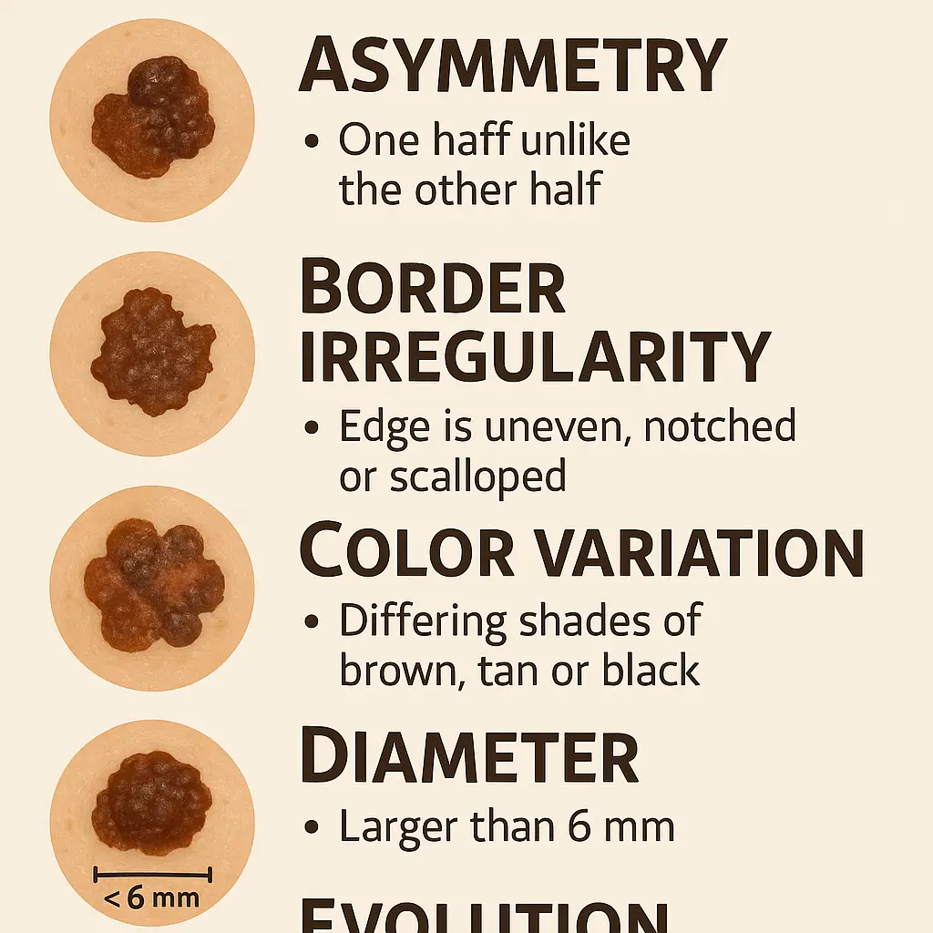
The ABCDE rule provides a systematic approach for evaluating crusty spots and other skin lesions:
A - Asymmetry: Benign spots are typically symmetrical, while concerning lesions often show asymmetrical growth patterns.
B - Border irregularity: Smooth, well-defined borders suggest benign conditions, while irregular or poorly defined borders raise concern.
C - Color variation: Uniform coloration is reassuring, while multiple colors within a single lesion warrant evaluation.
D - Diameter: Lesions larger than 6mm (pencil eraser size) require closer attention, though smaller lesions can also be significant.
E - Evolution: Any changing characteristics in size, shape, color, or symptoms should prompt medical evaluation.
For comprehensive evaluation of concerning skin conditions, individuals should consider consulting with specialists at facilities like The Minor Surgery Center, where experienced professionals can provide thorough assessment and appropriate treatment recommendations.
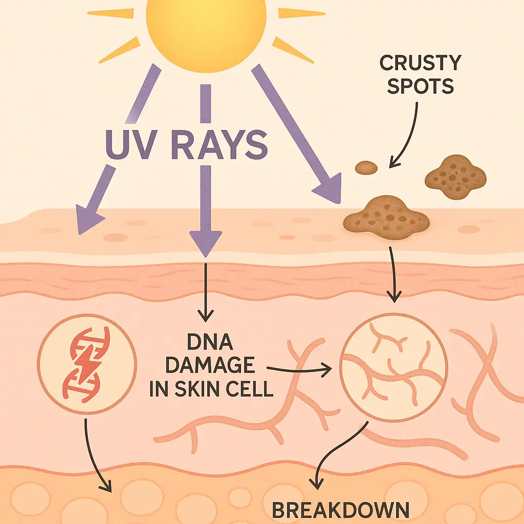
Ultraviolet radiation represents the primary environmental factor contributing to crusty spot development on the skin. Both UVA and UVB rays cause cumulative damage to skin cells, leading to various types of lesions visible in crusty spot on skin pictures.
Chronic sun exposure results in:
Individuals with fair skin, light hair, and light eyes face higher risk for developing sun-related crusty spots due to lower melanin protection. However, people of all skin types can develop these conditions with sufficient UV exposure.
Advancing age significantly increases the likelihood of developing various types of crusty skin spots. The natural aging process affects skin in multiple ways:
Most crusty spots become more common after age 40, with incidence continuing to increase with each decade of life. Regular skin monitoring becomes increasingly important as individuals age.
Family history plays a significant role in determining individual risk for developing certain types of crusty skin spots. Genetic factors influence:
Individuals with family histories of skin cancer or multiple skin lesions should maintain heightened awareness and pursue regular professional skin examinations.
Determining the appropriate timing for professional evaluation of crusty spots requires careful consideration of various factors. While not every crusty spot requires immediate medical attention, certain situations warrant prompt consultation.
Seek immediate evaluation for:
For routine evaluation of stable, benign-appearing spots, scheduling consultation within 1-2 months provides appropriate timing while ensuring thorough assessment.
Effective preparation enhances the value of medical consultations for crusty spot evaluation. Patients should consider:
Documentation strategies:
Questions to prepare:
Patients seeking comprehensive evaluation can find detailed information about available services at The Minor Surgery Center's conditions page, which outlines various dermatological conditions and treatment approaches.
Many crusty spots on skin respond well to non-invasive treatment approaches that preserve surrounding healthy tissue while effectively addressing the problematic areas.
Topical medications represent first-line treatment for many conditions:
Cryotherapy using liquid nitrogen provides effective treatment for many benign and precancerous crusty spots. This technique involves:
More aggressive or concerning crusty spots may require surgical intervention to ensure complete removal and accurate diagnosis.
Excisional surgery involves complete removal of the lesion with surrounding normal tissue margins. This approach provides:
Mohs micrographic surgery represents the gold standard for certain skin cancers, particularly those in cosmetically sensitive areas. This technique offers:
For individuals considering surgical treatment options, consulting with experienced professionals at The Minor Surgery Center's clinic ensures access to advanced techniques and comprehensive care.
Comprehensive sun protection represents the most effective strategy for preventing many types of crusty spots on skin. Effective protection requires multiple approaches:
Daily sunscreen application:
Protective clothing and accessories:
Monthly skin self-examinations enable early detection of new or changing crusty spots. Effective self-examination involves:
Systematic approach:
Key areas to examine:
Regular professional skin examinations complement self-examination efforts and provide expert evaluation of concerning findings.
Recommended screening frequency:
Individuals seeking professional screening can find experienced dermatological professionals through The Minor Surgery Center's team, who provide comprehensive evaluation and personalized care plans.
The discovery of crusty spots on skin often triggers significant emotional responses ranging from mild concern to severe anxiety. Understanding these reactions and developing healthy coping strategies supports overall well-being during evaluation and treatment processes.
Common emotional responses include:
Healthy coping strategies:
Individuals with crusty spots often benefit from implementing lifestyle changes that support skin health and reduce risk for future problems.
Beneficial modifications include:
Nutrition optimization:
Skin care routine enhancement:
No, not all crusty spots are dangerous. Many crusty spots represent benign conditions such as seborrheic keratoses or minor skin irritations that require no treatment. However, some crusty spots can indicate precancerous or cancerous conditions requiring professional evaluation and treatment.
The key lies in understanding which characteristics warrant concern and seeking appropriate medical evaluation when indicated. Regular monitoring and professional screening help distinguish between benign and potentially serious conditions.
Development timeframes vary significantly depending on the underlying condition causing the crusty spot. Some spots appear rapidly over days or weeks, while others develop gradually over months or years.
Typical development patterns:
Some crusty spots may resolve spontaneously, particularly those related to minor skin irritations, infections, or inflammatory conditions. However, most persistent crusty spots require some form of intervention for resolution.
Conditions that may self-resolve:
Conditions requiring treatment:
For comprehensive answers to additional questions about skin conditions and treatments, individuals can explore The Minor Surgery Center's FAQ section, which addresses common concerns and provides valuable information about various dermatological conditions.
Dermoscopy represents a significant advancement in evaluating crusty spots on skin, providing enhanced visualization of structures not visible to the naked eye. This non-invasive technique uses specialized magnification and lighting to reveal:
Digital dermoscopy adds computer-assisted analysis and storage capabilities, enabling:
When crusty spot on skin pictures and clinical examination cannot provide definitive diagnosis, biopsy procedures offer definitive histological analysis.
Types of biopsy procedures:
Shave biopsy:
Punch biopsy:
Excisional biopsy:
Emerging technologies are revolutionizing the evaluation of crusty spots and other skin lesions. Artificial intelligence systems trained on thousands of crusty spot on skin pictures can now:
Telemedicine platforms increasingly incorporate advanced imaging and analysis tools for skin condition evaluation. These systems enable:
Most benign crusty spots carry excellent long-term prognoses with appropriate management. Seborrheic keratoses, while permanent unless treated, remain stable and cause no health risks. Treatment, when desired for cosmetic reasons, typically provides excellent outcomes with minimal scarring.
Long-term considerations for benign spots:
Early-stage skin cancers and precancerous conditions generally carry excellent prognoses with appropriate treatment. Success rates for common skin cancers exceed 95% when detected and treated early.
Factors influencing outcomes:
For detailed information about specific conditions and their expected outcomes, patients can review comprehensive resources available at The Minor Surgery Center's blog, which provides current information about various dermatological conditions and treatments.
Effective management of crusty spots often involves coordination between multiple healthcare providers. A comprehensive team may include:
Knowledge empowers patients to participate actively in their care and make informed decisions about treatment options. Key educational components include:
Understanding condition specifics:
Self-advocacy skills:
Understanding crusty spots on skin through visual identification, professional evaluation, and appropriate treatment represents a crucial aspect of maintaining optimal skin health. The diversity of conditions that can present as crusty spots—from benign seborrheic keratoses to potentially serious skin cancers—underscores the importance of proper evaluation and management.
Crusty spot on skin pictures serve as valuable educational tools, helping individuals recognize concerning characteristics that warrant professional attention. However, these visual aids must be used in conjunction with, not as a replacement for, professional medical evaluation. The complexity of skin conditions and the potential for similar appearances among different diagnoses necessitates expert assessment for accurate diagnosis and appropriate treatment planning.
Prevention strategies, including comprehensive sun protection and regular skin monitoring, offer the most effective approach to reducing the risk of developing problematic crusty spots. When prevention efforts prove insufficient and spots do develop, early detection and intervention typically lead to excellent outcomes with minimal complications.
The emotional impact of discovering concerning skin changes should not be underestimated. Building a strong support network that includes knowledgeable healthcare providers, family members, and potentially other individuals who have faced similar challenges can significantly improve the overall experience and outcomes.
As medical technology continues to advance, new diagnostic and treatment options emerge regularly, offering improved outcomes and enhanced quality of life for individuals affected by various types of crusty spots. Staying informed about these developments while maintaining open communication with healthcare providers ensures access to the most current and effective care options available.
For individuals seeking comprehensive evaluation and treatment of crusty spots or other skin conditions, professional consultation provides the foundation for optimal outcomes. Expert assessment, personalized treatment planning, and ongoing monitoring support the best possible results while minimizing risks and complications.
Take action today by scheduling a comprehensive skin examination if you have noticed any concerning changes in your skin. Early detection and appropriate treatment offer the best opportunities for successful outcomes and maintained skin health. For professional evaluation and expert care, consider contacting The Minor Surgery Center to schedule a consultation with experienced dermatological specialists who can provide personalized assessment and treatment recommendations tailored to your specific needs.


