
Last updated: October 13, 2026
This article is general information, not medical advice. Speak with a physician or dermatologist about a specific lesion or diagnosis.
A skin biopsy removes a small sample of skin, or sometimes an entire spot, under local anesthetic so a pathologist can examine the tissue under a microscope. The three main types are shave biopsy, punch biopsy, and excisional biopsy, with incisional biopsy used for larger lesions. Most procedures take about 15 to 30 minutes, need little or no stitching, and results typically come back within one to two weeks.
A skin biopsy is a procedure that removes tissue from a suspicious mole, spot, or rash so a pathologist can study the cells and confirm a diagnosis. It is done whenever a visual exam alone cannot tell with certainty whether a lesion is harmless or cancerous.
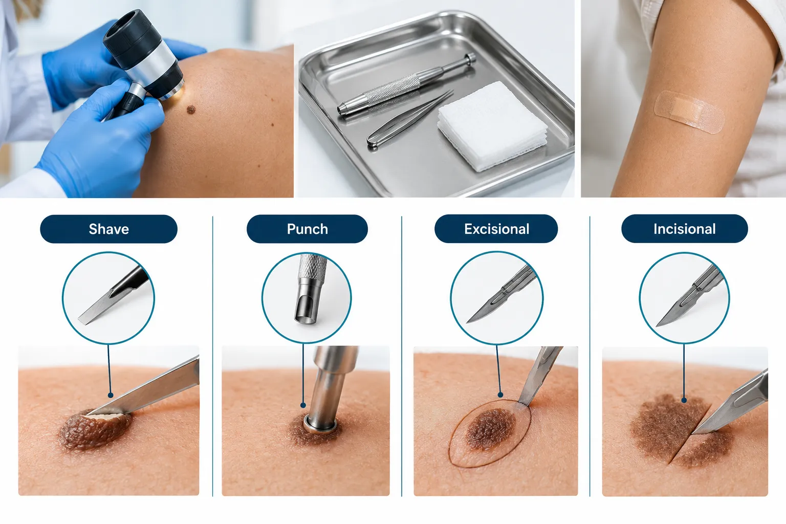
Doctors recommend a skin biopsy when a mole or patch shows features that raise concern during a clinical or dermoscopic exam: irregular borders, uneven color, recent growth, bleeding, or a new sore that will not heal. Family medicine and dermatology guidance is consistent on one point: a biopsy is required for a definitive diagnosis of suspected skin cancer, because no amount of visual inspection, even with magnification, replaces looking at the actual cells under a microscope [10]. The sample goes to a pathologist, a physician trained to identify disease patterns in tissue, who produces a written pathology report describing what the cells show.
A skin biopsy is not only for cancer screening. Biopsies can also help diagnose inflammatory skin conditions, chronic rashes, and in rarer cases, systemic diseases that show signs in the skin, researchers have even used skin biopsy tissue to detect markers of neurodegenerative conditions such as Parkinson's disease with high accuracy [9]. For most Ontario patients, though, the reason for referral is a single changing mole or an unusual new spot.
There are four standard techniques, and the choice depends on the size, depth, and suspected nature of the lesion rather than patient preference. A shave biopsy removes the top layers of skin, a punch biopsy removes a small cylindrical core, an excisional biopsy removes the entire lesion plus a margin of normal skin, and an incisional biopsy removes only part of a larger lesion.
TypeHow it's doneBest forStitches?Typical scarShave biopsyA thin blade slices off the raised portion of the lesionRaised spots thought to be superficial, such as suspected basal cell carcinomaUsually noneSmall flat markPunch biopsyA round, hollow tool cores a small cylinder of skinFlat or slightly raised lesions needing a full-thickness sampleOften one or twoSmall round scarExcisional biopsyThe entire lesion is cut out with a margin of surrounding skinSuspected melanoma, so the whole lesion and its depth can be assessedUsually yesThin line scarIncisional biopsyOnly a representative section of a large lesion is removedLarge patches or plaques too big to remove in one visitUsually yesThin line scar
For a lesion suspected to be melanoma, excisional biopsy is generally preferred because it preserves the lesion's full structure, allowing the pathologist to measure how deep the abnormal cells extend, a measurement called Breslow depth that drives staging and treatment decisions [10]. For most other suspicious moles, shave or punch biopsy is common and sufficient. A separate comparison of mole biopsy versus mole removal explains how these goals differ when a mole is removed for cosmetic reasons rather than diagnosis.
Newer micro-invasive tools are also being studied. A modified needle device described in 2026 research can collect a small, full-thickness core sample with less tissue disruption than a standard punch tool, aiming to reduce scarring while still giving the pathologist enough tissue for a full diagnosis [7]. These devices are not yet standard in most Ontario clinics.
Yes, a skin biopsy can be done for a mole specifically, in fact, a changing or atypical-looking mole is one of the most common reasons a biopsy is ordered in primary care and dermatology.
Dermoscopy is a non-invasive visual exam using a handheld magnifying device called a dermatoscope; a skin biopsy is a physical tissue sample sent for microscopic analysis. Dermoscopy helps decide whether a lesion looks concerning enough to biopsy, but it cannot provide a definitive diagnosis on its own.
A dermatoscope lets a clinician see pigment patterns, vessel structures, and borders invisible to the naked eye, which improves the accuracy of deciding who needs a biopsy and who can simply be monitored. Clinical suspicion, not dermoscopy findings alone, is what should trigger the decision to biopsy a lesion, since dermoscopy is a screening aid rather than a diagnostic test. Surveys of clinicians and patients also show growing interest in other non-invasive tools, such as reflectance confocal microscopy and electrical impedance spectroscopy, though biopsy remains the accepted gold standard for diagnosing melanocytic lesions today [3].
Decision rule: if dermoscopy shows a lesion with stable, symmetric, evenly colored features and no history of change, a clinician may recommend monitoring with photos instead of an immediate biopsy. If the lesion is asymmetric, has irregular borders, multiple colors, a diameter over 6mm, or has recently evolved, a biopsy is typically recommended regardless of how it looks under the dermatoscope. Readers wanting more detail on what a mole check involves can see the overview of a dermoscopy skin check.
A skin biopsy should not be painful during the procedure because the area is numbed first with a local anesthetic, usually lidocaine, injected just under the skin. Most people feel a brief sting or pinch from the needle, then pressure or tugging rather than pain while the sample is taken.
The injection itself is typically the most uncomfortable part, lasting a few seconds. Once the area is numb, patients commonly describe feeling pushing, scraping, or tugging sensations but not sharp pain. Mild soreness or tenderness at the site for a day or two afterward, once the anesthetic wears off, is normal and usually managed with acetaminophen if needed.
Scarring depends on the biopsy type and the body's healing response. Shave biopsies tend to leave a small, flat, light-colored mark. Punch and excisional biopsies that require stitches typically heal into a thin line scar. People prone to keloid or hypertrophic scarring should mention this to their clinician before the procedure, since technique or aftercare may be adjusted.
A skin biopsy performed to investigate a clinically suspicious skin lesion is covered by OHIP in Ontario, meaning there is no charge to the patient for the medically necessary procedure or the pathology analysis. Purely cosmetic removal of a lesion with no suspicious features, by contrast, is typically not covered and may carry a private fee.
For Ontario residents with a valid OHIP card, a physician-referred or physician-performed biopsy of a mole or spot that looks medically concerning is billed to OHIP, not the patient. In the Greater Toronto Area, clinics such as The Minor Surgery Center perform mole biopsies and skin lesion assessments without requiring a referral from a family doctor, which can shorten the wait between noticing a concerning spot and having it formally assessed.
Preparing for a skin biopsy mainly involves telling the clinician about medications, allergies, and bleeding history before the appointment. No fasting or special diet is required.
Before the procedure:
Common mistake: stopping a prescribed blood thinner without checking with the prescribing doctor first. For most minor skin biopsies, the bleeding risk is manageable without stopping the medication, and stopping it unsupervised can create other health risks.
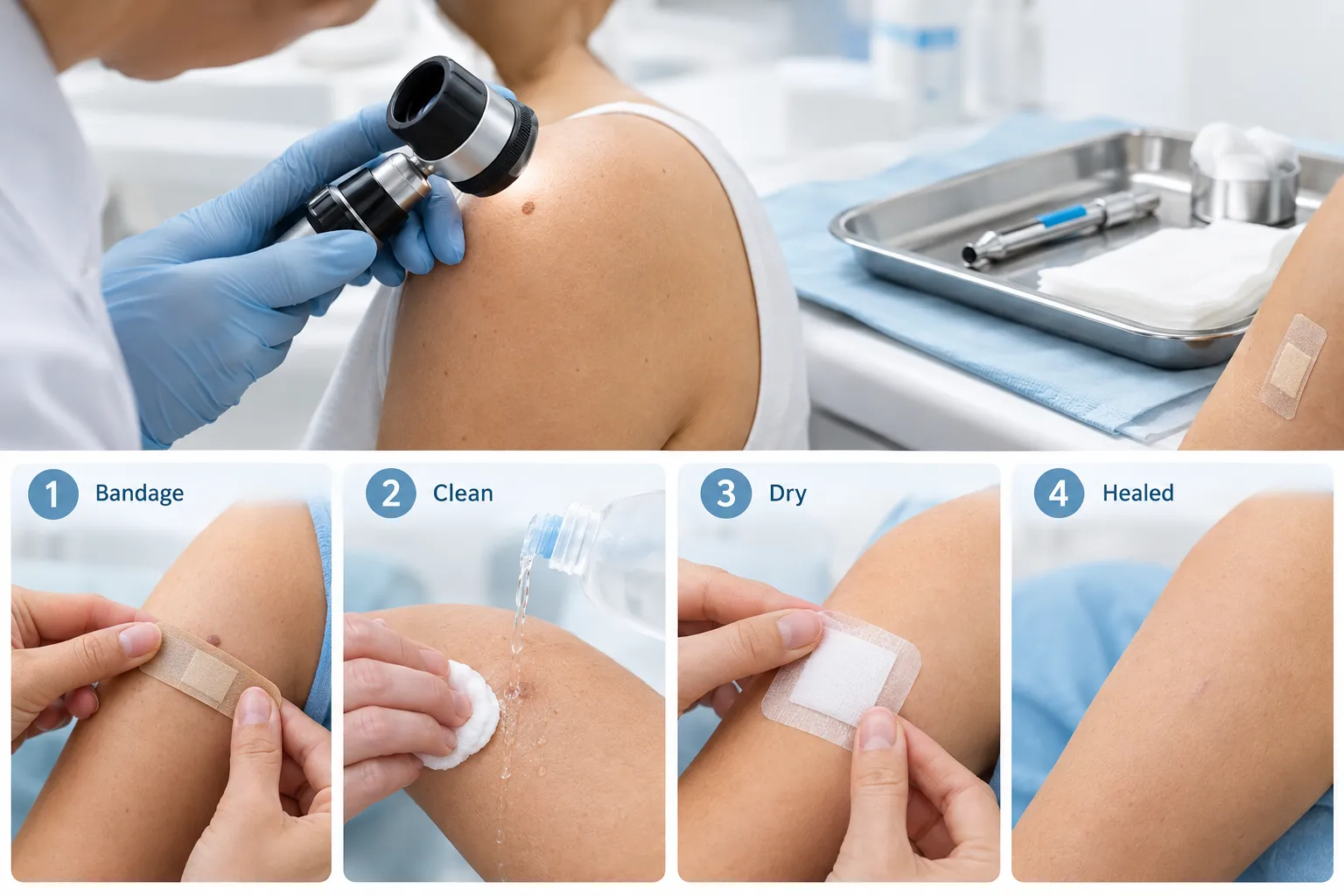
A skin biopsy procedure follows a short, predictable sequence: the area is cleaned, numbed, sampled, and dressed, typically within 15 to 30 minutes from start to finish.
A detailed walkthrough of this process, specific to moles, is available in the guide on what happens during a mole biopsy.
Biopsy site aftercare centers on keeping the wound clean, protected, and dry while it heals, with stitch removal timing depending on the body location. Following the clinician's specific instructions matters more than any general rule, since techniques and dressing types vary.
Edge case: biopsies on the lower leg or back tend to heal more slowly because of reduced blood flow or skin tension, so these sites may need longer dressing changes and a longer wait before resuming exercise.
Skin biopsy results in Ontario commonly take about one to two weeks to come back from the pathology lab, though exact timing varies by hospital, lab workload, and whether extra staining or a second specialist opinion is needed. Patients are usually asked to follow up with the ordering clinician rather than contact the lab directly.
Straightforward biopsies processed at community pathology labs tend to return results closer to the one-week mark, while cases needing immunohistochemistry stains, a second pathologist review, or referral to a specialized skin pathology centre can take longer. Clinics generally advise patients to call if they have not heard back within the expected window rather than assuming no news is good news. A detailed look at typical wait times and what affects them is available in the article on how long it takes to get biopsy results in Ontario.
A skin biopsy result generally falls into one of three categories: benign (no cancer, no concerning cell changes), atypical or dysplastic (abnormal but not cancerous, requiring monitoring or further excision), or malignant (cancer, such as melanoma, basal cell carcinoma, or squamous cell carcinoma, requiring further treatment).
The pathology report will describe the cell pattern seen, and for excised lesions, whether the margins, the edges of the removed tissue, are clear of abnormal cells. A dysplastic nevus is a mole with atypical but non-cancerous cells; depending on the degree of atypia, a doctor may recommend simply monitoring it or removing the remaining tissue with a wider margin to be safe. If the report confirms melanoma, basal cell carcinoma, or squamous cell carcinoma, the next step is usually a referral for complete removal with appropriate margins, and for some melanomas, further staging tests. Reading the specific wording on a report can be confusing; the plain-English breakdown in the pathology report explained guide covers terms like margins, atypia, and dysplastic nevus in detail.
Large-scale data illustrate why not every biopsied lesion turns out to be cancer: one nationwide analysis found an overall benign-to-malignant ratio of about 4 benign results for every 1 malignant result across all skin biopsies, improving to roughly 1.5 to 1 when biopsies were specifically ordered because of strong clinical suspicion of malignancy [8]. This is part of why clinicians use clinical judgment, sometimes supported by dermoscopy, to decide who truly needs a biopsy rather than biopsying every spot a patient is curious about.
A skin biopsy is a minor procedure with a low overall risk profile, but like any procedure that breaks the skin, it carries small risks of bleeding, infection, scarring, and rarely, a result that does not match the clinical picture.
Known risks include:
Emerging technology, including AI-assisted image analysis of biopsy slides, is being studied to reduce variability between pathologists reading the same sample, which may further improve diagnostic consistency over time [1]. For now, if a result seems inconsistent with how a lesion looked clinically, asking for a second opinion or repeat biopsy is a reasonable next step rather than something to avoid out of concern over "bothering" the system.
Can you biopsy skin at home, or do you need a doctor?
A skin biopsy must be performed by a physician or trained clinician because it involves local anesthetic, a sterile technique, and proper handling of tissue for pathology analysis. Attempting to remove or sample a mole at home carries infection risk and will not produce a usable diagnostic sample.
Can you get a skin biopsy for a mole that just looks odd, even without bleeding or pain?
Yes, a change in size, shape, color, or border is enough reason to request an assessment, since many early skin cancers cause no pain or bleeding at all.
Does a skin biopsy always leave a visible scar?
Most skin biopsies leave some mark, ranging from a barely visible flat spot after a shave biopsy to a thin line after a stitched excisional biopsy; the final appearance depends on technique, body location, and individual healing.
What happens if a skin biopsy comes back abnormal?
An abnormal result triggers a follow-up plan matched to the finding: monitoring for mild atypia, a second wider excision for a dysplastic nevus with significant atypia, or referral for full removal and possible staging if the result is melanoma, basal cell carcinoma, or squamous cell carcinoma.
Is a skin biopsy covered by OHIP in Ontario?
Yes, biopsy of a clinically suspicious skin lesion is covered under OHIP for Ontario residents with a valid health card; purely cosmetic removal of a non-suspicious lesion is generally not covered.
How soon can stitches come out after a skin biopsy?
Stitch removal timing depends on location: commonly around 5 to 7 days for facial sites and 10 to 14 days for the trunk or limbs, though the exact schedule is set by the clinician based on wound tension and healing.
A skin biopsy is a short, well-understood procedure that gives a definitive answer a visual exam alone cannot: whether a mole or spot is benign, a dysplastic nevus, or a skin cancer such as melanoma, basal cell carcinoma, or squamous cell carcinoma. For Ontario adults told by a doctor that a lesion needs sampling, the practical takeaways are straightforward: expect a 15 to 30 minute appointment, a numbing injection as the main discomfort, a simple dressing-change routine afterward, and results within roughly one to two weeks. The procedure is OHIP-covered when ordered for a clinically suspicious lesion, and options exist across the GTA, including clinics that accept patients without a referral, to get a concerning spot checked sooner rather than later. Anyone noticing a new or changing mole should book an assessment rather than wait and watch a spot that keeps changing.
[1] Full - https://www.frontiersin.org/journals/medicine/articles/10.3389/fmed.2025.1476685/full
[3] Pmc12482421 - https://pmc.ncbi.nlm.nih.gov/articles/PMC12482421/
[7] Pmc13348449 - https://pmc.ncbi.nlm.nih.gov/articles/PMC13348449/
[8] Pmc12188510 - https://pmc.ncbi.nlm.nih.gov/articles/PMC12188510/
[9] Pmc12924793 - https://pmc.ncbi.nlm.nih.gov/articles/PMC12924793/
[10] 564 Skin Cancer - https://www.aafp.org/fpe/2026/564-skin-cancer