Sun Spots vs Skin Cancer: Understanding the Critical Differences That Could Save Your Life

Every year, thousands of people notice new brown spots appearing on their skin and wonder: is this harmless sun damage or something more serious? With an estimated 112,000 new melanoma cases projected for the U.S. in 2026, representing a 6.7% increase from 2025 estimates, understanding the difference between sun spots vs skin cancer has never been more critical[3]. This comprehensive guide will help you distinguish between benign sun damage and potentially dangerous skin cancer, empowering you to take control of your skin health.

Key Takeaways

  • Sun spots (solar lentigines) are flat, uniformly colored brown spots caused by UV exposure, while skin cancer lesions often show irregular borders, multiple colors, and changes over time
  • The ABCDE criteria (Asymmetry, Border, Color, Diameter, Evolution) provides a reliable framework for identifying potentially cancerous lesions that require professional evaluation
  • Most skin cancer cases and deaths are caused by exposure to UV radiation, making sun protection the most effective prevention strategy[3]
  • Daily sunscreen use with SPF 15 or higher reduces melanoma risk by 50% when applied correctly[5]
  • Professional skin examinations are essential because some dangerous skin cancers can initially resemble harmless sun spots, making self-diagnosis unreliable

What Are Sun Spots? Understanding Solar Lentigines

Detailed landscape format (1536x1024) medical comparison infographic showing side-by-side visual characteristics of sun spots versus melanom

Sun spots, medically known as solar lentigines or age spots, are flat, brown patches that develop on sun-exposed areas of the skin. These benign (non-cancerous) lesions result from years of cumulative ultraviolet (UV) radiation exposure that triggers increased melanin production in localized areas.

Characteristics of Sun Spots

Sun spots typically display several distinctive features that help differentiate them from more concerning skin changes:

Visual Appearance:

  • ☀️ Uniform color: Usually tan, brown, or dark brown with consistent pigmentation throughout
  • 📏 Regular shape: Generally round or oval with smooth, well-defined edges
  • 📐 Flat surface: No raised texture or thickness variation
  • 📊 Size consistency: Typically range from a few millimeters to about 1 centimeter in diameter
  • 🔄 Stable appearance: Remain relatively unchanged over months and years

Common Locations:

  • Face, especially the cheeks and forehead
  • Backs of hands
  • Shoulders and upper back
  • Forearms
  • Décolletage (chest area)

These spots develop most commonly in individuals over 40 years old, though younger people with significant sun exposure can develop them earlier. Fair-skinned individuals with a history of sunburns face the highest risk of developing solar lentigines.

Why Sun Spots Develop

The formation of sun spots involves a complex biological process triggered by UV radiation. When skin absorbs UV rays, it stimulates melanocytes (pigment-producing cells) to create more melanin as a protective response. Over time, repeated UV exposure causes certain melanocytes to become overactive, producing concentrated areas of pigmentation.

Recent research published in Nature Communications reveals that UV exposure reduces levels of a protective protein called YTHDF2, which acts as a "gatekeeper" to prevent normal skin cells from becoming cancerous[1]. Scientists discovered that UV rays damage not only DNA but also RNA molecules, triggering inflammatory pathways linked to various skin conditions[1].

Understanding these mechanisms helps explain why sun protection remains critical even after sun spots have already formed. Continued UV exposure can worsen existing spots and increase the risk of developing new ones—or more serious conditions.

Understanding Skin Cancer: Types and Characteristics

Skin cancer encompasses several distinct types, each with unique characteristics and risk profiles. Understanding these differences is essential when evaluating suspicious skin lesions and determining when professional evaluation becomes necessary.

The Three Main Types of Skin Cancer

1. Basal Cell Carcinoma (BCC)

Basal cell carcinoma is the most common form of skin cancer, accounting for approximately 80% of all cases. These cancers develop in the basal cells that line the deepest layer of the epidermis.

BCC Characteristics:

  • Pearly or waxy bump appearance
  • Flat, flesh-colored or brown scar-like lesion
  • Bleeding or oozing sore that heals and returns
  • Slow-growing with minimal spreading risk
  • Rarely metastasizes but can cause local tissue damage if untreated

2. Squamous Cell Carcinoma (SCC)

Squamous cell carcinoma develops in the squamous cells that make up the middle and outer layers of the skin. SCC represents about 20% of skin cancer cases and carries a higher metastasis risk than BCC.

SCC Characteristics:

  • Firm, red nodule
  • Flat lesion with scaly, crusted surface
  • New sore or raised area on an old scar or ulcer
  • Rough, scaly patch on the lip that may evolve into an open sore
  • Red, raised patch or wart-like sore in the genital area

3. Melanoma

Melanoma is the most dangerous form of skin cancer, developing in melanocytes—the cells that produce skin pigment. Though less common than BCC or SCC, melanoma causes the majority of skin cancer deaths due to its tendency to spread rapidly.

Melanoma Characteristics:

  • Large brownish spot with darker speckles
  • Mole that changes in color, size, or feel
  • Mole that bleeds
  • Small lesion with irregular border and portions that appear red, pink, white, blue, or blue-black
  • Painful lesion that itches or burns
  • Dark lesions on palms, soles, fingertips, toes, or mucous membranes

For a comprehensive overview, explore our guide to the 4 types of skin cancer and learn about early stage skin cancer detection.

How Skin Cancer Differs From Sun Spots

The fundamental difference between sun spots vs skin cancer lies in cellular behavior. Sun spots represent a benign increase in melanin production without abnormal cell growth, while skin cancer involves uncontrolled, abnormal cell division that can invade surrounding tissues and potentially spread to other parts of the body.

Key Distinctions:

FeatureSun SpotsSkin CancerCell behaviorNormal cells with increased pigmentAbnormal, uncontrolled cell growthBorder appearanceSmooth, well-defined edgesOften irregular, jagged, or poorly definedColor uniformityConsistent throughoutMultiple colors or uneven pigmentationSize changesStable over timeOften grows or changesTextureFlat, smoothMay be raised, scaly, or crustySymptomsAsymptomaticMay itch, bleed, or hurtHealth riskCosmetic concern onlyPotentially life-threatening

Sun Spots vs Skin Cancer: The ABCDE Method for Identification

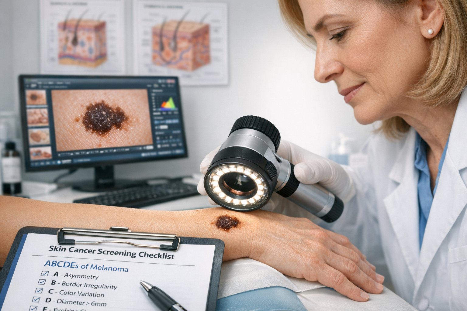
Medical professionals use a systematic approach called the ABCDE criteria to evaluate suspicious skin lesions. This method provides a structured framework for distinguishing between benign spots and potentially cancerous growths.

The ABCDE Criteria Explained

A - Asymmetry 🔄

Draw an imaginary line through the middle of a spot. If the two halves don't match, this asymmetry suggests possible malignancy. Sun spots typically display symmetrical, uniform shapes, while melanomas often appear asymmetrical.

B - Border 🔲

Examine the edges of the lesion. Sun spots have smooth, well-defined borders, while cancerous lesions often feature irregular, scalloped, or poorly defined edges that blend into surrounding skin.

C - Color 🎨

Assess color variation within the lesion. Benign sun spots maintain consistent tan or brown coloring throughout. Melanomas frequently display multiple colors including shades of brown, black, red, white, or blue within a single lesion.

D - Diameter 📏

Measure the size of the spot. While sun spots can vary in size, melanomas are often (though not always) larger than 6 millimeters (about the size of a pencil eraser). However, melanomas can be smaller, so size alone shouldn't determine whether to seek evaluation.

E - Evolution 📈

Monitor changes over time. This criterion is perhaps the most critical. Sun spots remain relatively stable, while skin cancers typically evolve—changing in size, shape, color, elevation, or developing new symptoms like bleeding, itching, or crusting.

When the ABCDE Method Indicates Concern

If a skin lesion displays any of the ABCDE warning signs, professional evaluation becomes essential. Don't wait for multiple criteria to be met—even one concerning feature warrants examination by a qualified healthcare provider.

Additional Warning Signs:

  • A sore that doesn't heal within three weeks
  • Spot that feels different from surrounding skin
  • New growth in an area of previously normal skin
  • Spot that looks significantly different from other spots on your body (the "ugly duckling" sign)
  • Lesion that bleeds easily or spontaneously

Understanding these cancer skin spots characteristics empowers individuals to identify concerning changes early, when treatment outcomes are most favorable.

Common Locations: Where Sun Spots vs Skin Cancer Typically Appear

Both sun spots and skin cancer develop most frequently on sun-exposed areas, though their specific distribution patterns can provide diagnostic clues.

Typical Sun Spot Locations

Sun spots concentrate on areas that receive the most cumulative UV exposure over a lifetime:

High-Risk Areas for Sun Spots:

  • Face: Particularly the cheeks, forehead, nose, and temples
  • Hands: Especially the backs of hands and fingers
  • Arms: Forearms and upper arms
  • Shoulders: Upper back and shoulder areas
  • Chest: Décolletage region
  • Scalp: In individuals with thinning hair or baldness

These locations correlate directly with chronic sun exposure patterns in daily life—areas that remain uncovered during routine outdoor activities.

Common Skin Cancer Sites

While skin cancer can develop anywhere on the body, including areas rarely exposed to sunlight, certain locations show higher incidence rates:

Basal Cell Carcinoma Locations:

  • Face (especially nose, forehead, and around eyes)
  • Ears
  • Neck
  • Scalp
  • Shoulders

Squamous Cell Carcinoma Locations:

  • Face
  • Ears
  • Neck
  • Lips
  • Backs of hands
  • Arms
  • Legs

Melanoma Locations:

Melanoma shows more varied distribution patterns:

  • Men: Often develops on the back, head, or neck
  • Women: Frequently appears on the lower legs
  • Both genders: Can occur on palms, soles, under nails, or in the mouth
  • Darker skin tones: More likely to develop on palms, soles, and under nails

This broader distribution pattern for melanoma underscores the importance of full-body skin examinations rather than focusing solely on obviously sun-exposed areas. Learn more about identifying age spots vs cancer spots in different locations.

The "Hidden" Skin Cancer Locations

Some skin cancers develop in unexpected places that receive minimal sun exposure:

  • Between toes
  • On the soles of feet
  • In the genital area
  • Under fingernails or toenails
  • Inside the mouth
  • On the scalp beneath hair

These locations highlight that while UV exposure represents the primary risk factor, skin cancer can develop anywhere on the body. This reality emphasizes the value of comprehensive professional skin examinations that include areas individuals might overlook during self-checks.

Risk Factors: Who Develops Sun Spots vs Skin Cancer?

Understanding personal risk factors helps individuals make informed decisions about prevention strategies and screening frequency. While sun spots and skin cancer share some common risk factors, important differences exist.

Shared Risk Factors

Both conditions share several fundamental risk factors related to UV exposure:

UV Exposure History:

  • ☀️ Chronic sun exposure over many years
  • 🔥 History of severe sunburns, especially during childhood
  • 🏖️ Frequent outdoor recreation without adequate protection
  • 💡 Regular tanning bed use
  • 🌍 Living in sunny climates or high altitudes

Skin Characteristics:

  • Fair skin that burns easily
  • Light-colored eyes (blue, green, or hazel)
  • Blonde or red hair
  • Tendency to freckle
  • Large number of moles (more than 50)

Age:

  • Risk increases with age for both conditions
  • Cumulative UV damage accumulates over decades
  • Most sun spots appear after age 40
  • Skin cancer risk increases significantly after age 50

Unique Skin Cancer Risk Factors

Beyond shared UV-related risks, several factors specifically increase skin cancer susceptibility:

Genetic and Family History:

  • Family history of melanoma or other skin cancers
  • Personal history of skin cancer
  • Inherited conditions like xeroderma pigmentosum or familial atypical multiple mole melanoma (FAMMM) syndrome
  • Fair skin with genetic variants affecting pigmentation

Medical History:

  • Weakened immune system (from organ transplantation, HIV/AIDS, or immunosuppressive medications)
  • Previous radiation therapy
  • Exposure to certain chemicals (arsenic, coal tar, paraffin)
  • Human papillomavirus (HPV) infection for certain SCC cases

Mole Characteristics:

  • Atypical (dysplastic) nevi
  • Congenital nevi (moles present at birth)
  • Large number of common moles

The American Cancer Society notes that "most skin cancer cases and deaths are caused by exposure to UV radiation"[3], emphasizing that modifiable behavioral factors play a crucial role in both conditions.

Population-Specific Considerations

Different populations face varying risk profiles:

Fair-Skinned Individuals:

  • Highest risk for both sun spots and all skin cancer types
  • Less natural melanin protection against UV damage
  • Require vigilant sun protection and regular screening

Individuals with Skin of Color:

  • Lower overall skin cancer incidence
  • Higher mortality rates when cancer develops due to later detection
  • Melanoma more likely in areas with less pigmentation (palms, soles, nail beds)
  • Often diagnosed at more advanced stages

Outdoor Workers:

  • Construction workers, farmers, landscapers, and others with occupational sun exposure
  • Significantly elevated risk for both conditions
  • Require workplace sun safety protocols

Understanding these risk factors enables personalized prevention strategies and appropriate screening schedules. Those with multiple risk factors should consider more frequent professional skin examinations at a best skin cancer clinic to ensure early detection.

Diagnosis: How Medical Professionals Distinguish Sun Spots vs Skin Cancer

Accurate diagnosis requires professional evaluation using specialized tools and techniques. While self-examination plays an important role in early detection, definitive diagnosis should always come from qualified healthcare providers.

The Clinical Examination Process

Visual Inspection:

Dermatologists begin with a thorough visual examination of the suspicious lesion and surrounding skin. They assess:

  • Overall appearance and symmetry
  • Border characteristics
  • Color uniformity and variations
  • Size and diameter
  • Texture and elevation
  • Presence of bleeding, crusting, or ulceration

Dermoscopy:

Dermoscopy (also called dermatoscopy) involves examining skin lesions using a specialized handheld device called a dermatoscope. This tool:

  • Magnifies the lesion 10-100 times
  • Uses polarized light to see beneath the skin surface
  • Reveals patterns invisible to the naked eye
  • Significantly improves diagnostic accuracy
  • Helps identify subtle features distinguishing benign from malignant lesions

Full-Body Skin Examination:

Comprehensive skin cancer screening includes examining the entire body surface, not just the area of concern. This thorough approach:

  • Identifies additional suspicious lesions
  • Establishes a baseline for future comparisons
  • Detects skin cancers in unexpected locations
  • Evaluates overall mole patterns
  • Documents concerning lesions for monitoring

When Biopsy Becomes Necessary

If visual examination and dermoscopy raise suspicion, a skin biopsy provides definitive diagnosis. Several biopsy techniques exist:

Shave Biopsy:

  • Removes the top layers of skin
  • Appropriate for suspected BCC or SCC
  • Quick outpatient procedure
  • Minimal scarring

Punch Biopsy:

  • Uses circular blade to remove deeper tissue sample
  • Captures full thickness of skin
  • Suitable for suspected melanoma
  • Provides more tissue for analysis

Excisional Biopsy:

  • Removes entire lesion plus margin of normal tissue
  • Preferred for suspected melanoma
  • Serves both diagnostic and therapeutic purposes
  • Requires stitches and longer healing time

Incisional Biopsy:

  • Removes portion of larger lesion
  • Used when complete removal isn't immediately feasible
  • Provides tissue for diagnosis before definitive treatment

The biopsy sample undergoes microscopic examination by a pathologist who evaluates:

  • Cell type and structure
  • Presence of abnormal cells
  • Depth of invasion (for cancers)
  • Margins (whether cancer extends to biopsy edges)
  • Specific cancer subtype and grade

For those seeking professional evaluation, finding a qualified provider is essential. Consider visiting a skin cancer clinic near you or exploring skin biopsy and screening services.

Advanced Diagnostic Technologies

Emerging technologies enhance diagnostic accuracy:

Digital Dermoscopy:

  • Captures high-resolution images
  • Enables precise comparison over time
  • Creates permanent records for monitoring
  • Facilitates telemedicine consultations

Confocal Microscopy:

  • Provides near-histologic resolution without biopsy
  • Examines skin at cellular level
  • Non-invasive imaging technique
  • Particularly useful for facial lesions where scarring is a concern

Artificial Intelligence (AI) Analysis:

  • Computer algorithms analyze dermoscopic images
  • Assists dermatologists in identifying suspicious features
  • Improves consistency in diagnosis
  • Particularly valuable in areas with limited specialist access

These technologies complement rather than replace clinical expertise, providing additional data points to support accurate diagnosis.

Prevention Strategies: Protecting Against Both Sun Spots and Skin Cancer

Prevention represents the most effective strategy for reducing both sun spots and skin cancer risk. While sun spots pose no health threat, preventing them requires the same UV protection measures that prevent skin cancer.

Comprehensive Sun Protection

Daily Sunscreen Application:

Daily sunscreen use with SPF 15 or higher reduces melanoma risk by 50% when applied as directed[5]. Experts recommend broad-spectrum sunscreen with SPF 30 or higher for daily use, combined with protective clothing[3][5].

Optimal Sunscreen Practices:

  • ☀️ Apply 15-30 minutes before sun exposure
  • 🤲 Use approximately one ounce (shot glass full) for full body coverage
  • 🔄 Reapply every two hours
  • 💧 Reapply immediately after swimming or heavy sweating
  • 📅 Use daily, even on cloudy days
  • ❄️ Apply year-round, including winter months

Sunscreen Selection Criteria:

  • Broad-spectrum protection (UVA and UVB)
  • SPF 30 or higher
  • Water-resistant formulation (40 or 80 minutes)
  • Non-comedogenic for facial use
  • Appropriate for skin type and sensitivity

Physical Sun Protection

Sunscreen alone provides insufficient protection. Comprehensive strategies include:

Protective Clothing:

  • 👕 Long-sleeved shirts and long pants
  • 👒 Wide-brimmed hats (at least 3-inch brim)
  • 🕶️ UV-blocking sunglasses (99-100% UVA and UVB protection)
  • 🧥 Clothing with UPF (Ultraviolet Protection Factor) rating

Behavioral Modifications:

  • Seek shade, especially during peak UV hours (10 AM - 4 PM)
  • Plan outdoor activities for early morning or late afternoon
  • Use umbrellas, trees, or structures for shade
  • Avoid tanning beds completely
  • Check UV index before outdoor activities

Environmental Awareness:

  • UV radiation reflects off water, snow, sand, and concrete
  • Higher altitudes increase UV exposure
  • Cloud cover reduces but doesn't eliminate UV radiation
  • Windows block UVB but not UVA rays

Emerging Preventive Approaches

Recent research has identified promising systemic (internal) prevention strategies:

Nicotinamide Supplementation:

A major VA study found nicotinamide reduces skin cancer incidence by up to 54% when started post-diagnosis, with a 14% overall reduction in broader populations involving over 30,000 patients[2]. Nicotinamide (vitamin B3) works by:

  • Enhancing DNA repair in UV-damaged cells
  • Reducing UV-induced immune suppression
  • Supporting cellular energy production
  • Preventing UV-induced inflammation

Vitamin D Considerations:

Recent evidence challenges earlier views: regular vitamin D supplementation has been linked to fewer melanoma cases, with a 2023 Finnish study showing reduced incidence among regular supplement users[2]. This finding requires balanced interpretation, as:

  • Vitamin D deficiency may increase cancer risk
  • Supplementation differs from sun exposure for vitamin D production
  • Optimal vitamin D levels support immune function
  • Sun exposure for vitamin D synthesis should be minimal and carefully controlled

Polypodium Leucotomos Extract:

Polypodium leucotomos extract demonstrates photoprotective effects, reducing UV-induced erythema and DNA damage[2]. This fern-derived supplement:

  • Provides antioxidant protection
  • Reduces UV-induced inflammation
  • Decreases DNA damage from UV exposure
  • Complements but doesn't replace topical sun protection

The Role of Diet in Skin Health

While no diet prevents skin cancer, certain nutritional strategies may support skin health:

Antioxidant-Rich Foods:

  • Colorful fruits and vegetables
  • Green tea
  • Dark chocolate (in moderation)
  • Foods rich in vitamins C and E

Omega-3 Fatty Acids:

  • Fatty fish (salmon, mackerel, sardines)
  • Walnuts and flaxseeds
  • May reduce inflammation

Carotenoid-Rich Foods:

  • Tomatoes (lycopene)
  • Carrots and sweet potatoes (beta-carotene)
  • May provide mild photoprotection

Learn more about diet and skin health and building a skin-healthy lifestyle.

Treatment Options: Managing Sun Spots vs Skin Cancer

Detailed landscape format (1536x1024) clinical dermatology examination scene showing dermatologist using dermatoscope to examine patient's s

Treatment approaches differ dramatically between benign sun spots and cancerous lesions, reflecting the fundamental difference in their health implications.

Sun Spot Treatment Options

Since sun spots pose no health risk, treatment remains entirely optional and cosmetic. Various approaches can reduce their appearance:

Topical Treatments:

  • Hydroquinone: Bleaching agent that lightens pigmentation over several months
  • Retinoids: Vitamin A derivatives that increase cell turnover and fade spots
  • Vitamin C serums: Antioxidant that inhibits melanin production
  • Kojic acid: Natural lightening agent from fungi
  • Azelaic acid: Reduces pigmentation and inflammation

Professional Procedures:

  • Cryotherapy: Freezing with liquid nitrogen destroys pigmented cells
  • Laser therapy: Targets melanin with specific light wavelengths
  • Intense Pulsed Light (IPL): Broad-spectrum light breaks up pigmentation
  • Chemical peels: Remove outer skin layers containing pigment
  • Microdermabrasion: Physical exfoliation of pigmented surface cells

Expected Outcomes:

  • Multiple treatments usually required
  • Results vary based on spot depth and skin type
  • New spots may develop without continued sun protection
  • Some treatments carry risks of hyperpigmentation or scarring

Skin Cancer Treatment Approaches

Skin cancer treatment depends on cancer type, size, location, depth, and patient factors. Options include:

Surgical Treatments:

Excisional Surgery:

  • Most common treatment for skin cancers
  • Removes tumor plus margin of healthy tissue
  • Margins examined to ensure complete removal
  • Requires stitches and healing time
  • Excellent cure rates for early-stage cancers

Mohs Micrographic Surgery:

  • Specialized technique for high-risk or cosmetically sensitive areas
  • Removes cancer layer by layer with immediate microscopic examination
  • Preserves maximum healthy tissue
  • Highest cure rates (up to 99% for primary BCC)
  • Preferred for facial cancers and recurrent tumors

Curettage and Electrodesiccation:

  • Scraping away cancer followed by electrical burning
  • Appropriate for small, superficial BCCs and SCCs
  • Quick office procedure
  • May leave white scar

Cryosurgery:

  • Freezing with liquid nitrogen
  • Limited to very small, superficial cancers
  • No tissue for pathologic examination
  • Lower cure rates than surgical excision

Non-Surgical Treatments:

Radiation Therapy:

  • Used when surgery isn't feasible
  • Appropriate for elderly patients or those with medical contraindications to surgery
  • Requires multiple treatment sessions
  • Effective for certain BCCs and SCCs

Topical Medications:

  • Imiquimod: Immune response modifier for superficial BCCs
  • 5-Fluorouracil: Chemotherapy cream for superficial skin cancers
  • Requires weeks of application
  • Suitable only for specific cancer types and locations

Photodynamic Therapy (PDT):

  • Applies photosensitizing agent followed by specific light exposure
  • Destroys cancer cells while preserving normal tissue
  • Used for superficial BCCs and precancerous lesions
  • Multiple sessions may be required

Melanoma-Specific Treatments:

Advanced melanoma requires more aggressive approaches:

  • Wide excision: Removes melanoma with wider margins than other skin cancers
  • Sentinel lymph node biopsy: Determines if melanoma has spread
  • Immunotherapy: Activates immune system to fight cancer cells
  • Targeted therapy: Drugs targeting specific genetic mutations
  • Chemotherapy: For advanced metastatic disease
  • Radiation therapy: Adjuvant treatment or for metastases

For detailed information about recovery expectations, see our guide on basal cell carcinoma recovery.

Treatment Success Rates

Prognosis varies significantly by cancer type and stage:

Basal Cell Carcinoma:

  • 5-year cure rate: >95% with appropriate treatment
  • Rarely metastasizes
  • Excellent prognosis with early detection

Squamous Cell Carcinoma:

  • 5-year cure rate: >95% for localized disease
  • 2-5% metastasis risk
  • Higher risk tumors require more aggressive treatment

Melanoma:

  • 5-year survival rate: 99% for localized melanoma
  • 5-year survival rate: 68% for regional spread
  • 5-year survival rate: 30% for distant metastases
  • Early detection dramatically improves outcomes

These statistics underscore the critical importance of early detection through regular skin examinations and prompt evaluation of suspicious lesions.

Self-Examination: Monitoring Your Skin for Changes

Regular self-examination enables early detection of new or changing lesions. While professional screening remains essential, self-checks between appointments can identify concerning changes promptly.

How to Perform a Skin Self-Exam

Preparation:

  • Choose a well-lit room with a full-length mirror
  • Have a hand mirror for viewing difficult areas
  • Enlist a partner to check hard-to-see locations
  • Keep a written or photographic record of existing spots
  • Schedule monthly self-exams on the same day

Systematic Examination Process:

Step 1: Face and Scalp

  • Examine face, ears, neck, and lips
  • Part hair to check scalp (use blow dryer to separate hair)
  • Check inside mouth and tongue

Step 2: Upper Body

  • Examine hands (palms, backs, between fingers, under nails)
  • Move up arms, examining all surfaces
  • Check underarms
  • Examine chest and abdomen
  • Women should check under breasts

Step 3: Lower Body

  • Sit to examine legs thoroughly
  • Check fronts, backs, and sides
  • Examine feet (tops, soles, between toes, under nails)
  • Use hand mirror for back of legs

Step 4: Back and Buttocks

  • Use hand mirror to view back in full-length mirror
  • Check shoulders, upper back, lower back
  • Examine buttocks and genital area

Step 5: Documentation

  • Note any new spots or changes to existing ones
  • Photograph concerning lesions for comparison
  • Record dates of observations
  • Track evolution over time

What to Look For During Self-Exams

The "Ugly Duckling" Sign:

Look for lesions that appear significantly different from other spots on your body. A mole or spot that stands out as unusual deserves professional evaluation, even if it doesn't meet other criteria.

Warning Signs Requiring Evaluation:

  • ⚠️ New growths or spots
  • 🔄 Changes in existing moles or spots
  • 💉 Sores that don't heal within 3 weeks
  • 🔴 Spots that itch, hurt, or bleed
  • 📈 Rapidly growing lesions
  • 🎨 Color changes in existing spots
  • 📏 Size increases in moles or spots

Limitations of Self-Examination

Self-exams provide valuable surveillance but have important limitations:

Coverage Gaps:

  • Difficult to examine scalp, back, and genital areas thoroughly
  • Easy to miss small or subtle changes
  • Limited ability to assess depth or texture accurately

Interpretation Challenges:

  • Distinguishing benign from malignant changes requires expertise
  • Some dangerous skin cancers lack obvious warning signs
  • Normal skin variations can cause unnecessary concern

False Reassurance:

  • Normal self-exam doesn't guarantee absence of skin cancer
  • Some melanomas develop quickly between exams
  • Professional examination uses specialized tools and expertise

These limitations emphasize that self-examination complements but never replaces professional screening. Individuals should maintain regular appointments with dermatologists regardless of self-exam findings.

When to Seek Professional Evaluation

Schedule an appointment with a dermatologist if you notice:

  • Any spot meeting ABCDE criteria
  • New growth that persists beyond 3-4 weeks
  • Sore that doesn't heal
  • Mole that looks different from others
  • Spot that bleeds spontaneously or with minimal trauma
  • Any concerning change, even if subtle

Don't wait for annual checkups if you identify worrying changes. Early evaluation provides peace of mind and, if cancer is present, significantly improves treatment outcomes. Consider consulting specialists at a skin cancer clinic in Toronto or Vaughan for expert assessment.

Living with Sun Damage: Long-Term Skin Health Strategies

Whether you have sun spots, a history of skin cancer, or simply want to prevent future damage, long-term skin health requires sustained commitment to protective behaviors and regular monitoring.

Post-Diagnosis Surveillance

Individuals diagnosed with skin cancer face elevated risk for developing additional skin cancers:

Recommended Follow-Up Schedule:

  • First year post-treatment: Every 3-6 months
  • Years 2-5: Every 6-12 months
  • After 5 years: Annually (or as recommended by dermatologist)
  • High-risk patients: More frequent monitoring

What Surveillance Includes:

  • Full-body skin examination
  • Lymph node palpation (for melanoma patients)
  • Discussion of new or changing lesions
  • Review of self-examination findings
  • Imaging studies if indicated

Sun Protection as a Lifestyle

Effective sun protection requires integration into daily routines:

Morning Routine:

  • Apply sunscreen before getting dressed
  • Choose protective clothing for the day's activities
  • Check UV index forecast
  • Prepare sun protection items (hat, sunglasses, umbrella)

Throughout the Day:

  • Reapply sunscreen every 2 hours
  • Seek shade during peak UV hours
  • Wear protective gear consistently
  • Remain vigilant even on cloudy days

Evening Routine:

  • Perform skin check during shower or before bed
  • Note any new or changing spots
  • Apply moisturizer to support skin barrier function
  • Plan next day's sun protection strategy

Addressing Existing Sun Damage

While prevention remains paramount, several approaches can improve existing sun damage:

Medical-Grade Skincare:

  • Retinoids to increase cell turnover
  • Antioxidants (vitamins C and E) to neutralize free radicals
  • Niacinamide to reduce inflammation
  • Alpha hydroxy acids for gentle exfoliation

Professional Treatments:

  • Chemical peels to remove damaged surface layers
  • Laser resurfacing to stimulate collagen and reduce pigmentation
  • IPL therapy for brown spots and redness
  • Microneedling to improve texture and tone

Preventive Measures:

  • Continue strict sun protection
  • Avoid additional UV damage
  • Support skin health through nutrition and hydration
  • Manage stress, which can impact skin health

Psychological Aspects of Skin Cancer Concern

Anxiety about skin cancer is common and understandable, particularly for those with previous diagnoses or high-risk factors:

Healthy Vigilance:

  • Regular self-exams without obsessive checking
  • Professional screenings at recommended intervals
  • Prompt evaluation of concerning changes
  • Balanced awareness without excessive worry

Managing Anxiety:

  • Focus on controllable factors (sun protection, screening)
  • Seek support from healthcare providers
  • Join support groups for skin cancer survivors
  • Practice stress-reduction techniques
  • Maintain perspective on actual risk levels

When to Seek Help:

  • Anxiety interfering with daily life
  • Excessive skin checking (multiple times daily)
  • Avoidance of normal outdoor activities
  • Persistent worry despite reassuring evaluations

Professional counseling can help individuals develop healthy coping strategies while maintaining appropriate vigilance.

Educating Family Members

Skin cancer risk often has genetic components, making family education important:

Share Information About:

  • Personal skin cancer diagnosis and type
  • Family risk factors
  • Importance of sun protection
  • Need for regular skin examinations
  • Self-examination techniques

Encourage Family Members To:

  • Establish care with a dermatologist
  • Practice sun protection
  • Perform monthly self-exams
  • Seek evaluation for suspicious lesions
  • Understand their personal risk factors

Children of skin cancer patients face elevated risk and benefit from early education about sun safety and the importance of skin health monitoring.

Advances in Skin Cancer Research and Future Directions

The field of dermatology and skin cancer research continues evolving, with promising developments offering hope for improved prevention, detection, and treatment.

Breakthrough Research in Prevention

Recent scientific discoveries are revealing new mechanisms of skin cancer development:

YTHDF2 Protein Discovery:

New research published in Nature Communications reveals that UV exposure reduces levels of a protective protein called YTHDF2, which acts as a "gatekeeper" to prevent normal skin cells from becoming cancerous[1]. When YTHDF2 is removed from skin cells, UV-triggered inflammation worsens, creating a vicious cycle[1].

This discovery opens potential therapeutic avenues:

  • Developing treatments that preserve or enhance YTHDF2 levels
  • Creating topical applications that protect this protein from UV damage
  • Identifying individuals with naturally lower YTHDF2 levels who may need enhanced protection

RNA Damage Understanding:

Scientists discovered that UV rays damage not only DNA but also RNA molecules, triggering inflammatory pathways linked to cancer development[1]. This expanded understanding suggests:

  • New targets for preventive interventions
  • Potential for RNA-protective compounds
  • Better understanding of inflammation's role in cancer development

Emerging Diagnostic Technologies

Diagnostic accuracy continues improving through technological innovation:

Artificial Intelligence Applications:

  • Machine learning algorithms achieving dermatologist-level accuracy
  • Smartphone apps enabling preliminary lesion assessment
  • Computer-aided diagnosis supporting clinical decision-making
  • Potential for increased access in underserved areas

Non-Invasive Imaging:

  • Reflectance confocal microscopy providing cellular-level imaging
  • Optical coherence tomography visualizing skin structure
  • Multispectral imaging detecting subtle color variations
  • Reduced need for diagnostic biopsies

Biomarker Development:

  • Blood tests detecting circulating tumor DNA
  • Genetic profiling of high-risk lesions
  • Molecular markers predicting cancer progression
  • Personalized risk assessment tools

Novel Treatment Approaches

Treatment options continue expanding, particularly for advanced skin cancers:

Immunotherapy Advances:

  • Checkpoint inhibitors (pembrolizumab, nivolumab) for advanced melanoma
  • Combination immunotherapy regimens
  • Adjuvant immunotherapy for high-risk melanoma
  • Expanding applications to advanced SCC

Targeted Therapies:

  • BRAF and MEK inhibitors for BRAF-mutant melanoma
  • Hedgehog pathway inhibitors for advanced BCC
  • Combination targeted therapy approaches
  • Personalized treatment based on tumor genetics

Topical Innovations:

  • New formulations of existing medications
  • Novel immune-modulating agents
  • Combination topical therapies
  • Improved delivery systems for better penetration

Public Health Initiatives

Broader efforts aim to reduce skin cancer incidence at the population level:

Sun Safety Education:

  • School-based sun protection programs
  • Public awareness campaigns
  • Workplace safety initiatives for outdoor workers
  • Community screening events

Policy Interventions:

  • Regulations on tanning bed access
  • Shade requirements in public spaces
  • Sunscreen provision in schools and workplaces
  • UV index reporting and warnings

Access Improvements:

  • Telemedicine skin cancer screening
  • Mobile screening units for underserved communities
  • Training primary care providers in skin cancer detection
  • Insurance coverage for preventive screenings

These multi-level approaches hold promise for reducing the burden of skin cancer across diverse populations.

Frequently Asked Questions About Sun Spots vs Skin Cancer

Q: Can sun spots turn into skin cancer?

A: Sun spots themselves do not transform into skin cancer. They represent benign accumulations of melanin without abnormal cell growth. However, sun spots indicate significant UV exposure history, which increases skin cancer risk. Additionally, skin cancer can develop near or within areas of sun damage, potentially being mistaken for a sun spot initially.

Q: How quickly does skin cancer develop?

A: Development timelines vary by cancer type. Basal cell and squamous cell carcinomas typically develop slowly over months to years. Melanoma can develop more rapidly, sometimes appearing and growing within weeks to months. Some melanomas arise from existing moles, while others develop in previously normal skin.

Q: Are sun spots permanent?

A: Without treatment, sun spots typically persist indefinitely. However, they can fade somewhat with strict sun protection and may lighten with topical treatments or professional procedures. New sun spots will continue developing with ongoing UV exposure.

Q: Can skin cancer be completely cured?

A: When detected early, most skin cancers can be completely cured with appropriate treatment. Cure rates exceed 95% for basal cell and squamous cell carcinomas. Localized melanoma has a 99% five-year survival rate. Advanced or metastatic skin cancers are more challenging to cure but increasingly treatable with modern therapies.

Q: How often should I have professional skin examinations?

A: Recommendations vary based on risk factors. Average-risk individuals should have annual skin examinations. Those with previous skin cancer, multiple atypical moles, or strong family history may need examinations every 3-6 months. Discuss appropriate screening frequency with a dermatologist based on your personal risk profile.

Q: Can people with dark skin get skin cancer?

A: Yes, though at lower rates than fair-skinned individuals. People with darker skin can develop all types of skin cancer, often in areas with less pigmentation like palms, soles, and nail beds. Unfortunately, skin cancer in darker-skinned individuals is often diagnosed at later stages, leading to worse outcomes. Everyone, regardless of skin tone, should practice sun protection and skin monitoring.

Q: Do sunscreens prevent vitamin D production?

A: While sunscreen can reduce vitamin D synthesis, most people get adequate vitamin D through diet and incidental sun exposure. The skin cancer prevention benefits of sunscreen far outweigh concerns about vitamin D deficiency. Those concerned about vitamin D levels should discuss supplementation with their healthcare provider rather than increasing unprotected sun exposure.

Conclusion: Taking Control of Your Skin Health

Understanding the differences between sun spots vs skin cancer empowers individuals to make informed decisions about skin health, recognize warning signs, and seek appropriate care when needed. While sun spots represent cosmetic concerns resulting from cumulative UV exposure, skin cancer poses serious health risks requiring prompt professional evaluation and treatment.

The distinction between these conditions often comes down to careful observation using the ABCDE criteria, awareness of personal risk factors, and commitment to regular self-examination and professional screening. With melanoma cases projected to reach 112,000 in the U.S. in 2026—a 6.7% increase from the previous year—vigilance has never been more important[3].

Your Action Plan for Skin Health

Immediate Steps:

  1. Schedule a baseline skin examination with a qualified dermatologist, especially if you've never had one or it's been more than a year
  2. Perform a thorough self-examination this week, documenting any concerning spots
  3. Evaluate your sun protection habits and identify areas for improvement
  4. Purchase broad-spectrum SPF 30+ sunscreen if you don't already use it daily
  5. Assess your UV exposure history and discuss risk factors with your healthcare provider

Ongoing Commitments:

  1. Apply sunscreen daily as part of your morning routine, regardless of weather
  2. Conduct monthly self-examinations on the same day each month
  3. Wear protective clothing including wide-brimmed hats and UV-blocking sunglasses
  4. Seek shade during peak UV hours (10 AM - 4 PM)
  5. Maintain regular professional screenings at intervals recommended by your dermatologist
  6. Stay informed about new research and recommendations in skin cancer prevention

When to Seek Immediate Evaluation:

  • Any spot meeting ABCDE criteria
  • New growth persisting beyond 3-4 weeks
  • Sore that doesn't heal
  • Spot that bleeds easily or spontaneously
  • Any concerning change in existing moles or spots

The Bottom Line

The difference between sun spots vs skin cancer can literally be life-saving knowledge. While sun spots signal UV damage and cosmetic concerns, they don't threaten health. Skin cancer, conversely, requires prompt diagnosis and treatment for optimal outcomes. The good news: most skin cancers are highly curable when detected early, and many cases are entirely preventable through consistent sun protection.

Your skin tells the story of your UV exposure history. Whether that story includes sun spots, skin cancer, or fortunately neither, the next chapter remains unwritten. The choices you make today—applying sunscreen, seeking shade, performing self-exams, and maintaining professional screening—determine your future skin health.

Don't wait for concerning changes to prioritize skin health. Prevention and early detection offer the best outcomes, and the tools for both are readily available. Take control of your skin health today by implementing the strategies outlined in this guide and consulting qualified healthcare professionals for personalized recommendations.

For expert evaluation and treatment, consider visiting a comprehensive skin cancer clinic where experienced professionals can assess your individual situation and provide evidence-based care.

Remember: your skin is your body's largest organ and deserves the same attention and care you give to your overall health. By understanding sun spots vs skin cancer and taking proactive steps toward prevention and early detection, you're investing in a healthier future.

References

[1] Skin Cancer Prevention Mechanism - https://www.womenshealthmag.com/beauty/a69620573/skin-cancer-prevention-mechanism/

[2] Systemic Photoprotection Innovative Approaches For Preventing Melanoma And Non Melanoma Skin Cancer In 2026 - https://www.skincarenetwork.co.uk/skin-cancer-news/systemic-photoprotection-innovative-approaches-for-preventing-melanoma-and-non-melanoma-skin-cancer-in-2026/

[3] Melanoma Cases 2026 Skin Cancer Risk Prevention - https://www.newbeauty.com/melanoma-cases-2026-skin-cancer-risk-prevention/

[4] Watch - https://www.youtube.com/watch?v=sYnUPxG7ONk

[5] A Sun Smart Resolution - https://www.skincancer.org/blog/a-sun-smart-resolution/

February 3, 2026
🇨🇦 Our clinic currently provides care to patients within Canada only. We apologize for any inconvenience this may cause.