Early Stage Skin Cancer on Ear Pictures: Complete Guide to Recognition and Treatment

The ear is one of the most commonly overlooked areas when people check their skin for cancer. This small but vital body part receives significant sun exposure throughout life, making it a prime location for skin cancer development. Understanding what early stage skin cancer on ear pictures reveal can be the difference between successful treatment and more serious health complications.

Skin cancer on the ear accounts for approximately 6-8% of all skin cancers, yet many people remain unaware of the warning signs. The ear's complex structure, with its curves, folds, and hard-to-see areas, makes self-examination challenging. However, recognizing the early signs through visual identification can lead to prompt medical attention and better outcomes.

Early detection remains the most critical factor in skin cancer treatment success. When skin cancer is identified in its initial stages, treatment options are typically less invasive, more effective, and associated with excellent prognosis. This comprehensive guide explores what early stage skin cancer on ear pictures show, helping readers understand the various types, warning signs, and when to seek medical attention.

Key Takeaways

Early detection is crucial: Skin cancer on the ear has a 95% cure rate when caught early, making visual recognition skills essential for everyone

A hyper-realistic image of a A highly detailed, photo-reali...

Three main types affect ears: Basal cell carcinoma, squamous cell carcinoma, and melanoma each present with distinct visual characteristics that can be identified in early stages

Regular self-examination saves lives: Monthly ear inspections using mirrors and proper lighting can help identify suspicious changes before they become serious

Professional evaluation is necessary: While pictures and self-examination are valuable tools, any suspicious lesion requires immediate medical assessment for proper diagnosis

Prevention strategies work: Simple measures like wearing hats, using sunscreen, and avoiding peak sun hours can significantly reduce ear skin cancer risk

Understanding Skin Cancer on the Ear

Why the Ear Is Vulnerable to Skin Cancer

The ear's prominent position on the head makes it particularly susceptible to ultraviolet (UV) radiation damage. Unlike other body parts that can be easily covered or shaded, the ears often receive direct sun exposure throughout the day. The ear's cartilaginous structure and thin skin provide minimal natural protection against harmful UV rays.

Several factors contribute to the ear's vulnerability:

  • Consistent sun exposure during outdoor activities
  • Lack of natural hair coverage on most ear surfaces
  • Forgotten area during sunscreen application
  • Complex anatomy that creates shadows and missed spots during protection efforts
  • Cumulative damage from years of unprotected exposure

Anatomy of the Ear and Cancer Risk Areas

Understanding ear anatomy helps identify high-risk areas where skin cancer commonly develops. The ear consists of several distinct regions, each with varying cancer risk levels:

Outer Ear (Auricle) Components:

  • Helix: The outer rim, most exposed to sun
  • Antihelix: The inner curved ridge
  • Concha: The bowl-shaped central area
  • Tragus: The small projection near the ear canal
  • Antitragus: The projection opposite the tragus
  • Earlobe: The soft, lower portion

High-Risk Areas:

  1. Helix rim - receives maximum sun exposure
  2. Upper ear cartilage - prominent and often unprotected
  3. Ear canal entrance - frequently missed during skin checks
  4. Behind the ear - hidden area with significant sun exposure

Types of Skin Cancer Found on Ears

Basal Cell Carcinoma (BCC)

Basal cell carcinoma represents the most common type of skin cancer affecting the ear, accounting for approximately 60-70% of ear skin cancers. This cancer type develops from the basal cells in the skin's deepest layer and typically grows slowly.

Early Stage BCC Characteristics:

  • Pearly or waxy appearance with raised edges
  • Pink, red, or flesh-colored bumps
  • Central depression that may bleed or crust
  • Translucent quality allowing blood vessels to show through
  • Slow growth over months or years

Visual Identification Tips:

  • Look for small, shiny bumps that don't heal
  • Notice recurring sores that heal and reopen
  • Watch for pink patches with slightly raised borders
  • Observe scar-like areas without previous injury

Early stage skin cancer on ear pictures of BCC often show small, pearl-like bumps that might be mistaken for harmless skin conditions. The key distinguishing feature is persistence - these lesions don't disappear with time and may slowly increase in size.

Squamous Cell Carcinoma (SCC)

Squamous cell carcinoma is the second most common ear skin cancer, representing about 20-30% of cases. SCC develops from squamous cells in the skin's upper layers and tends to grow more aggressively than BCC.

Early Stage SCC Features:

  • Rough, scaly patches that persist
  • Red, raised bumps that may be tender
  • Open sores that don't heal properly
  • Wart-like growths with irregular surfaces
  • Crusty or bleeding lesions

Distinguishing Characteristics:

  • Rapid growth compared to BCC
  • Rough texture rather than smooth
  • More likely to bleed when touched
  • Irregular borders and surface texture

Early stage skin cancer on ear pictures showing SCC typically display rougher, more irregular surfaces compared to the smooth, pearly appearance of BCC. These lesions often have a more aggressive appearance even in early stages.

Melanoma

Although less common on the ear (5-10% of ear skin cancers), melanoma represents the most serious type due to its potential for rapid spread. Melanoma can develop anywhere on the ear, including areas with minimal sun exposure.

Early Melanoma Warning Signs (ABCDE Rule):

  • Asymmetry: One half doesn't match the other
  • Border irregularity: Edges are ragged or blurred
  • Color variation: Multiple colors within the lesion
  • Diameter: Larger than 6mm (pencil eraser size)
  • Evolving: Changes in size, shape, or color

Melanoma Locations on Ear:

  • Helix and antihelix - most common sites
  • Concha - bowl-shaped area
  • Behind the ear - often overlooked location
  • Ear canal - rare but possible

Early stage skin cancer on ear pictures of melanoma may show subtle color variations or asymmetry that becomes more pronounced over time. Unlike BCC and SCC, melanoma can appear as a dark spot, mole, or pigmented lesion.

Recognizing Early Warning Signs

Visual Characteristics to Monitor

Successful early detection requires understanding what normal ear skin looks like versus suspicious changes. Regular self-examination helps establish baseline appearance and identify new or changing lesions.

Normal Ear Skin Features:

  • Consistent color matching surrounding skin
  • Smooth texture without persistent roughness
  • Symmetrical appearance on both ears
  • No spontaneous bleeding or discharge
  • Stable moles or spots without recent changes

Suspicious Changes to Watch:

  • New growths appearing after age 30
  • Changing existing moles or spots
  • Persistent sores that don't heal within 2-3 weeks
  • Unusual bleeding without obvious injury
  • Itching or tenderness in specific areas

The ABCDE Method for Ear Examination

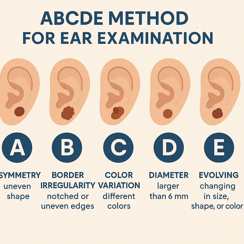
The ABCDE method provides a systematic approach for evaluating suspicious lesions on the ear. This method, originally developed for melanoma detection, applies to all skin cancer types.

Detailed ABCDE Assessment:

Asymmetry (A):

  • Draw an imaginary line through the center of the lesion
  • Compare both halves for shape and appearance
  • Benign lesions typically show symmetrical patterns
  • Cancerous lesions often display irregular, asymmetrical shapes

Border Irregularity (B):

  • Examine the edges of any spots or growths
  • Normal moles have smooth, even borders
  • Suspicious lesions show jagged, blurred, or irregular edges
  • Pay attention to areas where the lesion meets normal skin

Color Variation (C):

  • Look for multiple colors within a single lesion
  • Normal moles are typically uniform in color
  • Suspicious lesions may contain black, brown, red, white, or blue areas
  • Color changes over time are particularly concerning

Diameter (D):

  • Measure or estimate the size of suspicious lesions
  • Lesions larger than 6mm (pencil eraser size) require attention
  • However, early melanomas can be smaller than 6mm
  • Focus on changes in size rather than absolute measurements

Evolving (E):

  • Monitor changes in size, shape, color, or symptoms
  • Document changes with photos when possible
  • Any evolution in appearance warrants medical evaluation
  • New symptoms like itching, bleeding, or tenderness are significant

When to Seek Medical Attention

Immediate Consultation Indicators

Certain warning signs require prompt medical evaluation, regardless of the lesion's size or apparent severity. Early intervention can prevent progression and improve treatment outcomes.

Urgent Medical Attention Needed:

  • Rapidly growing lesions (noticeable growth within weeks)
  • Spontaneous bleeding without trauma
  • Ulceration or open sores that don't heal
  • Significant color changes in existing moles
  • New symptoms like pain, itching, or tenderness

Schedule Routine Evaluation:

  • New growths appearing after age 30
  • Persistent rough patches lasting more than a month
  • Asymmetrical or irregular lesions discovered during self-examination
  • Any concerns about skin changes, even if subtle

Professional Examination Benefits

Medical professionals possess specialized training and tools for accurate skin cancer diagnosis. While early stage skin cancer on ear pictures provide valuable reference points, professional evaluation remains essential for proper diagnosis and treatment planning.

Advantages of Professional Assessment:

  • Dermoscopy examination reveals details invisible to naked eye
  • Biopsy capabilities for definitive diagnosis
  • Staging assessment to determine cancer extent
  • Treatment planning based on specific cancer type and location
  • Follow-up monitoring to prevent recurrence

Patients seeking professional evaluation can find specialized care at facilities like The Minor Surgery Center, where experienced professionals provide comprehensive skin cancer assessment and treatment services.

Diagnostic Procedures and Imaging

Clinical Examination Process

Professional skin cancer evaluation follows a systematic approach designed to identify suspicious lesions and determine appropriate next steps. Understanding this process helps patients prepare for appointments and know what to expect.

A hyper-realistic image of a A conceptual infographic illus...

Initial Assessment Steps:

  1. Medical history review including sun exposure patterns
  2. Visual examination of the entire ear and surrounding areas
  3. Dermoscopy evaluation using specialized magnification tools
  4. Photography documentation for monitoring changes
  5. Risk assessment based on patient factors and lesion characteristics

Dermoscopy Advantages:

  • 10x magnification reveals microscopic details
  • Pattern recognition helps distinguish benign from malignant lesions
  • Vascular pattern analysis identifies abnormal blood vessel growth
  • Color assessment detects subtle variations invisible to naked eye

Biopsy Procedures

When clinical examination suggests possible skin cancer, biopsy provides definitive diagnosis. Several biopsy techniques are available, with selection based on lesion characteristics and location.

Common Biopsy Types:

Shave Biopsy:

  • Technique: Removes surface and shallow layers
  • Best for: Suspected BCC or SCC
  • Advantages: Quick procedure with minimal scarring
  • Limitations: May not provide adequate depth for melanoma

Punch Biopsy:

  • Technique: Circular tool removes full-thickness skin sample
  • Best for: Suspected melanoma or deeper lesions
  • Advantages: Provides complete tissue architecture
  • Considerations: Requires sutures but excellent diagnostic yield

Excisional Biopsy:

  • Technique: Complete lesion removal with margins
  • Best for: Small lesions or high melanoma suspicion
  • Advantages: Diagnostic and potentially curative
  • Considerations: Larger procedure but comprehensive treatment

For patients requiring biopsy procedures, facilities specializing in minor surgical procedures offer expert care. More information about available services can be found through The Minor Surgery Center's conditions page.

Treatment Options for Early Stage Ear Skin Cancer

Surgical Treatments

Early stage skin cancer treatment focuses on complete lesion removal while preserving ear function and appearance. Several surgical approaches are available, with selection based on cancer type, size, location, and patient factors.

Mohs Micrographic Surgery:

  • Technique: Layer-by-layer removal with immediate microscopic examination
  • Advantages: 99% cure rate with maximum tissue preservation
  • Best for: Recurrent cancers, large lesions, or cosmetically sensitive areas
  • Process: Same-day procedure with real-time margin assessment

Wide Local Excision:

  • Technique: Removal of cancer with surrounding healthy tissue margin
  • Margins: Typically 4-6mm for BCC, 6-10mm for SCC
  • Advantages: Single procedure with excellent cure rates
  • Considerations: May require reconstruction for larger defects

Reconstruction Options:

  • Primary closure: Direct suturing for smaller defects
  • Skin grafts: Transplanted skin from other body areas
  • Local flaps: Repositioned adjacent tissue
  • Cartilage grafts: For structural support when needed

Non-Surgical Treatments

Certain early stage skin cancers may be candidates for non-surgical treatment approaches. These options are typically reserved for specific circumstances and cancer types.

Topical Chemotherapy:

  • Agents: 5-fluorouracil (5-FU) or imiquimod
  • Mechanism: Destroys abnormal cells while sparing normal tissue
  • Best for: Superficial BCC or pre-cancerous lesions
  • Duration: Several weeks of daily application

Radiation Therapy:

  • Technique: Focused radiation beams target cancer cells
  • Advantages: Non-invasive with good cosmetic outcomes
  • Best for: Elderly patients or those unable to undergo surgery
  • Considerations: Multiple treatment sessions required

Cryotherapy:

  • Technique: Liquid nitrogen freezes and destroys abnormal tissue
  • Advantages: Quick office procedure
  • Best for: Small, superficial lesions
  • Limitations: Cannot assess complete removal margins

Prevention Strategies

Sun Protection Measures

Prevention remains the most effective strategy against ear skin cancer. Implementing comprehensive sun protection measures significantly reduces cancer risk and prevents future lesions.

Essential Protection Strategies:

Physical Barriers:

  • Wide-brimmed hats providing ear coverage (minimum 4-inch brim)
  • UV-protective clothing with tightly woven fabrics
  • Sunglasses to reduce reflected UV exposure
  • Shade seeking during peak UV hours (10 AM - 4 PM)

Sunscreen Application:

  • Broad-spectrum SPF 30+ applied to all ear surfaces
  • Reapplication every 2 hours or after swimming/sweating
  • Year-round use regardless of weather conditions
  • Adequate amount - most people apply too little sunscreen

Lifestyle Modifications:

  • UV index awareness - check daily UV forecasts
  • Outdoor activity timing - avoid peak sun hours when possible
  • Regular skin examinations - monthly self-checks plus annual professional exams
  • Protective habits - making sun protection automatic and routine

Risk Factor Management

Understanding and managing personal risk factors helps reduce skin cancer likelihood. Some factors cannot be changed, but awareness allows for enhanced surveillance and protection.

Modifiable Risk Factors:

  • Sun exposure patterns - reduce cumulative UV damage
  • Tanning bed use - eliminate artificial UV exposure
  • Sunburn frequency - prevent burns through adequate protection
  • Occupational exposure - use protective equipment for outdoor work

Non-Modifiable Risk Factors Requiring Vigilance:

  • Fair skin complexion - increased UV sensitivity
  • Family history - genetic predisposition to skin cancer
  • Previous skin cancer - higher risk of developing new lesions
  • Age over 50 - cumulative sun damage effects
  • Male gender - statistically higher ear cancer rates

Self-Examination Techniques

Monthly Ear Inspection Protocol

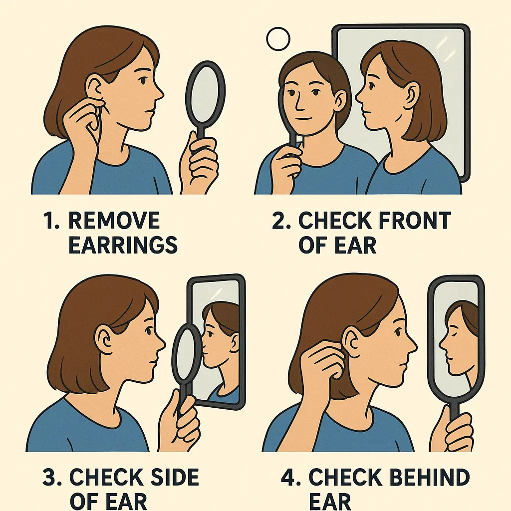
Regular self-examination enables early detection of suspicious changes. Establishing a monthly routine helps identify new lesions or changes in existing spots before they become serious.

Required Equipment:

  • Hand mirror and wall mirror for different angles
  • Good lighting - natural light or bright LED lamps
  • Magnifying glass for detailed inspection
  • Camera or smartphone for documentation
  • Examination log to track findings

Step-by-Step Examination Process:

Preparation:

  1. Choose consistent timing - same day each month
  2. Ensure adequate lighting - avoid shadows
  3. Remove earrings and clean ears gently
  4. Have documentation tools ready

Systematic Inspection:

  1. Front view examination - inspect visible ear surfaces
  2. Side angle assessment - use mirrors to see ear profile
  3. Behind-ear inspection - check hidden areas carefully
  4. Inner ear examination - look into concha and canal entrance
  5. Comparison check - compare both ears for symmetry

Documentation Process:

  • Record findings in examination log
  • Photograph suspicious areas for comparison
  • Note any changes from previous examinations
  • Schedule medical evaluation for concerning findings

Technology-Assisted Monitoring

Modern technology offers tools to enhance self-examination effectiveness and documentation accuracy. These tools supplement but don't replace professional medical evaluation.

Smartphone Applications:

  • Skin cancer detection apps provide risk assessment
  • Photo documentation features track changes over time
  • Reminder systems for regular examinations
  • Educational resources for learning recognition skills

Photography Guidelines:

  • Consistent lighting conditions for accurate comparison
  • Multiple angles to capture all ear surfaces
  • Close-up details of suspicious areas
  • Regular intervals - monthly or as recommended

When Technology Indicates Concern:

  • High-risk app assessments warrant professional evaluation
  • Documented changes in photos require medical attention
  • New lesions identified through systematic photography
  • Any uncertainty about findings should prompt consultation

For individuals seeking professional guidance on skin examination techniques or concerning findings, expert consultation is available through The Minor Surgery Center's team of experienced specialists.

Living with Early Stage Skin Cancer Diagnosis

Emotional and Psychological Support

Receiving a skin cancer diagnosis, even in early stages, can provoke anxiety and concern. Understanding that early stage ear skin cancer has excellent treatment outcomes helps patients maintain perspective and focus on recovery.

Common Emotional Responses:

  • Initial shock and disbelief at diagnosis
  • Anxiety about treatment procedures and outcomes
  • Fear of cancer spread or recurrence
  • Relief when early detection is confirmed
  • Motivation to improve prevention habits

Coping Strategies:

  • Education about condition and treatment options
  • Support groups for skin cancer patients
  • Open communication with healthcare providers
  • Family and friend involvement in care process
  • Professional counseling if anxiety becomes overwhelming

Follow-Up Care and Monitoring

Successful early stage skin cancer treatment requires ongoing surveillance to monitor for recurrence and detect new lesions. Follow-up schedules vary based on cancer type, treatment method, and individual risk factors.

Typical Follow-Up Schedule:

  • First year: Examinations every 3-4 months
  • Second year: Examinations every 6 months
  • Subsequent years: Annual examinations for low-risk patients
  • High-risk patients: More frequent monitoring as recommended

Follow-Up Examination Components:

  • Treatment site inspection for recurrence signs
  • Complete skin examination for new lesions
  • Photography documentation of healing progress
  • Patient education on self-examination techniques
  • Risk factor reassessment and prevention counseling

Patients requiring ongoing follow-up care can access comprehensive services and support through The Minor Surgery Center's clinic, where specialized teams provide continuity of care.

Frequently Asked Questions

Common Concerns About Ear Skin Cancer

Patients often have similar questions and concerns about ear skin cancer. Addressing these common inquiries helps reduce anxiety and provides practical guidance.

A hyper-realistic image of a A step-by-step diagram depicti...

"How quickly does ear skin cancer grow?" Growth rates vary by cancer type. BCC typically grows slowly over months to years, while SCC may grow more rapidly over weeks to months. Melanoma can grow quickly, making early detection crucial. Any noticeable growth warrants prompt medical evaluation.

"Can ear skin cancer spread to other parts of the body?" Early stage skin cancer on the ear rarely spreads when detected and treated promptly. BCC almost never metastasizes, SCC has low metastatic potential in early stages, and melanoma metastasis risk depends on thickness and other factors. Early detection and treatment prevent spread.

"Will treatment affect my hearing?" Most ear skin cancer treatments don't affect hearing since they involve outer ear structures rather than inner ear components. Extensive reconstructive procedures might temporarily impact hearing, but specialists work to preserve function while ensuring complete cancer removal.

"What are the chances of recurrence?" Recurrence rates for properly treated early stage ear skin cancer are very low. Mohs surgery has 99% cure rates, while standard excision has 95-98% success rates for early lesions. Regular follow-up monitoring helps detect any recurrence early.

For additional questions and detailed information, patients can access comprehensive resources through The Minor Surgery Center's FAQ section.

Advanced Prevention and Risk Assessment

Genetic and Environmental Factors

Understanding personal risk factors enables targeted prevention strategies and appropriate surveillance schedules. Both genetic predisposition and environmental exposures contribute to skin cancer development.

Genetic Risk Factors:

  • Family history of skin cancer increases personal risk
  • Fair skin, light eyes, and blonde/red hair indicate higher UV sensitivity
  • Multiple atypical moles suggest genetic predisposition
  • Previous skin cancer significantly increases future risk
  • Certain genetic syndromes like xeroderma pigmentosum

Environmental Risk Assessment:

  • Geographic location - higher UV exposure near equator and at altitude
  • Occupational exposure - outdoor workers face increased risk
  • Recreational activities - water sports, skiing increase UV exposure
  • Childhood sunburn history - severe burns increase lifetime risk
  • Tanning bed use - artificial UV exposure multiplies cancer risk

Comprehensive Protection Strategies

Effective skin cancer prevention requires multi-faceted approaches addressing various exposure scenarios and risk factors.

Daily Protection Routine:

  • Morning sunscreen application to all exposed areas including ears
  • Protective clothing selection based on daily activities
  • Hat wearing whenever outdoors, regardless of duration
  • Shade seeking during peak UV hours
  • UV-protective eyewear to reduce reflected exposure

Activity-Specific Protection:

  • Water activities: Water-resistant sunscreen with frequent reapplication
  • Winter sports: High-altitude UV protection with reflective snow consideration
  • Gardening/outdoor work: Long sleeves, hats, and frequent sunscreen reapplication
  • Beach/pool time: Umbrellas, protective clothing, and regular shade breaks
  • Driving: UV-protective window films and consistent sunscreen use

Future Developments and Research

Emerging Diagnostic Technologies

Advances in diagnostic technology continue to improve early detection accuracy and reduce the need for invasive procedures.

Innovative Diagnostic Tools:

  • Artificial intelligence imaging analyzes lesion patterns with high accuracy
  • Confocal microscopy provides real-time cellular-level imaging
  • Optical coherence tomography creates detailed tissue cross-sections
  • Molecular testing identifies genetic markers of malignancy
  • Smartphone dermoscopy makes professional-quality imaging accessible

Research Directions:

  • Blood-based screening tests for early cancer detection
  • Genetic risk profiling for personalized prevention strategies
  • Telemedicine platforms for remote skin cancer screening
  • Wearable UV monitors providing real-time exposure feedback
  • Improved imaging algorithms enhancing diagnostic accuracy

Treatment Innovations

Ongoing research continues to develop less invasive treatment options with improved outcomes and reduced side effects.

Emerging Treatments:

  • Targeted therapy drugs for advanced cases
  • Immunotherapy approaches harnessing body's natural defenses
  • Photodynamic therapy using light-activated medications
  • Nanotechnology delivery systems for precise drug targeting
  • Gene therapy approaches for high-risk genetic variants

Conclusion and Next Steps

Early stage skin cancer on ear pictures serve as valuable educational tools, but they cannot replace professional medical evaluation for suspicious lesions. The ear's prominent position and frequent sun exposure make it a common site for skin cancer development, yet early detection offers excellent treatment outcomes with cure rates exceeding 95%.

Understanding the visual characteristics of different skin cancer types - from the pearly appearance of basal cell carcinoma to the rough texture of squamous cell carcinoma and the asymmetrical features of melanoma - empowers individuals to recognize concerning changes. The ABCDE method provides a systematic approach for evaluating suspicious lesions, while regular self-examination establishes familiarity with normal ear appearance.

Prevention remains the most effective strategy against ear skin cancer. Comprehensive sun protection, including wide-brimmed hats, broad-spectrum sunscreen, and UV-avoidant behaviors, significantly reduces cancer risk. For those with increased risk factors, enhanced surveillance and professional monitoring provide additional protection.

When suspicious changes are identified, prompt medical evaluation ensures accurate diagnosis and appropriate treatment. Modern treatment options, from Mohs surgery to topical therapies, offer excellent outcomes while preserving ear function and appearance. Follow-up care and ongoing monitoring help prevent recurrence and detect new lesions early.

Immediate Action Steps: 🔍 Examine your ears monthly using proper lighting and mirrors

☀️ Implement daily sun protection including hat wearing and sunscreen application

📅 Schedule professional skin examination if you haven't had one recently

📱 Document suspicious areas with photos for monitoring changes

🏥 Seek immediate medical attention for rapidly changing or bleeding lesions

The journey from recognition to recovery begins with awareness and action. Early stage skin cancer on ear pictures provide important reference points, but personal vigilance and professional care ensure the best possible outcomes. Take charge of your skin health today - your future self will thank you.

For comprehensive skin cancer evaluation and treatment services, consider consulting with specialists who understand the unique challenges of ear skin cancer. Professional guidance ensures accurate diagnosis, appropriate treatment, and ongoing support throughout the healing process.

Remember: Early detection saves lives, and effective treatment preserves both health and quality of life. Don't wait to address concerns about skin changes - prompt action leads to better outcomes and peace of mind.

June 6, 2025