
Last updated: October 14, 2026
Seborrheic keratoses are common, harmless, waxy "stuck-on" growths that appear with age and are not cancer; they never need removal for medical reasons. Removal in Ontario is usually elective and paid out of pocket, done in one short visit with cryotherapy, curettage, or shave excision. Any spot that changes quickly, bleeds, or looks different from the others should be assessed by a physician before assuming it is a seborrheic keratosis.
This article is general information, not medical advice. See a physician for diagnosis and treatment decisions.
Seborrheic keratosis is a benign (non-cancerous) skin growth made of a buildup of normal skin cells on the outer layer of skin, and it is one of the most common skin lesions in adults past midlife. People consider removal mainly for cosmetic reasons, or because a lesion catches on clothing, becomes itchy, or bleeds after irritation.
A seborrheic keratosis forms when keratinocytes (skin surface cells) accumulate in a flat or slightly raised patch rather than shedding normally. Multiple seborrheic keratoses are common, and a sudden eruption of many new ones is sometimes called the Leser-Trélat sign, which has been linked in rare cases to an underlying internal condition and warrants a medical review rather than self-diagnosis. A related, usually smaller and darker variant that appears mostly on the face and neck in people with darker skin tones is known as dermatosis papulosa nigra; readers comparing the two conditions can review the difference between DPN and seborrheic keratosis moles.
Reasons people pursue seborrheic keratosis removal include:
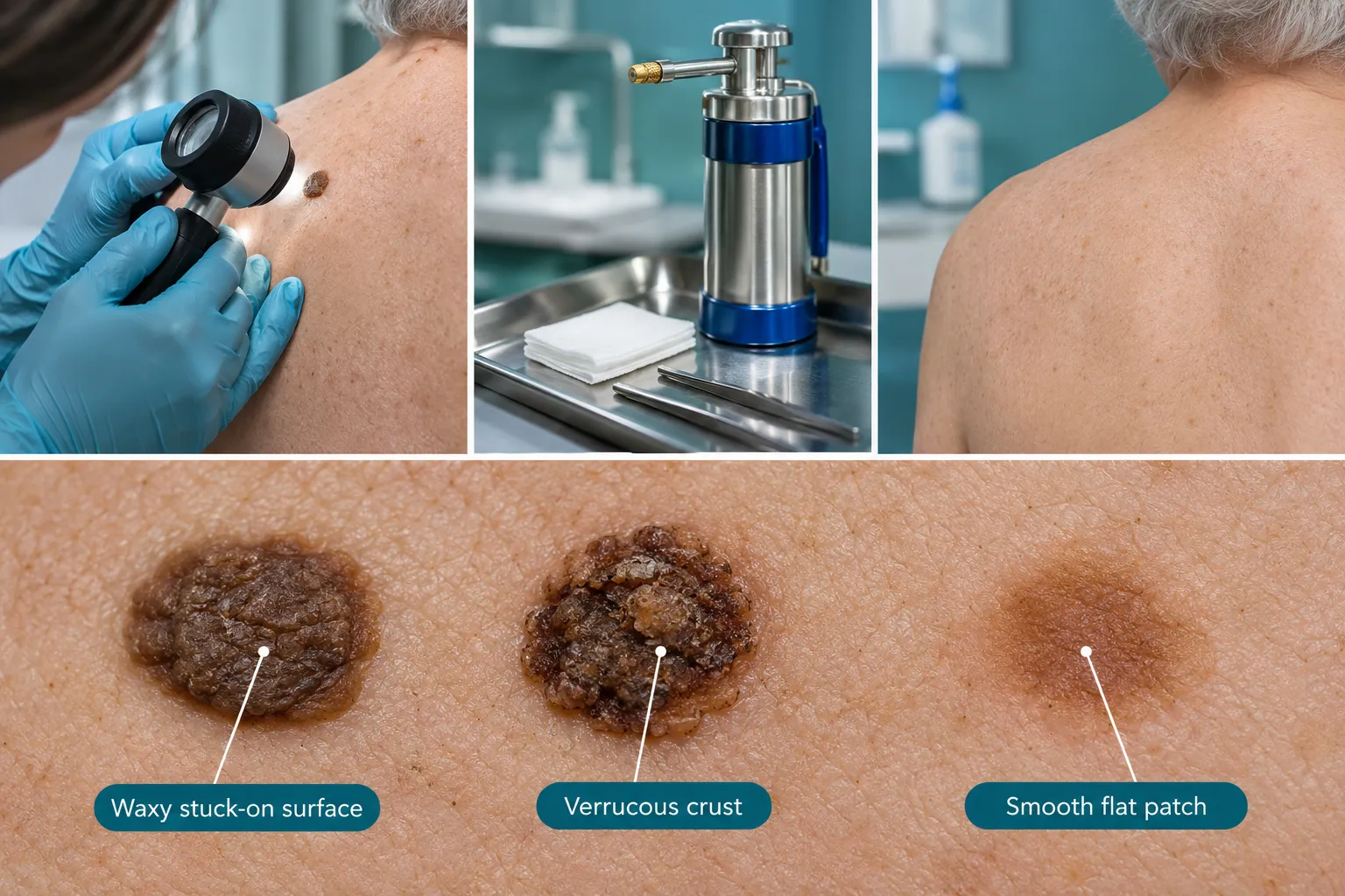
A seborrheic keratosis typically looks waxy, slightly scaly, and "stuck on" the skin surface, as if it could be peeled off with a fingernail, though it should not actually be picked at. Color ranges from light tan to dark brown or black, and the surface often has a rough, warty, or crumbly texture.
Common visual features include:
Because the waxy texture and dark colour can resemble other lesions, people often confuse seborrheic keratosis with a wart or with a sun-related brown spot. For a side-by-side breakdown, see seborrheic keratosis vs wart and seborrheic keratosis vs solar lentigo.
The exact cause of seborrheic keratosis is not fully understood, but age and genetics are the strongest known factors. The lesions become more common after age 40 and tend to run in families, and unlike age spots, they are not primarily driven by cumulative sun exposure.
Factors associated with seborrheic keratosis include:
Seborrheic keratosis is not caused by a virus, and it is not contagious, which separates it clearly from warts. Readers who want a focused comparison against sun-related pigment changes can see age spots vs cancer spots.
Seborrheic keratosis is not cancer and does not become cancer; it is a benign growth of skin cells with no malignant potential. The clinical concern is not that a seborrheic keratosis will turn into cancer, but that certain melanomas and other skin cancers can visually resemble one, which is why atypical-looking lesions need professional evaluation.
Warning signs that a spot should be checked promptly, rather than assumed to be a routine seborrheic keratosis, include:
Dermoscopy, a handheld magnification technique, helps physicians distinguish seborrheic keratosis from melanoma and other pigmented lesions without biopsy in many cases; when a lesion remains uncertain after examination, a shave or excisional biopsy allows histopathologic confirmation.[9][10] A full comparison of features is available at seborrheic keratosis vs skin cancer, and readers interested in sun-driven precancerous lesions specifically can review actinic keratosis to SCC.
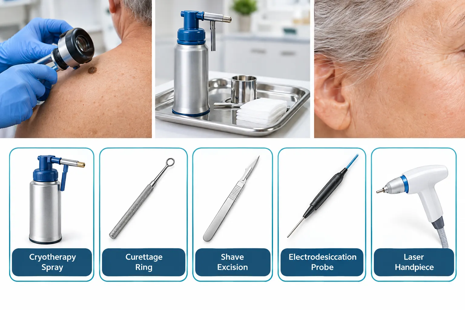
Seborrheic keratosis removal is typically done with cryotherapy, curettage, shave excision, electrodesiccation, or laser, chosen based on lesion thickness, location, and whether tissue needs to be sent for pathology. Cryotherapy using liquid nitrogen remains the most frequently used method, especially for flat or thin lesions, because it is fast and does not require anesthesia injection, but it does not allow histologic confirmation of the diagnosis.[7]
MethodHow it worksTypical sessionsScarring / pigment riskBest suited forCryotherapy (liquid nitrogen)Freezes and destroys surface cellsOften 1, sometimes repeatedRisk of permanent hypopigmentation, especially in darker skinFlat or thin lesions, low suspicion of malignancyCurettageScrapes off the lesion with a curetteUsually 1Low scarring risk; tissue can be sent for pathologyThicker, hyperkeratotic lesionsShave excisionShaves lesion flush with skin using a bladeUsually 1Low to moderate; allows histologyRaised lesions where diagnosis confirmation mattersElectrodesiccationUses electrical current to dry and destroy tissueUsually 1Possible pigment changeCombined with curettage for thicker lesionsAblative laser (CO2/Er:YAG)Vaporizes lesion layer by layerOften 1 for ablative lasersTransient redness/swelling; less hyperpigmentation than cryotherapy in some reportsCosmetic priority, pigment preservationTopical 40% hydrogen peroxideChemical application that dries the lesionMultiple applicationsMild irritationSelect superficial lesions under clinician supervision
A 2026 PRISMA-guided review of 22 laser studies covering 1,492 patients found ablative CO2 and Er:YAG lasers cleared up to 90% of lesions in a single session, while pigment-selective non-ablative lasers generally needed one to three sessions; recurrence after laser was reported at 0-6% within 12 months, with satisfaction ranging from 64-95%.[1] A 2025 randomized study comparing curettage, 532 nm laser, and cryotherapy across 123 lesions found curettage achieved the highest objective clearance (87.5%) compared with laser (55%) and cryotherapy (50%), though patients rated laser healing as more complete (90%) than curettage (87%) or cryotherapy (53%).[2][4] Electrodesiccation is most often paired with curettage for thicker lesions, and erbium:YAG laser has been reported to cause less post-treatment hyperpigmentation than cryotherapy, which matters most for people with darker skin tones.
Most seborrheic keratosis removal procedures cause brief discomfort rather than significant pain, since local anesthetic or the short duration of freezing limits sensation. Cryotherapy can cause a stinging or burning sensation during treatment and mild soreness afterward, while curettage and shave excision use local anesthetic injection so the procedure itself is typically not painful, though the injection pinch is felt briefly.
Side effect patterns reported across studies include:
Decision rule: choose curettage or shave excision if tissue confirmation matters or the lesion is thick; choose cryotherapy if the lesion is thin, flat, and clinically clear-cut; consider laser if cosmetic outcome and pigment preservation are the priority and the provider has the equipment and experience.[1][7]
Most people heal from seborrheic keratosis removal within one to three weeks, depending on the method and lesion size. Cryotherapy sites typically form a blister or crust that sheds within one to two weeks, while curettage or shave sites usually take about two to three weeks to fully re-epithelialize.
During healing, it helps to:
Family physicians, dermatologists, and minor surgery clinics in Ontario all remove seborrheic keratoses, and a referral is not always required to book a cosmetic removal consultation. The Minor Surgery Center removes seborrheic keratoses in its GTA clinics without a referral; details on the conditions page for seborrheic keratoses and current pricing are available directly. Anyone choosing a provider should confirm the clinician's experience with skin lesion assessment, since distinguishing a seborrheic keratosis from an atypical pigmented lesion before removal is part of safe practice.
Removing seborrheic keratosis at home is not recommended, and over-the-counter creams marketed for "mole and skin tag removal" are not proven to safely or completely clear these lesions. Picking, cutting, or applying acidic over-the-counter products risks infection, scarring, and skin discoloration, and it prevents a clinician from confirming the diagnosis beforehand.
Relevant context on emerging and non-recommended approaches:
Edge case: a lesion that looks exactly like a long-standing seborrheic keratosis but suddenly changes should never be treated at home with any cream, peel, or freezing device; it should be examined by a physician first.
Seborrheic keratosis removal cost in Ontario depends on the number and size of lesions, the technique used, and whether the visit is billed privately or through OHIP. TMSC's published starting price for seborrheic keratosis removal is $600, available on its pricing page; other clinics and dermatology offices set their own private-pay fees, which can vary based on lesion count, size, and chosen method.
Cost generally depends on:
For context on related elective skin-lesion procedures and pricing structures, see mole removal cost 2026 and skin lesion removal in Etobicoke.
Seborrheic keratosis removal is usually considered a cosmetic procedure in Ontario and is not covered by OHIP when done purely for appearance. A lesion that is irritated, bleeding, rapidly changing, or diagnostically uncertain may be assessed by the physician as medically necessary, in which case OHIP coverage could apply depending on clinical findings and billing criteria at the time of assessment.
Practical implications:
A removed seborrheic keratosis does not typically regrow at the exact same spot once fully treated, but new seborrheic keratoses commonly appear elsewhere on the skin over time because the underlying tendency toward these growths continues with age. Recurrence specifically at the treated site is uncommon when the lesion was fully cleared.
Recurrence and outcome data:
If removal appears to go wrong (the treated area will not heal after three to four weeks, develops increasing redness, swelling, pain, discharge, or the lesion regrows looking different from before), the person should return to the treating clinician promptly for reassessment rather than waiting or self-treating. A changed appearance at a treated site occasionally warrants a biopsy to rule out an atypical process, since removal methods like cryotherapy do not provide tissue for pathology.[7][9]
Is seborrheic keratosis contagious?
No. Seborrheic keratosis is not caused by a virus and cannot spread from person to person or from one area of skin to another through contact.
Can seborrheic keratosis turn into melanoma?
No, seborrheic keratosis does not transform into melanoma. The concern is that melanoma can sometimes look similar on the surface, which is why atypical lesions need professional evaluation rather than self-diagnosis.
What is the fastest way to remove a seborrheic keratosis?
Cryotherapy with liquid nitrogen is usually the quickest in-office method for thin, flat lesions, often completed in one short visit, though curettage and shave excision are comparably fast for thicker lesions.[7]
Does insurance or OHIP ever pay for removal?
OHIP typically does not cover removal done for cosmetic reasons, but a lesion judged medically necessary because of irritation, bleeding, or diagnostic uncertainty may be covered based on the physician's assessment.
How many sessions does laser removal usually take?
Ablative lasers often clear a lesion in a single session, while pigment-selective lasers typically require one to three sessions depending on lesion depth and colour.[1]
Should every seborrheic keratosis be removed?
No. Removal is optional for most people and is generally pursued for cosmetic reasons or physical irritation; a stable, typical-looking seborrheic keratosis does not need treatment for health reasons.[10]
Seborrheic keratosis is a benign, common skin growth, not a form of skin cancer, and most people never need it removed. When removal is wanted for comfort or appearance, cryotherapy, curettage, shave excision, electrodesiccation, and increasingly laser are the established options, each with different trade-offs in healing time, scarring, and pigment change that a clinician can match to lesion type and skin tone. Because melanoma and other skin cancers can occasionally resemble a seborrheic keratosis, any lesion that changes quickly, bleeds without obvious cause, or looks different from a person's other spots should be examined before assuming it is benign. Next steps: book an in-person skin check for any uncertain lesion, confirm whether a proposed removal is being billed privately or through OHIP, and avoid at-home creams, pens, or freezing kits on pigmented lesions that have not been professionally assessed.
[1] pubmed.ncbi.nlm.nih.gov - https://pubmed.ncbi.nlm.nih.gov/40937913/
[2] onlinelibrary.wiley - https://onlinelibrary.wiley.com/doi/full/10.1002/lsm.70042
[3] Antioxidant Keratolytic Hydrating Treatment Seborrheic Keratoses S1545961625p7977x - https://jddonline.com/articles/antioxidant-keratolytic-hydrating-treatment-seborrheic-keratoses-S1545961625P7977X/
[4] onlinelibrary.wiley - https://onlinelibrary.wiley.com/doi/10.1002/lsm.70042
[5] Nct07334600 - https://clinicaltrials.gov/study/NCT07334600
[6] Pmc12719391 - https://pmc.ncbi.nlm.nih.gov/articles/PMC12719391/
[7] Pmc13587691 - https://pmc.ncbi.nlm.nih.gov/articles/PMC13587691/
[9] Seborrheic Keratosis - https://www.mypathologyreport.ca/diagnosis-library/seborrheic-keratosis/
[10] Diagnosing Benign Skin Tumors - https://www.aafp.org/afp/2024/1000/diagnosing-benign-skin-tumors