You've noticed a new brown spot on your skin. It wasn't there last year—or maybe you just didn't notice it. Now you're wondering: is this something harmless, or should you be concerned?
If you're comparing seborrheic keratosis vs solar lentigo, you're already asking the right questions. Both are common skin growths that appear as we age, and both can look surprisingly similar at first glance. But they're actually quite different in how they form, what they mean for your health, and how they're treated.
Understanding the difference between seborrheic keratosis and solar lentigo isn't just about satisfying curiosity—it's about making informed decisions for your skin health. While neither is typically dangerous, knowing which one you're dealing with helps you and your healthcare provider create the right plan moving forward.
Let's break down everything you need to know about these two common skin conditions, so you can move forward with confidence and clarity.
Seborrheic keratosis (SK) is one of the most common benign skin growths in adults. Think of it as your skin's way of showing its age—harmless, but sometimes unwelcome.
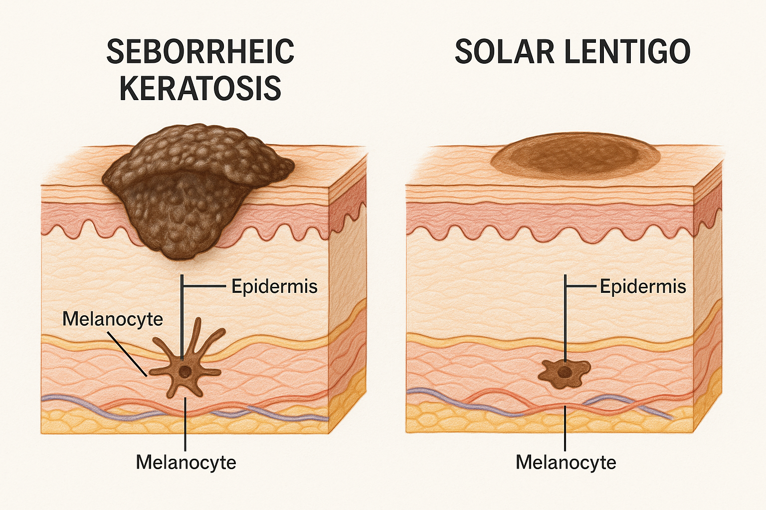
These growths typically appear as brown, black, or tan patches that look like they've been stuck onto the skin's surface. The "stuck-on" appearance is actually one of the hallmark features dermatologists look for when diagnosing SK.
Seborrheic keratoses have several distinctive features that set them apart:
Appearance: They range in color from light tan to dark brown or black. Some can even appear pink or flesh-colored, especially in their early stages.
Texture: The surface is typically waxy, scaly, or crusty. When you run your finger over one, it feels raised and rough—almost like a barnacle on your skin.
Size: They can be as small as a few millimeters or grow to several centimeters in diameter. Most fall somewhere in the half-inch to one-inch range.
Location: SK can appear almost anywhere on the body except the palms and soles. They're most common on the chest, back, face, and shoulders.
Number: Some people develop just one or two, while others accumulate dozens over their lifetime. It's not unusual to see multiple seborrheic keratoses clustered in the same area.
The exact cause of seborrheic keratosis remains somewhat mysterious, but we know several contributing factors:
Genetics play a significant role. If your parents or siblings have them, you're more likely to develop them too. Some families seem particularly prone to developing multiple SKs.
Age is the biggest risk factor. These growths are rare in people under 30 but become increasingly common after age 40. By age 60, most people have at least one.[1]
Sun exposure may contribute, though the relationship isn't as direct as with solar lentigines. SKs can appear on both sun-exposed and protected areas.
Unlike many skin conditions, seborrheic keratoses are not contagious and not cancerous. They don't transform into skin cancer, which is reassuring for many patients.
Here's the good news: seborrheic keratoses are completely benign. They pose no health risk whatsoever.
However, they can occasionally cause minor issues:
The main challenge with SK is distinguishing them from other, potentially serious skin conditions. Some melanomas can mimic the appearance of seborrheic keratoses, which is why professional evaluation matters.
If you notice a growth that's changing rapidly, bleeding without trauma, or looks different from your other spots, it's worth having it checked by experienced specialists who can provide accurate diagnosis.
Solar lentigo—commonly called an age spot, sun spot, or liver spot—is a flat, darkened patch of skin that results from years of sun exposure.
Unlike seborrheic keratoses, which are raised growths, solar lentigines (the plural form) are completely flat. They're essentially concentrated areas of melanin, the pigment that gives skin its color.
Solar lentigines have their own distinctive features:
Appearance: They're typically tan, brown, or dark brown. The color is usually uniform throughout the spot, though some may have slightly irregular borders.
Texture: Completely flat and smooth. If you close your eyes and run your finger over a solar lentigo, you wouldn't feel it—it's just a color change in the skin.
Size: Usually range from a few millimeters to about a centimeter in diameter. They're generally smaller and more uniform than seborrheic keratoses.
Location: Solar lentigines appear almost exclusively on sun-exposed areas: face, hands, arms, shoulders, and upper back. If you're seeing spots on areas that rarely see sunlight, they're probably not solar lentigines.
Number: People often develop multiple solar lentigines, especially on the backs of their hands and across their cheeks and forehead.
The cause of solar lentigo is straightforward: ultraviolet (UV) radiation from sun exposure.
Here's what happens: Over years of sun exposure, certain skin cells called melanocytes become overactive in specific areas. They produce excess melanin, creating those characteristic brown spots.[2]
Think of solar lentigines as your skin's permanent record of sun exposure. They're essentially a visible marker of cumulative UV damage over your lifetime.
Risk factors include:
Unlike seborrheic keratoses, solar lentigines are entirely preventable with consistent sun protection throughout life.
Solar lentigines themselves are benign and harmless. They don't turn into cancer.
However, they serve as an important warning sign: they indicate significant sun damage to your skin. And sun damage is the primary risk factor for skin cancer, including melanoma.[3]
People with multiple solar lentigines have a higher risk of developing skin cancer—not because the spots themselves are dangerous, but because they indicate a history of UV exposure that also increases cancer risk.
This is why dermatologists pay close attention when they see solar lentigines. It's a signal to:
If you have solar lentigines, it's wise to establish a relationship with a skin cancer screening specialist who can monitor your skin health over time.
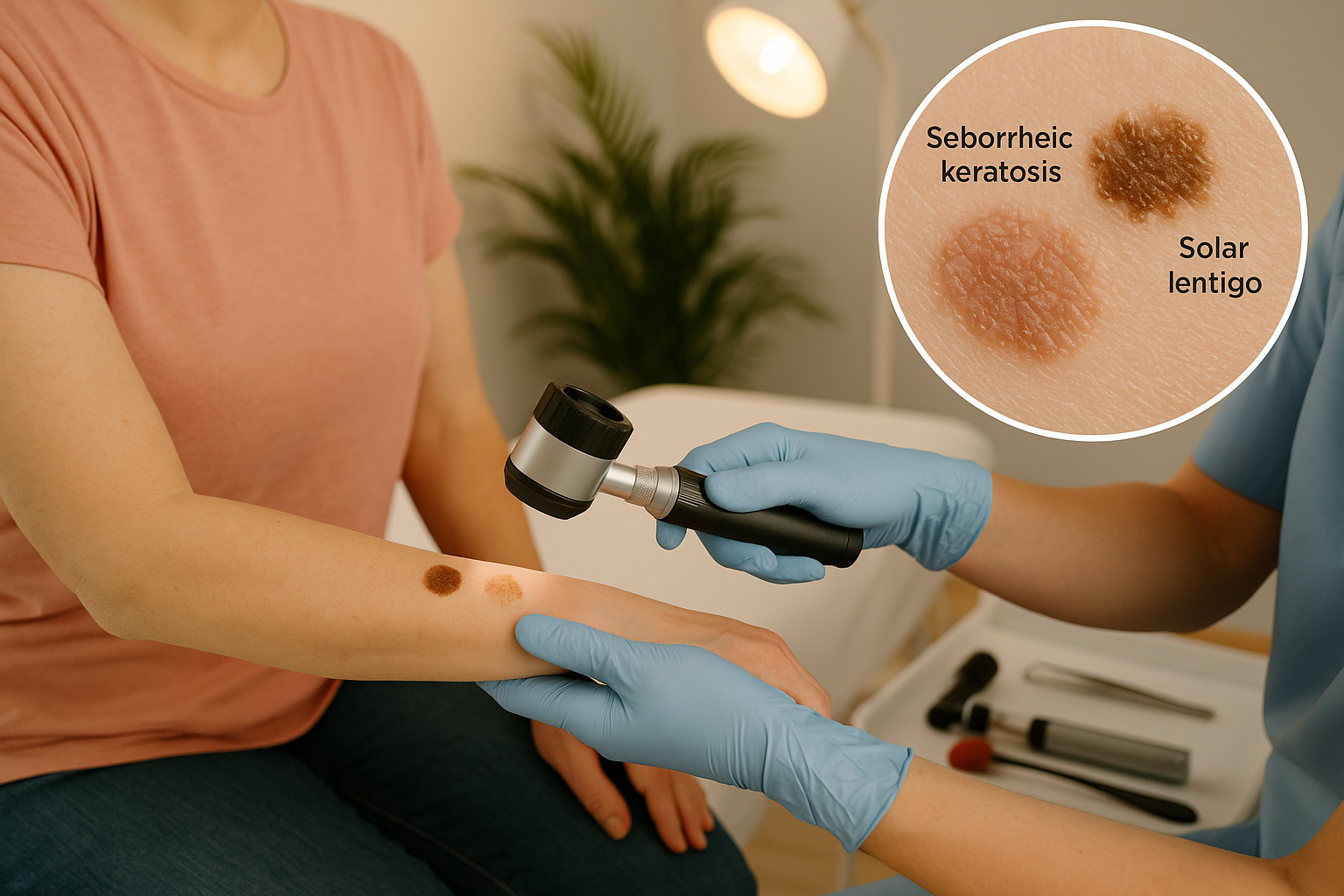
Now that we've explored each condition individually, let's put them side by side. Understanding the differences between seborrheic keratosis vs solar lentigo helps you recognize what you're seeing on your own skin—and know when professional evaluation is needed.
The most obvious difference is texture:
FeatureSeborrheic KeratosisSolar LentigoSurfaceRaised, waxy, "stuck-on"Completely flatFeelRough, scaly, bumpySmooth, no texture changeBordersOften irregular, may be well-definedUsually well-defined, regularColorTan, brown, black, sometimes pinkTan to dark brown, uniformThicknessNoticeably raised above skinLevel with surrounding skin
The "touch test" is remarkably helpful: Close your eyes and run your finger gently over the spot. If you can feel it clearly, it's likely a seborrheic keratosis. If you can't feel any change in texture, it's probably a solar lentigo.
Seborrheic keratoses can appear virtually anywhere on the body:
Solar lentigines are much more predictable:
If you're seeing spots on your chest that's usually covered by clothing, they're more likely seborrheic keratoses. Spots on the backs of your hands? Probably solar lentigines.
Seborrheic keratoses:
Solar lentigines:
This is where the seborrheic keratosis vs solar lentigo comparison becomes particularly important for your overall health:
Seborrheic keratoses result from:
Solar lentigines result from:
Understanding this difference helps explain why your healthcare provider might react differently to these two types of spots. Solar lentigines warrant a more comprehensive skin examination because they indicate sun damage.
Let's be clear about this critical distinction:
Seborrheic keratoses:
Solar lentigines:
Neither condition is cancerous, but they have different implications for your skin cancer risk profile. This is one reason why self-diagnosis isn't recommended—you want an experienced professional to evaluate any new or changing skin lesions.
For comprehensive evaluation of any concerning spots, consider visiting specialists who understand the full spectrum of skin lesions and can provide accurate diagnosis.
When you visit a healthcare provider concerned about a spot on your skin, they use several methods to distinguish between seborrheic keratosis and solar lentigo—and to rule out more serious conditions.
The first step is always a visual and tactile examination. Your doctor will:
Look closely at the lesion's characteristics:
Feel the lesion to assess:
Ask questions about:
For experienced providers, the clinical examination alone is often sufficient to distinguish between seborrheic keratosis and solar lentigo. The textural difference is usually quite obvious upon touch.
Dermoscopy (also called dermatoscopy) is a non-invasive technique that provides a magnified, illuminated view of skin structures beneath the surface.
Using a handheld device called a dermatoscope, your provider can see patterns invisible to the naked eye:
Seborrheic keratosis patterns:
Solar lentigo patterns:
Dermoscopy significantly improves diagnostic accuracy and helps providers confidently differentiate benign lesions from potentially dangerous ones.[4]
In most cases, a biopsy isn't necessary for typical seborrheic keratoses or solar lentigines. However, your doctor might recommend a skin biopsy if:
A biopsy involves removing a small sample of the lesion for microscopic examination. It's a quick, minimally invasive procedure typically done right in the office.
The results provide definitive diagnosis and can rule out skin cancer with certainty. If you're in the Toronto area and need a biopsy, experienced surgical teams can perform this procedure efficiently with a comfort-first approach.
Part of the diagnostic process involves ruling out other conditions that might look similar:
Conditions that can mimic seborrheic keratosis:
Conditions that can mimic solar lentigo:
This is precisely why professional evaluation matters. What looks like a harmless age spot could occasionally be something requiring treatment. Conversely, a scary-looking growth might be completely benign.
Your provider's expertise in distinguishing these conditions—and knowing when further testing is needed—is invaluable for your peace of mind and health.
Here's an important truth: neither seborrheic keratoses nor solar lentigines require treatment for medical reasons. Both are benign conditions that pose no health threat.
However, many people choose to have them removed for cosmetic reasons or because they're causing irritation. The good news? Several effective treatment options exist for both conditions.
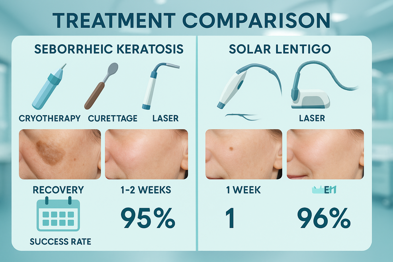
Since seborrheic keratoses are raised growths, removal techniques focus on physically eliminating the lesion.
Cryotherapy uses liquid nitrogen to freeze the growth, causing it to fall off within a few weeks.
Advantages:
Considerations:
Curettage involves numbing the area and using a special instrument to scrape off the growth.
Advantages:
Considerations:
Electrocautery uses electrical current to burn off the growth after numbing.
Advantages:
Considerations:
Laser treatment can vaporize seborrheic keratoses with minimal damage to surrounding tissue.
Advantages:
Considerations:
Solar lentigines are flat pigmented spots, so treatment focuses on lightening or removing the pigmentation.
Prescription lightening creams containing ingredients like hydroquinone, tretinoin, or kojic acid can gradually fade solar lentigines over several months.
Advantages:
Considerations:
Similar to seborrheic keratosis treatment, liquid nitrogen can be used to freeze solar lentigines.
Advantages:
Considerations:
Laser treatments (particularly Q-switched lasers and IPL) specifically target melanin in solar lentigines.
Advantages:
Considerations:
Chemical peels use acids to remove the top layers of skin, including pigmented cells.
Advantages:
Considerations:
The best treatment for you depends on several factors:
At The Minor Surgery Center, our experienced surgical team takes a personalized approach to treatment planning. We'll discuss your specific situation, explain your options clearly, and help you choose the approach that best fits your needs and lifestyle.
Whether you're dealing with a single bothersome spot or multiple lesions, we offer safe and efficient surgery with minimally invasive solutions designed to help you get back to your life quickly.
When it comes to prevention, seborrheic keratosis and solar lentigo are quite different. One is largely unavoidable, while the other is highly preventable.
Unfortunately, there's no proven way to prevent seborrheic keratoses. Since they're primarily related to genetics and aging, they're not something you can avoid through lifestyle changes.
However, you can:
Monitor your skin regularly for new growths or changes in existing ones. Early detection of anything unusual is always beneficial.
Protect existing lesions from irritation. If you have seborrheic keratoses in areas where clothing or jewelry rubs, consider having them removed to prevent bleeding or discomfort.
Avoid picking or scratching at them, which can cause bleeding, infection, or inflammation.
Think of seborrheic keratoses as similar to gray hair—they're a natural part of aging that happens to most people eventually. There's no shame in having them, and they don't reflect anything you did wrong.
Solar lentigines, on the other hand, are highly preventable because they result directly from UV exposure. The key is comprehensive sun protection throughout your life.
Daily sunscreen use is your first line of defense:
Protective clothing provides physical barriers:
Behavioral modifications reduce exposure:
Regular skin checks help catch changes early:
Even if you already have solar lentigines, improving your sun protection habits now still matters. It can:
Think of it like this: past sun damage is done, but you control what happens from this point forward. Every day of good sun protection is an investment in your future skin health.
For comprehensive guidance on protecting your skin and monitoring for concerning changes, our blog offers evidence-based information on various skin health topics.
While both seborrheic keratoses and solar lentigines are typically harmless, certain situations warrant professional evaluation. Knowing when to seek medical attention can provide peace of mind—and occasionally catch something serious early.
See a healthcare provider if you notice:
🚩 Rapid changes in size, shape, or color
🚩 Bleeding without trauma
🚩 Irregular borders
🚩 Multiple colors within one lesion
🚩 Asymmetry
🚩 Diameter larger than 6mm (about the size of a pencil eraser)
🚩 Evolution or change over time
These are components of the ABCDE rule for melanoma detection, which applies to any pigmented lesion:[5]
Even if your spot seems to match the description of seborrheic keratosis or solar lentigo perfectly, self-diagnosis has limitations.
Here's why professional evaluation matters:
Accuracy: Trained providers see hundreds of skin lesions and can spot subtle differences you might miss.
Peace of mind: Uncertainty about a spot can cause significant anxiety. A definitive diagnosis lets you stop worrying.
Early detection: If a lesion turns out to be something concerning, early identification dramatically improves outcomes.
Comprehensive assessment: Providers examine your entire skin health picture, not just the spot that brought you in.
Documentation: Professional photos and notes create a baseline for tracking changes over time.
Treatment planning: If removal is desired, providers can recommend the best approach for your specific situation.
Understanding what happens during a skin evaluation can reduce anxiety about scheduling that appointment:
Medical history: Your provider will ask about the lesion's timeline, symptoms, and your sun exposure history.
Full skin examination: Even if you came in for one spot, a thorough provider will check your entire skin surface. Melanomas often appear in unexpected places.
Dermoscopy: Non-invasive magnified examination of concerning lesions.
Photography: Documentation for future comparison.
Discussion: Clear explanation of findings and recommendations.
Biopsy if needed: Quick, minimally uncomfortable procedure if diagnosis is uncertain.
Treatment planning: If removal is desired, discussion of options, timing, and expectations.
The entire visit typically takes 15-30 minutes, and you'll leave with clear answers and a plan forward.
At The Minor Surgery Center, we pride ourselves on clear communication and a comfort-first approach. We understand that skin concerns can be stressful, and we're here to provide reassurance along with expert care.
Beyond addressing specific concerns, establishing a relationship with a skin health provider for regular monitoring is wise, especially if you:
Annual skin checks can catch problems early when they're most treatable. Think of it like dental cleanings—preventive care that protects your long-term health.
For those in the Toronto area seeking expert evaluation, our team provides comprehensive skin cancer screening with personalized treatment plans when needed.
If you've been diagnosed with seborrheic keratoses or solar lentigines, you're in good company. These are among the most common skin changes in adults, and most people develop at least a few as they age.
Let's be honest: these spots can be frustrating, especially when they appear on visible areas like your face or hands.
You have options:
Embrace them: Many people choose to accept these spots as natural signs of aging. They're not dangerous, and they don't define you.
Camouflage them: Makeup can effectively conceal spots that bother you, especially solar lentigines. Dermatologists can recommend products designed for this purpose.
Remove them: If spots genuinely impact your confidence or quality of life, removal is a reasonable choice. It's not vain to want to feel comfortable in your skin.
Selective removal: You don't have to remove all of them—just the ones that bother you most.
The decision is entirely personal. What matters is that you feel good about your appearance and comfortable in your own skin.
Skin changes can affect how we feel about ourselves, and those feelings are valid.
Some people experience:
If these feelings are significant, talking with a healthcare provider can help. They can:
Remember: seeking treatment for cosmetic concerns is perfectly legitimate. Your emotional well-being matters just as much as your physical health.
Living with these conditions doesn't require major lifestyle changes, but a few adjustments can help:
For seborrheic keratoses:
For solar lentigines:
For both:
Here's an important perspective: while these spots might annoy you, they're also reminders to pay attention to your skin health.
Solar lentigines, in particular, serve as visible markers that sun protection matters. They're your skin's way of telling you to be more careful going forward.
Seborrheic keratoses remind us that bodies change as we age, and that's completely normal. They're not a reflection of anything you did wrong—just a natural part of the aging process.
Both conditions offer opportunities to:
Understanding the difference between seborrheic keratosis vs solar lentigo empowers you to make informed decisions about your skin health.
You now know:
✓ What each condition is and how to recognize it ✓ Why they develop and what they mean for your health ✓ When to seek professional evaluation ✓ What treatment options exist ✓ How to prevent future sun damage
This knowledge helps you move forward with confidence rather than anxiety. You're equipped to recognize normal aging changes, identify concerning signs, and seek appropriate care when needed.
And remember: whether you choose to treat these conditions or simply monitor them, you're making the right choice for you. There's no single "correct" approach—only what works best for your individual situation, values, and goals.
The comparison of seborrheic keratosis vs solar lentigo reveals two common, benign skin conditions that share some similarities but differ in important ways.
Seborrheic keratoses are raised, waxy growths that appear as we age—harmless barnacles on the skin that require no treatment unless they bother you. They're genetic, unavoidable, and carry no cancer risk.
Solar lentigines are flat, pigmented spots that result from cumulative sun exposure. While benign themselves, they signal UV damage and remind us that sun protection matters for skin cancer prevention.
The key differences—texture, location, and underlying cause—help distinguish between them. But professional evaluation remains essential for accurate diagnosis and peace of mind.
If you're concerned about spots on your skin:
1. Schedule an evaluation with a qualified provider who can examine your skin and provide definitive diagnosis.
2. Discuss your concerns openly, including both health worries and cosmetic preferences.
3. Explore treatment options if you're interested in removal, keeping in mind that treatment is optional for both conditions.
4. Commit to sun protection going forward to prevent new solar lentigines and reduce skin cancer risk.
5. Establish regular monitoring through self-checks and professional skin examinations.
At The Minor Surgery Center, we understand that skin concerns can be stressful. Whether you're worried about a changing spot or simply want cosmetic improvement, our experienced surgical team provides expert outpatient care with a personalized treatment plan tailored to your needs.
We offer minimally invasive solutions delivered with clear communication, compassion, and efficiency. Our goal is simple: help you get back to your life with confidence and peace of mind.
Ready to address your skin concerns? Contact The Minor Surgery Center today to schedule a consultation. We're here to provide the answers you need and the care you deserve.
Your skin health matters. Let's work together to keep it healthy, address your concerns, and help you feel comfortable and confident in your own skin.
[1] Hafner C, Vogt T. Seborrheic keratosis. Journal der Deutschen Dermatologischen Gesellschaft. 2008;6(8):664-677.
[2] Ortonne JP, Bissett DL. Latest insights into skin hyperpigmentation. Journal of Investigative Dermatology Symposium Proceedings. 2008;13(1):10-14.
[3] American Academy of Dermatology Association. Sunscreen FAQs. Updated 2025. Accessed January 2025.
[4] Argenziano G, Soyer HP. Dermoscopy of pigmented skin lesions—a valuable tool for early diagnosis of melanoma. The Lancet Oncology. 2001;2(7):443-449.
[5] American Cancer Society. Signs and Symptoms of Melanoma Skin Cancer. Updated 2025. Accessed January 2025.


