
Last updated: October 14, 2026
A pyogenic granuloma is a benign, fast-growing red bump made of small, tightly packed blood vessels. It bleeds easily with even minor contact, usually appears on fingers, lips, the face, or gums, and almost never resolves without treatment. Most are removed in a short clinic procedure, shave excision with cautery, curettage, or full excision, and sent for pathology to confirm the diagnosis and rule out lookalike conditions such as amelanotic melanoma.
A pyogenic granuloma is a benign overgrowth of small blood vessels in the skin or mucous membrane, medically known as lobular capillary hemangioma. It is not caused by bacteria and is not a true granuloma in the infectious sense, despite the misleading name.
The lesion forms when capillaries proliferate rapidly in a small area, creating a soft, raised, vascular nodule. It most often develops on:
Major medical references classify pyogenic granuloma as a benign vascular skin tumor with a well-documented clinical course [1][2]. Oral and gingival lesions have distinct drivers, including hormonal change and chronic local irritation, as outlined in recent reviews of oral pyogenic granuloma [3].
A pyogenic granuloma typically appears as a small, dome-shaped, bright red to purplish-red bump with a shiny, moist surface that may look like raw tissue. It grows noticeably over days to a few weeks and bleeds easily, even from light touch or clothing friction.
Typical physical features include:
Because the lesion is fragile and vascular, it bleeds out of proportion to its size when bumped, scratched, or rubbed against a surface. This bleeding pattern, combined with rapid growth, is one of the main reasons people seek assessment. For a focused look at distinguishing it from similar-looking red spots, see this comparison of cherry angiomas and pyogenic granulomas.
Pyogenic granulomas form after an exaggerated vascular healing response, most often triggered by minor local injury, hormonal change, or certain medications. The exact cause is multifactorial, and in many cases no single trigger is identified.
Recognized contributing factors include:
A practical example: a small kitchen cut on a fingertip that seems to heal normally but then develops a growing, easily bleeding red bump over the following two to three weeks is a classic presentation pattern seen in clinical reviews [1][5].
A pyogenic granuloma bleeds heavily because it is made almost entirely of small, thin-walled blood vessels packed close to the skin surface, with minimal protective tissue covering them. Even gentle friction can rupture these vessels and cause disproportionate bleeding for the lesion's size.
Practical ways to manage bleeding at home before a medical assessment:
A common mistake is treating a bleeding bump like a typical cut by scrubbing it or applying antiseptic ointment repeatedly. This irritates the fragile vessels further and can prolong bleeding episodes. Lesions that bleed repeatedly over several weeks are generally a signal that removal, rather than ongoing home management, is the appropriate next step.
A pyogenic granuloma itself is benign and is not a form of skin cancer. However, because it can resemble more serious lesions on visual inspection alone, pathology confirmation after removal is considered standard practice.
The main lookalike of clinical concern is amelanotic melanoma, a form of melanoma that lacks typical dark pigment and can appear as a pink, red, or skin-colored nodule that grows and bleeds in a similar pattern. Other conditions in the differential diagnosis include:
Diagnostic guidance consistently recommends histopathologic examination of excised tissue to confirm the diagnosis and exclude malignancy, particularly for lesions that are atypical in appearance, recur after treatment, or arise without a clear trigger [1][3]. Visual presentations that mimic melanoma, including nodular and amelanotic subtypes, are discussed further in this overview of nodular melanoma appearance. Understanding what a pathology report actually shows can also help interpret results once a specimen is reviewed; see this plain-English guide to pathology reports.
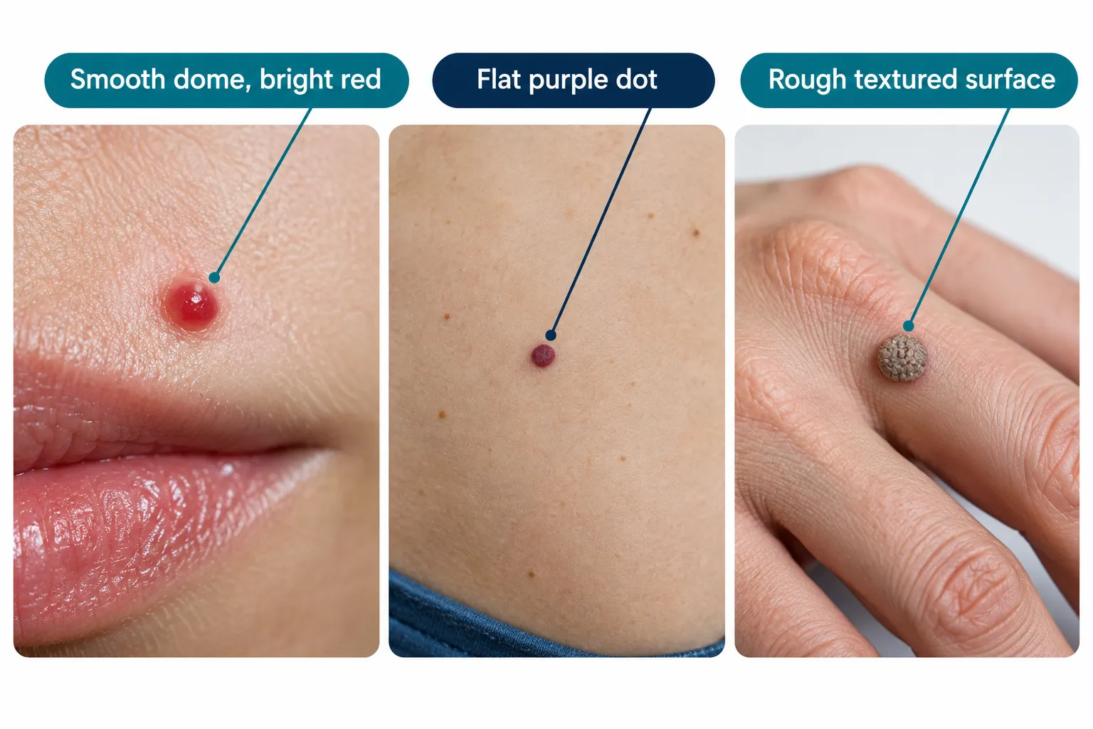
A pyogenic granuloma grows rapidly and bleeds easily, a cherry angioma grows slowly and rarely bleeds, and a wart has a rough, dry surface caused by a virus rather than blood vessel overgrowth. Growth speed, surface texture, and bleeding tendency are the three most useful features for telling them apart at home before a clinical check.
FeaturePyogenic granulomaCherry angiomaWartLookShiny, moist, red to purple domeSmooth, flat or slightly raised, bright red/purple dotRough, dry, grey or skin-coloured bumpGrowth speedFast (days to weeks)Slow (months to years)Slow, can spread locallyBleedingBleeds easily and oftenRarely bleeds unless scratchedBleeds only if picked or cutTypical locationFingers, lips, face, gumsTrunk, arms (common with age)Hands, feet, fingersCauseVascular overgrowth, injury, hormonesBenign vascular proliferation, agingHPV viral infectionTreatmentShave excision, curettage, or excisionCautery, laser, or excision if desiredCryotherapy, salicylic acid, excision
A side-by-side look at the vascular lesions specifically is covered in this guide comparing cherry angiomas and pyogenic granulomas, and a broader overview of red skin spots is available in this article on red spots on skin.
A pyogenic granuloma is usually removed using shave excision combined with electrocautery, curettage with cautery, or full surgical excision, depending on lesion size, location, and recurrence history. The removed tissue is routinely sent to pathology to confirm the diagnosis.
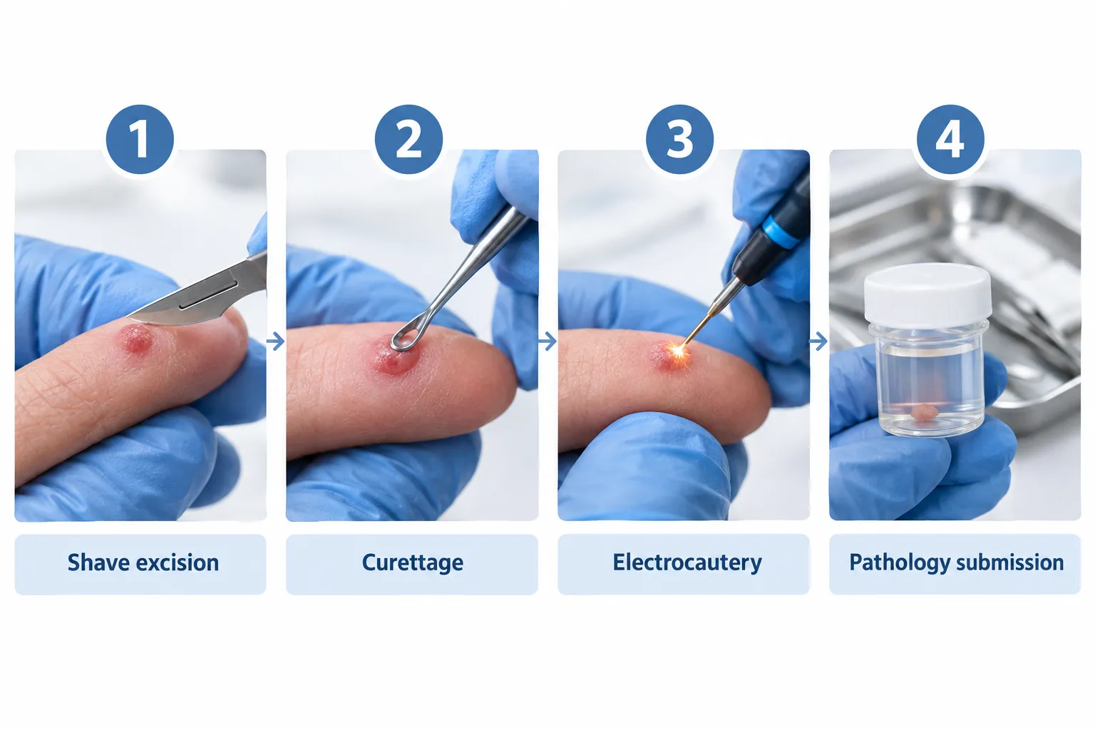
MethodWhat it involvesRegrowth riskScarringBest forShave excision + cauteryLesion shaved flush with skin, base cauterized to stop bleedingLow to moderateMinimal, flat scarMost finger, face, and lip lesionsCurettage + cauteryLesion scooped out with a curette, base cauterizedModerateSmall, round scarSmaller or superficial lesionsFull surgical excisionEntire lesion and margin of surrounding tissue removed, wound stitchedLowestLinear scar, usually fine over timeRecurrent, deep, or diagnostically uncertain lesionsLaser ablationVascular laser targets and destroys blood vesselsModerateMinimalSmaller lesions, cosmetically sensitive sitesSilver nitrateChemical cauterization applied topicallyHigher for larger lesionsMinimalVery small, early lesions, especially in children
Pediatric cases are increasingly managed with conservative, tissue-sparing protocols that favour the least invasive option likely to succeed, reserving full excision for lesions that fail simpler treatment or recur [3]. For stubborn or recurrent oral lesions, recent protocols describe combining surgical removal with adjunct measures such as correcting local irritants, which supports a multimodal approach rather than relying on one technique alone [3][6]. Conjunctival (eye-area) pyogenic granulomas require specialized ophthalmic technique and follow-up distinct from skin lesions, as described in region-specific clinical data [3].
Standard surgical excision continues to show high success rates in practical reviews of treatment outcomes, making it the preferred option when a lesion has recurred after a more conservative method [5].
Yes, a pyogenic granuloma can recur, particularly if the original treatment did not remove the full base of the lesion. Recurrence is less common after full surgical excision with margin control compared with surface-level treatments like curettage or silver nitrate alone.
Factors that influence recurrence risk:
A practical decision rule: choose full excision over shave or curettage if a lesion has already regrown once, if it sits in a high-recurrence site like the lip or gumline, or if the diagnosis remains uncertain after an initial superficial treatment.
Pyogenic granulomas that develop during pregnancy, most often on the gums, are specifically called granuloma gravidarum and are linked to pregnancy hormone changes. These lesions often shrink or resolve partially after delivery, but persistent or bleeding lesions may still need removal.
Key points for pregnancy-associated cases:
Spontaneous resolution is uncommon in adults, and most pyogenic granulomas continue to grow and bleed until actively treated. Some pregnancy-related oral lesions are an exception, as they can shrink after delivery once hormone levels normalize [3].
Outside of that pregnancy-related pattern, waiting for a pyogenic granuloma to disappear without treatment usually means continued bleeding episodes, gradual size increase, and a longer window during which a lookalike condition like amelanotic melanoma cannot be confidently ruled out. This is the main reason clinical guidance favours removal and pathology review rather than observation alone for most lesions [1][2].
There is no proven home remedy that reliably shrinks or clears a pyogenic granuloma, and over-the-counter treatments are not a substitute for clinical removal. Home care should focus only on protecting the area and controlling bleeding until it can be assessed.
Reasonable home measures include:
Attempting to cut, tie off, or burn the lesion at home is not recommended, since it increases infection risk, causes uncontrolled bleeding, and removes the opportunity for pathology confirmation.
Pyogenic granulomas can affect anyone but are more frequently reported in children and young adults, pregnant women, and people on certain medications or with specific skin or mucosal injuries. No single risk group accounts for all cases, since the underlying trigger is often multifactorial [3].
Groups more frequently represented in clinical literature:
A pyogenic granuloma that bleeds repeatedly, interferes with daily function, or cannot be confidently distinguished from a more serious lesion is commonly treated as medically necessary, which can make removal eligible for OHIP coverage. The final determination of medical necessity and coverage is made by the treating physician at the time of assessment.
Lesions removed primarily for cosmetic reasons, without bleeding or diagnostic concern, may not meet the same criteria. A clinician typically evaluates the lesion's behaviour, bleeding history, and appearance before confirming whether it qualifies as medically necessary.
The Minor Surgery Center removes pyogenic granulomas in Greater Toronto Area clinics without requiring a referral; details on the assessment and removal process are outlined on its pyogenic granuloma page. General information on accessing minor skin surgery without a referral in Ontario is also available in this overview of no-referral skin surgery in Toronto.
Is a pyogenic granuloma an infection?
No. Despite the name, a pyogenic granuloma is not caused by bacteria and is not an infection. It is a benign overgrowth of small blood vessels, medically termed lobular capillary hemangioma [1].
How fast does a pyogenic granuloma grow?
Most pyogenic granulomas reach noticeable size, often 0.5 cm to 2 cm, within days to a few weeks of first appearing. Rapid growth combined with frequent bleeding is a hallmark feature used in clinical assessment [1][5].
Does removing a pyogenic granuloma leave a scar?
Minor scarring is common after removal, and the size and texture of the scar depend on the technique used. Shave excision and curettage typically leave a small, flat mark, while full surgical excision leaves a thin linear scar that usually fades over time.
Can a pyogenic granuloma turn into melanoma?
A pyogenic granuloma does not transform into melanoma. The concern is diagnostic, not transformative: amelanotic melanoma can visually mimic a pyogenic granuloma, which is why removed tissue is routinely checked by pathology [1][3].
Why do pyogenic granulomas form on the gums during pregnancy?
Elevated estrogen and progesterone during pregnancy can trigger an exaggerated vascular response in gum tissue, producing a lesion known as granuloma gravidarum. These lesions often shrink after delivery once hormone levels return to baseline [3].
What happens if a pyogenic granuloma is left untreated?
Without treatment, most pyogenic granulomas continue to grow slowly and bleed intermittently, sometimes for months. Long-term observation also delays ruling out lookalike conditions, which is why removal and pathology review are generally recommended over watching and waiting [1][2].
A pyogenic granuloma is a benign but disruptive vascular growth that rarely improves without treatment and often continues to bleed with minor contact. The practical path forward for most adults and parents noticing a fast-growing red bump is straightforward: protect the area, control bleeding with direct pressure, and arrange a clinical assessment rather than waiting it out. A clinician can confirm the diagnosis, choose an appropriate removal method based on size and location, and send the tissue for pathology to rule out lookalike conditions such as amelanotic melanoma. For anyone in the Greater Toronto Area looking for an assessment without a referral, the pyogenic granuloma condition page outlines how removal is typically arranged.
This article provides general information and is not a substitute for individualized medical advice. Consult a qualified physician for diagnosis and treatment specific to your situation.
Select a lesion trait to see common discussion points for your doctor. Not a diagnosis.
Small, first occurrenceHas regrown beforeOn lip or faceDiagnosis unclear
Select an option above to see typical talking points for a clinic visit.
[1] Pyogenic Granulomas - https://www.merckmanuals.com/professional/dermatologic-disorders/benign-skin-tumors-growths-and-vascular-lesions/pyogenic-granulomas
[2] Pyogenic Granulomas - https://www.merckmanuals.com/home/skin-disorders/noncancerous-skin-growths/pyogenic-granulomas
[3] Pmc12941191 - https://pmc.ncbi.nlm.nih.gov/articles/PMC12941191/
[5] Pyogenic Granuloma Diagnosis And Management A.43 - https://drjeffreyjanis.com/wp-content/uploads/2025/06/Pyogenic_granuloma_diagnosis_and_management__a.43.pdf
[6] academic.oup - https://academic.oup.com/jscr/article/2026/7/rjag554/8725720?searchresult=1
[7] academic.oup - https://academic.oup.com/jscr/article/2026/7/rjag560/8729456