You've noticed a small red bump on your skin. It appeared seemingly out of nowhere, and now you're wondering: What is this? Should I be worried? Red skin growths can be alarming, especially when you're not sure what you're dealing with. Two of the most common culprits are cherry angiomas and pyogenic granulomas—both appear red, both can grow quickly, but they're actually quite different in nature, cause, and treatment.
Understanding the difference between cherry angiomas and pyogenic granulomas isn't just about satisfying your curiosity. It's about knowing when to watch and wait versus when to seek professional care. While cherry angiomas are typically harmless and slow-growing, pyogenic granulomas can bleed easily and may require prompt attention. Let's break down everything you need to know about these two common skin conditions so you can make informed decisions about your skin health.
Cherry angiomas, also called senile angiomas or Campbell de Morgan spots, are small, benign skin growths made up of clustered blood vessels. They get their name from their bright cherry-red color, though they can sometimes appear purple or even blue-black, especially in people with darker skin tones.
These little spots are incredibly common—so common, in fact, that most adults over 30 have at least one. By age 70, nearly everyone has several cherry angiomas scattered across their body. They're one of those things that simply come with aging, like gray hair or reading glasses.
Here's what makes cherry angiomas distinctive:
Cherry angiomas are not painful, don't itch, and don't typically cause any symptoms beyond their appearance. They're purely vascular growths—essentially a cluster of dilated capillaries that have decided to set up shop in one spot.
The exact cause of cherry angiomas remains somewhat mysterious, but researchers have identified several contributing factors:
Age 🎂
The biggest risk factor is simply getting older. Cherry angiomas are rare in children and young adults but become increasingly common after age 30.
Genetics
If your parents or siblings have cherry angiomas, you're more likely to develop them too. They tend to run in families.
Hormonal Changes
Pregnancy and certain hormonal conditions may trigger the development of cherry angiomas, suggesting hormones play a role in their formation.
Chemical Exposure
Some studies have linked exposure to certain chemicals, including bromides and mustard gas, to increased cherry angioma development.
Climate
Interestingly, people living in tropical climates seem to develop more cherry angiomas, though the reason isn't entirely clear.
The good news? Cherry angiomas are completely benign. They don't turn into cancer, and they don't indicate any underlying health problem. They're simply a cosmetic concern for most people.
Despite their intimidating medical name, pyogenic granulomas aren't actually infections (which "pyogenic" might suggest) and they're not true granulomas. Instead, they're rapidly growing vascular lesions that develop in response to minor trauma, hormonal changes, or certain medications.
Think of a pyogenic granuloma as your skin's overenthusiastic response to an injury. Instead of healing normally, the blood vessels in that area proliferate wildly, creating a small, mushroom-shaped growth that bleeds at the slightest touch.
Pyogenic granulomas have several distinctive features that set them apart:
Unlike cherry angiomas, pyogenic granulomas can be problematic. They bleed easily, can interfere with daily activities, and may become secondarily infected if they're constantly traumatized.
Pyogenic granulomas develop when something triggers excessive blood vessel growth in a localized area. Common triggers include:
Physical Trauma 🤕
Minor cuts, burns, insect bites, or even aggressive skin picking can trigger a pyogenic granuloma. This is why they're common on the hands and fingers.
Pregnancy
Hormonal changes during pregnancy can trigger pyogenic granulomas, particularly in the mouth (where they're called "pregnancy tumors"—though they're not actually tumors at all).
Medications
Certain medications, including retinoids (like isotretinoin for acne), protease inhibitors, and some chemotherapy drugs, can trigger pyogenic granuloma development.
Underlying Skin Conditions
People with acne, rosacea, or other inflammatory skin conditions may be more prone to developing pyogenic granulomas.
Port-Wine Stains
Pyogenic granulomas sometimes develop within existing port-wine stain birthmarks.
Unlike cherry angiomas, pyogenic granulomas rarely resolve on their own. Without treatment, they typically persist and continue to bleed intermittently, which is why professional removal is usually recommended.
Let's put these two conditions side by side so you can see the key differences at a glance:
FeatureCherry AngiomaPyogenic GranulomaGrowth RateSlow (months to years)Rapid (days to weeks)Size1-5mm typically5-10mm or largerAppearanceSmooth, dome-shapedRaw, raspberry-like, often on a stalkColorBright red to purpleBright red to reddish-brownBleedingRarely bleedsBleeds easily and frequentlyAge GroupAdults 30+Any age, including childrenCommon TriggersAge, geneticsTrauma, pregnancy, medicationsSymptomsNone (asymptomatic)Bleeding, occasional painSelf-ResolutionNever disappears on ownRarely resolves without treatmentCancer RiskNoneNoneUrgencyCan be left aloneUsually requires treatment
This table makes it clear: while both are benign vascular growths, they behave quite differently and require different approaches to management.
.webp)
Most of the time, an experienced healthcare provider can diagnose cherry angiomas and pyogenic granulomas simply by looking at them. The clinical appearance is usually distinctive enough that no testing is needed.
During your appointment at The Minor Surgery Center, your provider will:
Sometimes, your provider may use a dermoscope—a specialized magnifying device with a light—to examine the lesion more closely. This can help distinguish between different types of vascular lesions and rule out other conditions.
In most cases, a biopsy isn't necessary. However, your provider might recommend one if:
A biopsy involves removing all or part of the lesion and sending it to a pathology lab for microscopic examination. This provides a definitive diagnosis.
Important Note: While cherry angiomas and pyogenic granulomas are benign, not every red bump on your skin is harmless. Some serious conditions, including certain skin cancers, can mimic benign vascular lesions. That's why professional evaluation is always recommended for any new or changing skin growth.
If you're concerned about a red spot or growth on your skin, the expert team at The Minor Surgery Center can provide a thorough evaluation and clear answers.
The truth about cherry angiomas? You don't have to treat them at all. Since they're completely harmless, many people choose to leave them alone, especially if they're small and in areas that aren't visible.
That said, you might want to have cherry angiomas removed if they:
Several effective methods can remove cherry angiomas with minimal scarring:
Laser Therapy 💡
Pulsed dye lasers target the blood vessels in the angioma, causing them to collapse and be absorbed by the body. This method is precise, causes minimal damage to surrounding skin, and typically leaves no scar. Multiple sessions may be needed for larger angiomas.
Electrocautery (Electrodesiccation)
This technique uses electrical current to burn away the angioma. It's quick, effective, and can be done in a single office visit. A small scab forms and falls off within a week or two.
Cryotherapy
Liquid nitrogen freezes the angioma, causing it to fall off. This works well for smaller cherry angiomas but may require multiple treatments.
Excision
For larger angiomas, surgical removal with a scalpel may be the best option. The area is numbed with local anesthetic, the angioma is cut out, and the wound is closed with a small stitch or left to heal on its own.
All of these procedures are quick, relatively painless, and can be performed in an outpatient setting. At The Minor Surgery Center, we specialize in these types of minor procedures, making the process simple and stress-free.
Recovery from cherry angioma removal is typically straightforward:
Most people return to normal activities immediately after the procedure.
Unlike cherry angiomas, pyogenic granulomas usually require treatment. They rarely go away on their own, and their tendency to bleed makes them more than just a cosmetic nuisance.
Several treatment approaches are effective for pyogenic granulomas:
Surgical Excision ✂️
The gold standard for pyogenic granulomas is complete surgical removal. The lesion is numbed with local anesthetic, then carefully cut out along with a small margin of surrounding tissue to reduce recurrence risk. The wound is then sutured closed or allowed to heal by secondary intention (from the bottom up).
Shave Excision with Cautery
For pedunculated (stalked) pyogenic granulomas, the lesion can be shaved off at the base, and the base is then cauterized to destroy any remaining abnormal tissue. This method is quick but has a slightly higher recurrence rate than full excision.
Curettage and Cautery
The pyogenic granuloma is scraped away with a curette (a spoon-shaped instrument), and the base is cauterized. This is effective but may leave a small scar.
Laser Therapy
Pulsed dye or CO2 lasers can destroy pyogenic granulomas with good cosmetic results, especially on the face. Multiple treatments may be needed.
Chemical Cautery
Silver nitrate or other chemicals can be applied to smaller pyogenic granulomas to destroy them. This is less invasive but often requires repeated applications.
Topical Medications
In some cases, prescription medications like timolol (a beta-blocker) or imiquimod (an immune modulator) can be applied topically to shrink pyogenic granulomas, though this approach is less reliable than surgical removal.
Here's something important to know: pyogenic granulomas have a recurrence rate of 15-40% if not completely removed. That's because any remaining abnormal tissue can regrow. This is why complete excision, including the base of the lesion, is usually recommended.
The experienced surgeons at The Minor Surgery Center use meticulous technique to ensure complete removal while minimizing scarring—giving you the best possible outcome.
Recovery depends on the removal method used:
If you notice any signs of infection (increasing redness, warmth, pus, or fever), contact your healthcare provider immediately.
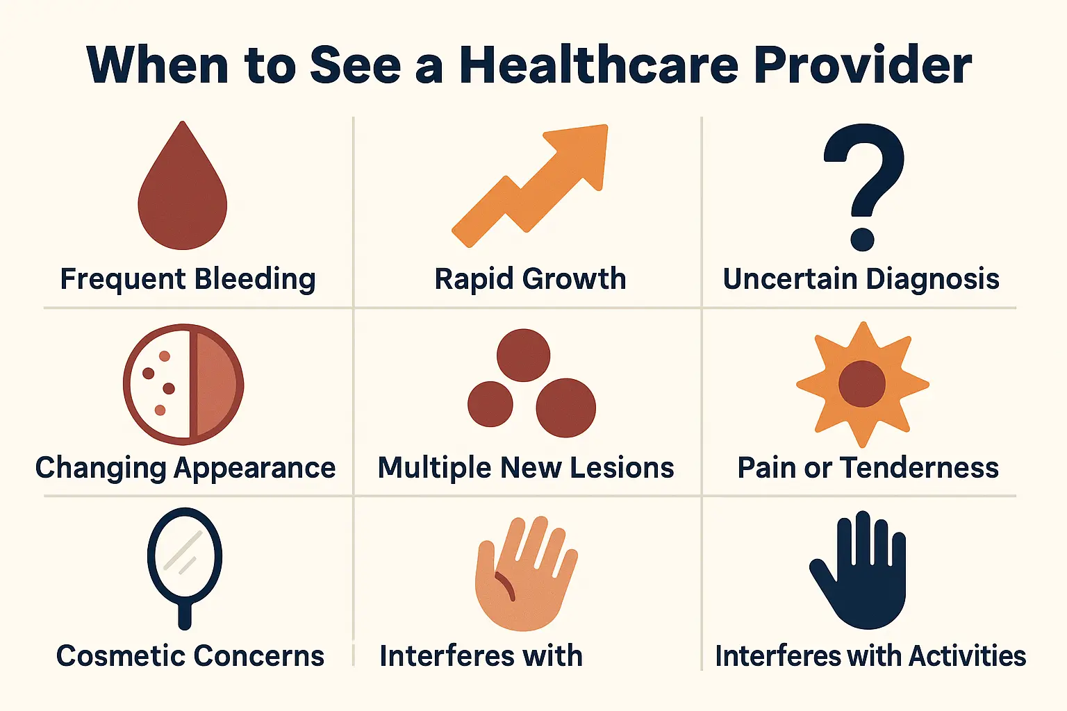
While both cherry angiomas and pyogenic granulomas are benign, certain situations warrant professional evaluation:
✅ A red bump bleeds frequently or heavily – This is especially common with pyogenic granulomas and indicates treatment is needed
✅ A lesion grows rapidly – Sudden growth over days or weeks should always be evaluated
✅ You're unsure what the bump is – Not all red skin lesions are benign; some serious conditions can look similar
✅ A lesion changes in appearance – Changes in color, size, shape, or texture should be checked
✅ You have multiple new lesions appearing – While cherry angiomas commonly appear in multiples, sudden appearance of many vascular lesions could indicate other conditions
✅ A lesion is painful or tender – Neither cherry angiomas nor pyogenic granulomas are typically painful
✅ You want cosmetic removal – Even benign lesions can be removed if they bother you
✅ A lesion interferes with daily activities – For example, a pyogenic granuloma on a finger that catches on things
The team at The Minor Surgery Center is here to help. We understand that skin concerns can be worrying, and we're committed to providing clear answers and expert care without long wait times or unnecessary hassle.
Part of why professional diagnosis matters is that several other conditions can mimic cherry angiomas or pyogenic granulomas. Here are a few look-alikes:
Petechiae and Purpura
These are tiny red or purple spots caused by bleeding under the skin. Unlike cherry angiomas, they don't blanch (turn white) when you press on them, and they're flat rather than raised.
Spider Angiomas
These have a central red dot with tiny blood vessels radiating outward like spider legs. They're common in pregnancy and liver disease.
Angiokeratomas
These dark red or purple bumps have a warty, scaly surface, unlike the smooth surface of cherry angiomas.
Amelanotic Melanoma ⚠️
This is a serious skin cancer that lacks the typical dark pigmentation of most melanomas. It can appear as a pink or red bump that bleeds easily—very similar to a pyogenic granuloma. This is why biopsy is sometimes necessary.
Basal Cell Carcinoma
This common skin cancer can sometimes appear as a pearly pink or red bump that bleeds easily.
Kaposi Sarcoma
This rare cancer, associated with immune system problems, can appear as purple or red nodules.
Hemangioma
These benign vascular tumors are most common in infants but can occur in adults. They're usually larger and deeper than cherry angiomas.
Bacillary Angiomatosis
This rare bacterial infection in immunocompromised people can create red, pyogenic granuloma-like lesions.
The bottom line? If you're not sure what a skin lesion is, get it checked. Most of the time, it'll turn out to be something harmless like a cherry angioma or pyogenic granuloma. But occasionally, what looks benign could be something more serious that requires prompt treatment.
You can learn more about various skin conditions we treat at The Minor Surgery Center.
The short answer is: not really. Both cherry angiomas and pyogenic granulomas develop for reasons that are largely beyond your control.
Since cherry angiomas are primarily related to aging and genetics, there's no proven way to prevent them. However, some dermatologists suggest:
Honestly, though, cherry angiomas are so common and harmless that prevention isn't really a concern for most people.
Since pyogenic granulomas often develop after trauma, you can potentially reduce your risk by:
That said, many pyogenic granulomas develop without any identifiable trigger, so prevention isn't always possible.
For most people, these vascular lesions are minor inconveniences rather than major health concerns. Here's how to manage them:
Many people have cherry angiomas and never give them a second thought.
Let's talk about something that doesn't get discussed enough: the emotional toll that visible skin lesions can take. Whether it's a cluster of bright red cherry angiomas on your chest or a bleeding pyogenic granuloma on your face, these lesions can affect your self-confidence and quality of life.
You might feel:
Here's what you need to know: Your concerns are valid. Even if a lesion is medically benign, if it bothers you, that's reason enough to seek treatment. At The Minor Surgery Center, we understand that skin concerns aren't just medical issues—they're personal issues that affect how you feel about yourself.
"You deserve to feel confident in your skin. That's why we make removal procedures fast, simple, and clear—without the wait."
Don't let anyone dismiss your concerns or make you feel like you're being vain for wanting treatment. Your comfort and confidence matter.
.webp)
One of the most common questions people have about cherry angioma and pyogenic granuloma removal is: Will insurance cover it?
The answer depends on why you're having the lesion removed:
Medically Necessary Removal ✅
Insurance typically covers removal if the lesion:
Cosmetic Removal ❌
If you're removing a lesion purely for cosmetic reasons (which is common with cherry angiomas), insurance usually won't cover it. However, the cost is typically modest, especially for small lesions.
At The Minor Surgery Center, we believe in transparency—no hidden fees, no surprises. During your consultation, we'll:
Don't let cost concerns prevent you from seeking the care you need. We're happy to discuss your options and help you find a solution that works for your budget. You can contact us to discuss your specific situation.
No. Cherry angiomas are completely benign and do not turn into cancer. However, if a cherry angioma changes significantly in appearance, it's worth having it checked to make sure it's still just a cherry angioma and not something else.
Yes, probably. Cherry angiomas tend to increase in number as you age. Removing existing ones doesn't prevent new ones from developing elsewhere.
The key differences are growth rate and bleeding. Cherry angiomas develop slowly over months, are smooth, and rarely bleed. Pyogenic granulomas appear rapidly (days to weeks), have a raw surface, and bleed easily with minimal trauma.
Rarely. While it's possible for small pyogenic granulomas to resolve spontaneously, the vast majority persist and continue to bleed until treated. Waiting for spontaneous resolution usually isn't the best approach.
No. Both cherry angiomas and pyogenic granulomas are removed under local anesthesia, so you won't feel pain during the procedure. You might feel a small pinch from the anesthetic injection, and there may be mild discomfort for a day or two afterward, but the procedures themselves are not painful.
Most removals take just 15-30 minutes from start to finish. It's a quick, in-office procedure that you can easily fit into your lunch break.
With proper technique, scarring is typically minimal. Cherry angiomas often leave no visible scar at all. Pyogenic granulomas may leave a small scar, but skilled removal minimizes this. The scar is almost always less noticeable than the original lesion.
We strongly advise against this. While you might see DIY removal methods online, attempting to remove skin lesions at home can lead to infection, scarring, and incomplete removal. Plus, you need a professional diagnosis to be certain the lesion is actually a cherry angioma and not something more serious.
For more answers to common questions, visit our FAQs page.
When you're dealing with a skin concern like a cherry angioma or pyogenic granuloma, you want expert care delivered with compassion and efficiency. That's exactly what you'll find at The Minor Surgery Center.
🎯 Expert Surgeons
Our team has extensive experience with skin lesion removal. We use precise techniques to ensure complete removal with minimal scarring.
⚡ No Long Wait Times
We know you're busy. We've streamlined everything from booking to post-op care so you can get the treatment you need without the wait.
💙 Patient-Focused Care
We treat every patient with dignity, compassion, and respect. Your concerns matter to us, and we take the time to answer all your questions.
🔍 Transparent Pricing
No hidden fees, no surprises. We'll tell you exactly what to expect before any procedure.
🏥 Safe, Welcoming Environment
Our facility is designed to make you feel comfortable and at ease, not anxious and intimidated.
📋 No Referrals Needed
You can schedule directly with us—no need to jump through hoops or wait for referrals.
Whether you're dealing with a bleeding pyogenic granuloma that needs prompt attention or a cluster of cherry angiomas that make you self-conscious, we're here to help. Learn more about our clinic and what makes us different.
Let's bring it all together:
Cherry angiomas are those common, harmless red spots that appear as we age. They grow slowly, rarely cause problems, and don't require treatment unless they bother you cosmetically or get irritated. Think of them as the skin equivalent of gray hair—a natural part of aging that's completely benign.
Pyogenic granulomas are a different story. These rapidly growing vascular lesions bleed easily, rarely resolve on their own, and typically require professional removal. They're not dangerous, but they're problematic enough that treatment is usually recommended.
The key differences:
Both conditions are benign and treatable. Neither turns into cancer. But both should be professionally evaluated to confirm the diagnosis and determine the best treatment approach.
If you have a red bump or growth on your skin and you're not sure what it is, don't wait and worry. Get it checked.
At The Minor Surgery Center, we make the process simple:
You can contact us today to schedule your appointment. No long wait times, no runaround—just expert care delivered with compassion.
For more information about the conditions we treat and the procedures we offer, visit our blog for helpful articles and resources.
Understanding the difference between cherry angiomas and pyogenic granulomas empowers you to make informed decisions about your skin health. While both are benign vascular growths that appear red, their behavior, implications, and treatment needs are quite different.
Cherry angiomas are those harmless red spots that accumulate as we age—common, benign, and requiring treatment only if they bother you. Pyogenic granulomas are rapidly growing lesions that bleed easily and typically need professional removal to resolve.
The most important takeaway? Professional evaluation matters. What looks like a simple red bump could occasionally be something more serious, and even benign lesions can affect your quality of life and deserve proper treatment.
At The Minor Surgery Center, we're here to help you navigate these concerns with expertise, compassion, and efficiency. Whether you need a quick evaluation for peace of mind or professional removal of a bothersome lesion, we make the process simple and stress-free.
Your skin health matters. Your confidence matters. And you deserve expert care that treats both with equal importance.
Don't let uncertainty or anxiety about a skin lesion linger. Reach out to The Minor Surgery Center today and take the first step toward clear answers and confident skin. We're here to help—no referrals needed, no long waits, just expert care when you need it.


