
Last updated: September 29, 2026
Keratoacanthoma and squamous cell carcinoma (SCC) can look nearly identical on the skin, but they behave differently: keratoacanthoma typically grows fast for a few weeks, then stabilizes and often shrinks on its own, while SCC keeps growing and can spread if untreated. Because the two are so hard to separate by eye alone, most dermatologists biopsy and treat keratoacanthoma as if it were SCC, even though pathology and current classification systems list them as distinct entities.[1][9]
Keratoacanthoma is a fast-growing, dome-shaped skin bump made of squamous cells, and it usually looks like a small volcano with a crusty, keratin-filled center. Squamous cell carcinoma is a cancer built from the same cell type, but it grows without a built-in stopping point and can spread to nearby tissue or, less often, to lymph nodes.[1][10]
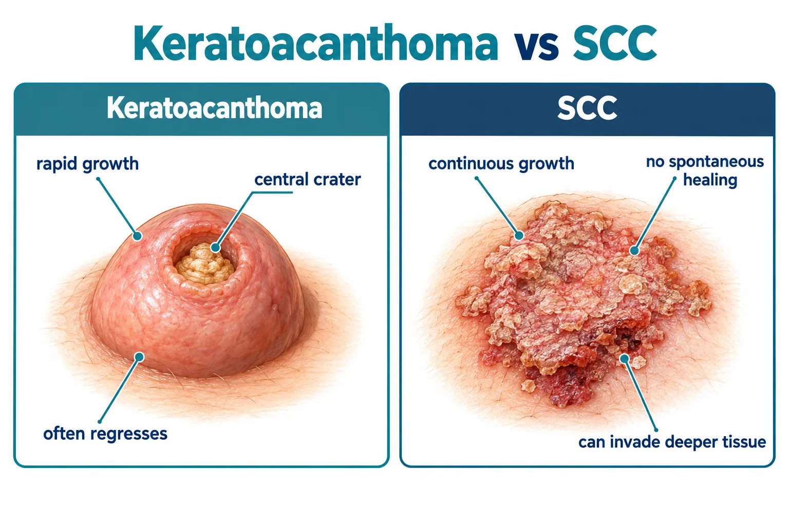
Both lesions arise from keratinocytes, the cells that make up the outer layer of skin. Under a microscope early on, they can look strikingly alike, which is part of why the keratoacanthoma vs SCC question comes up so often in dermatology clinics.
Key structural differences include:
That same 2024 review explicitly states that terminology combining the two names, such as "squamous cell carcinoma, keratoacanthoma-type," is not recommended, because it blurs the line between two lesions that current classification treats as separate entities.[1] Despite that, the practical reality in most clinics is that a fast-growing keratinized bump gets biopsied and treated as if it could be cancer, because the two conditions cannot be told apart with total confidence just by looking at them.
If a bump on the skin doubles in size within a month and develops a central crusted plug, choose evaluation over watching, since that growth speed is a hallmark of keratoacanthoma but also overlaps with aggressive SCC. A clinician who can compare it directly with the skin cancer screening protocols used during a full-body mole check is better positioned to flag it early.
Keratoacanthoma sits in a genuinely gray zone: some pathologists call it benign, some call it a low-grade or well-differentiated variant of SCC, and a smaller number treat it as SCC outright. There is no single universal answer, and that disagreement is documented in current medical literature rather than being a gap in this article.[9]
The Merck Manual, updated in March 2026, states plainly that opinion on keratoacanthoma remains divided among specialists, describing it as variably classified as a benign lesion, a variant of well-differentiated SCC, or a borderline entity.[9] That same source notes that unlike most SCCs, keratoacanthomas have a tendency to regress or involute on their own, though rare aggressive cases have been reported.[9]
Separately, dermatologic oncology sources increasingly favor treating keratoacanthoma as a subtype of well-differentiated squamous cell carcinoma, which is now described as the most commonly accepted classification in that field.[9] Patient-facing health resources reflect this split. Cleveland Clinic and MedlinePlus describe keratoacanthoma as usually noncancerous or a mild type of squamous cell cancer, while still consistently advising that it be treated as if it were skin cancer.[6][10]
What this means practically: whether a specific keratoacanthoma is "benign" or "a mild SCC" often depends on which classification framework a pathologist uses, not on anything different happening in the patient's skin. Because rare keratoacanthomas do behave aggressively and because misclassification carries a real cost, nearly every clinician removes and tests these lesions rather than assuming they are harmless.[9]
Can keratoacanthoma turn into skin cancer, or is it already skin cancer? The honest answer is that it may already meet the technical definition of SCC by some standards, so the safer framing for patients is: treat it exactly as you would treat SCC, regardless of which label ends up on the pathology report.
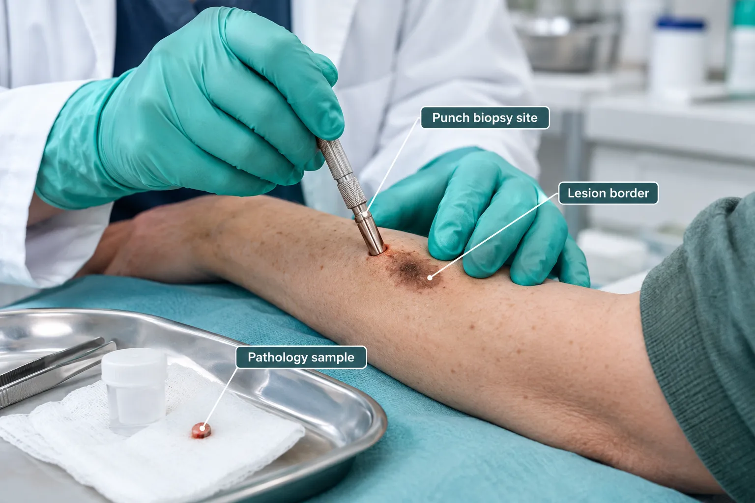
Doctors distinguish keratoacanthoma from SCC using a combination of growth history, physical exam, and a tissue biopsy, since visual inspection alone is not reliable enough to separate the two conditions. A biopsy remains the deciding test in almost every case.[8][4]
Clinical clues that favor keratoacanthoma:
Clinical clues that favor SCC:
What pathologists look for on biopsy:
A biopsy sample gets examined under a microscope for specific architectural features. Pathologists assess:
Even with all of these tools, some biopsy reports come back ambiguous, sometimes worded as "keratoacanthoma, cannot exclude SCC." When that happens, the typical next step is complete surgical removal with margins, so the entire lesion can be examined rather than just a small sample. The American Academy of Family Physicians' clinical guidance on skin cancer diagnosis supports this cautious, biopsy-first approach for any new or changing keratinized skin growth.[4][5]
Decision rule: If a biopsy report is inconclusive between keratoacanthoma and SCC, treat the lesion as SCC for management purposes. This is the standard, conservative approach used in most dermatology and skin cancer clinics.
Keratoacanthoma can regress spontaneously, but nearly every dermatologist still recommends treating it rather than waiting, because there is no reliable way to be certain in advance that a specific lesion is not SCC. Waiting without a diagnosis is the main mistake to avoid.[1][9]
How long does keratoacanthoma take to go away without treatment?
When keratoacanthoma does resolve on its own, the typical timeline is:
That said, this natural course is not a treatment plan. A few important caveats apply:
Choose observation only if: a biopsy has already confirmed keratoacanthoma, the lesion is small, and a clinician is actively monitoring it on a defined schedule. Outside of that specific situation, biopsy and removal are the standard of care.
Common mistake: assuming a shrinking bump means it is safe to ignore. Keratoacanthomas do get smaller before they disappear, but some SCCs also temporarily plateau in size before resuming growth. Size changes alone are not diagnostic.
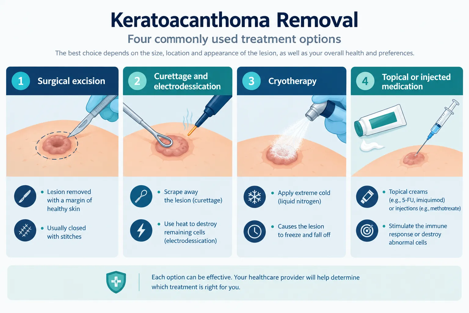
Keratoacanthoma removal typically costs less than treatment for advanced skin cancer and is usually covered when billed as medically necessary skin lesion removal, though exact pricing depends on lesion size, location, and the method used. The most common treatment options are surgical excision, curettage with electrodessication, cryotherapy, and, less often, topical or injected medications.[8]
Treatment options compared:
MethodBest ForNotesSurgical excisionMost cases, especially if SCC cannot be ruled outRemoves full lesion with a margin; sample sent for complete pathology reviewCurettage and electrodessicationSmaller, clearly benign-appearing lesionsScrapes and cauterizes the lesion; faster but less tissue for pathologyCryotherapyVery small, low-risk, biopsy-confirmed lesionsFreezes tissue; not always ideal when diagnosis is uncertainTopical or injected medicationSelect cases where surgery is not preferredUsed selectively; requires close follow-up
Choosing between methods:
Recovery after excision is generally short, with most people back to normal activity within days and a healing scar over several weeks. For guidance on minimizing visible scarring after any skin lesion removal, see this comparison of scar care methods after mole or lesion removal. Patients considering where to have a suspicious growth assessed and removed can review what a proper evaluation involves at a dedicated skin cancer clinic or compare options at a leading skin cancer clinic in Toronto.
Keratoacanthoma is most strongly linked to cumulative ultraviolet (UV) sun exposure, and it develops most often in older adults with fair skin and a long history of sun damage. Other contributing factors include immune suppression, prior skin injury, and certain genetic syndromes.[8]
Main risk factors:
Who is not typically affected: Keratoacanthoma is uncommon in children and young adults without significant sun exposure history or immune compromise. A fast-growing bump in a younger person still warrants evaluation, but the statistical likelihood shifts toward other diagnoses.
Edge case: People who have had one keratoacanthoma are more likely than the general population to develop another, particularly if sun exposure habits have not changed. Anyone with a personal history of skin cancer or keratoacanthoma benefits from routine surveillance, similar to the follow-up recommended after conditions like Bowen's disease, another precancerous squamous skin condition.
Keratoacanthoma recurrence after proper treatment is uncommon but not impossible, and separately, many people who develop one keratoacanthoma go on to develop additional, unrelated ones at different times or sites over their lifetime.[8]
It helps to separate two different situations:
Decision rule: If a bump returns at the exact site of a prior removed keratoacanthoma within weeks to months, treat it as a possible incomplete excision and have it re-evaluated and re-biopsied rather than assuming it is simply a "repeat" keratoacanthoma.
Reducing future risk:
Anyone with a pattern of repeated skin growths benefits from a structured monitoring plan. A comprehensive skin check on a set schedule catches new lesions earlier, when they are smaller and simpler to treat.
Keratoacanthoma and warts can both appear as raised, rough bumps on the skin, but they have completely different causes: warts come from a viral infection (HPV), while keratoacanthoma is a squamous skin growth linked to sun damage, not a virus. This difference matters because warts are contagious and keratoacanthoma is not.
FeatureKeratoacanthomaCommon WartCauseSquamous skin cell growth, linked to UV damageHuman papillomavirus (HPV) infectionGrowth speedFast, reaches full size in weeksSlow, develops over monthsTypical sizeOften larger, can exceed 1-2 cmUsually smaller, under 1 cmSurfaceSmooth dome with central keratin craterRough, cauliflower-like textureContagiousNoYes, spreads by skin contactNeeds biopsy to rule out cancerYes, alwaysRarely, unless atypical in appearance
Common mistake: Treating a fast-growing bump at home with over-the-counter wart removal products because it "looks like a wart." Wart treatments target viral tissue and will not address a squamous growth correctly, and delaying proper evaluation risks missing SCC.
Quick example: A 68-year-old with a rough bump on the back of the hand that appeared over three weeks and has a crusted center is far more consistent with keratoacanthoma than a wart, given the age, growth speed, and location on sun-exposed skin. A person in their 20s with a small, slow-growing rough bump on the hand is more likely dealing with a common viral wart.
A keratoacanthoma on the face deserves prompt medical evaluation, not panic, because facial lesions are treatable but also sit in a cosmetically and functionally sensitive area where early, precise removal matters more than on the trunk or limbs. The bigger concern with a facial lesion is confirming it is not SCC before it grows larger or requires more extensive repair.[1][8]
Reasons facial keratoacanthoma warrants faster action:
When to seek care urgently:
When it is reasonable to book a routine (not emergency) visit:
For anyone in the Toronto area weighing where to have a facial or body lesion assessed, a dedicated clinic experienced with both cosmetic and cancer-focused skin evaluation, such as the skin cancer clinic in York Mills, or a broader skin biopsy and skin cancer screening service in Mississauga, can provide both the biopsy and the cosmetic planning in one visit. Patients concerned about a lesion that might be melanoma rather than a squamous growth should also review guidance from melanoma specialists in Toronto, since melanoma requires a different urgency and treatment path.
Is keratoacanthoma the same as squamous cell carcinoma?
No. Current classification treats keratoacanthoma and SCC as separate diagnoses, though many specialists consider keratoacanthoma a well-differentiated, less aggressive variant of SCC. Because the two can look identical and behave similarly in some cases, most clinicians treat keratoacanthoma with the same urgency as SCC.[1][9]
Can keratoacanthoma go away without any treatment?
Yes, keratoacanthoma can spontaneously shrink and resolve over roughly two to six months, but this should never be assumed without a confirmed biopsy, since SCC does not reliably do the same and can be mistaken for keratoacanthoma early on.[1][9]
How painful is keratoacanthoma removal?
Removal is done under local anesthetic, so the procedure itself is not painful. Mild soreness at the site for a few days afterward is common, similar to recovery after other minor skin lesion excisions.
What does a keratoacanthoma biopsy actually show?
A biopsy shows whether the cells are well-organized and symmetric, consistent with keratoacanthoma, or irregular and invasive, consistent with SCC. Pathologists also assess depth of growth and cell differentiation to help finalize the diagnosis.[8][1]
Does insurance or provincial health coverage pay for keratoacanthoma removal?
When a lesion is removed because it is suspicious for skin cancer, removal is typically classified as medically necessary rather than cosmetic, which usually affects coverage. Confirming coverage details directly with the treating clinic before the procedure avoids surprises.
Can keratoacanthoma spread to other parts of the body?
Classic keratoacanthoma does not spread the way advanced SCC can. However, because some lesions labeled keratoacanthoma may actually represent an aggressive SCC variant, complete removal and pathology confirmation remain the standard precaution.[9][1]
Is a keratoacanthoma dangerous if left on the skin for months?
The main danger is not the keratoacanthoma itself but the possibility that the lesion is actually SCC growing under a similar appearance. Leaving any fast-growing skin bump unevaluated for months removes the chance to catch a true SCC early.
What is the difference between keratoacanthoma and a cyst?
A cyst is a fluid- or keratin-filled sac beneath the skin that grows slowly and feels soft or fluctuant, while keratoacanthoma is a solid squamous growth that grows quickly and feels firm with a hard central plug. If uncertain, a clinician can distinguish the two on exam or with a simple biopsy, similar to how a sebaceous cyst is evaluated and confirmed.
Keratoacanthoma vs SCC is less about spotting a difference by eye and more about confirming the diagnosis with a biopsy, because the two conditions can look nearly identical while carrying different risk profiles. Keratoacanthoma often grows fast and then fades on its own, while SCC keeps growing and carries clearer malignant potential, but current classification and clinical practice both treat any uncertain, fast-growing squamous bump with the same seriousness.[1][9]
Practical next steps:
Acting early keeps treatment simple and scarring minimal, whether the final diagnosis turns out to be a self-limited keratoacanthoma or a true squamous cell carcinoma requiring closer follow-up.
[1] Pmc11503433 - https://pmc.ncbi.nlm.nih.gov/articles/PMC11503433/
[2] nature - https://www.nature.com/articles/3800063
[3] Modpathol20155 - https://www.nature.com/articles/modpathol20155
[4] P601 - https://www.aafp.org/afp/2015/1001/p601
[5] P339 - https://www.aafp.org/pubs/afp/issues/2020/0915/p339.pdf
[6] Keratoacanthoma - https://my.clevelandclinic.org/health/diseases/keratoacanthoma
[7] Side Effect Central Cutaneous Squamous Cell Carcinoma And Keratoacanthoma - https://www.aimatmelanoma.org/side-effect-central-cutaneous-squamous-cell-carcinoma-and-keratoacanthoma/
[8] Keratoacanthoma - https://dermnetnz.org/topics/keratoacanthoma
[9] Keratoacanthoma - https://www.merckmanuals.com/professional/oncology/cancers-of-the-skin/keratoacanthoma
[10] medlineplus.gov - https://medlineplus.gov/ency/article/000829.htm
Meta Title: Keratoacanthoma vs SCC: Key Differences Explained
Meta Description: Learn how keratoacanthoma vs SCC compares in growth, biopsy findings, treatment, cost, and recurrence risk, and when to see a doctor for a fast-growing bump.
Tags: keratoacanthoma, squamous cell carcinoma, skin cancer diagnosis, skin biopsy, skin lesion removal, dermatology, skin cancer screening, sun damage skin, precancerous skin growths, skin cancer treatment options, keratoacanthoma recurrence, cSCC