

Last updated: September 29, 2026
Pigmented BCC vs melanoma is a distinction that can only be confirmed with a biopsy, because both conditions can look nearly identical to the eye and even under a dermatoscope. Pigmented basal cell carcinoma is a slow-growing, rarely fatal skin cancer that happens to contain dark pigment, while melanoma is an aggressive cancer of pigment-producing cells with a much higher risk of spreading. The two share overlapping colors and shapes often enough that misdiagnosis happens in both directions, which is why dermatologists rely on dermoscopy plus tissue sampling rather than appearance alone[4][6].
Pigmented basal cell carcinoma (BCC) is a variant of the most common skin cancer that contains melanin, the pigment that gives it a brown, blue-gray, or black color instead of the usual pink or flesh-toned appearance. Melanoma is a completely different type of cancer that starts in melanocytes, the pigment-making cells themselves, and it behaves far more aggressively than any form of BCC.
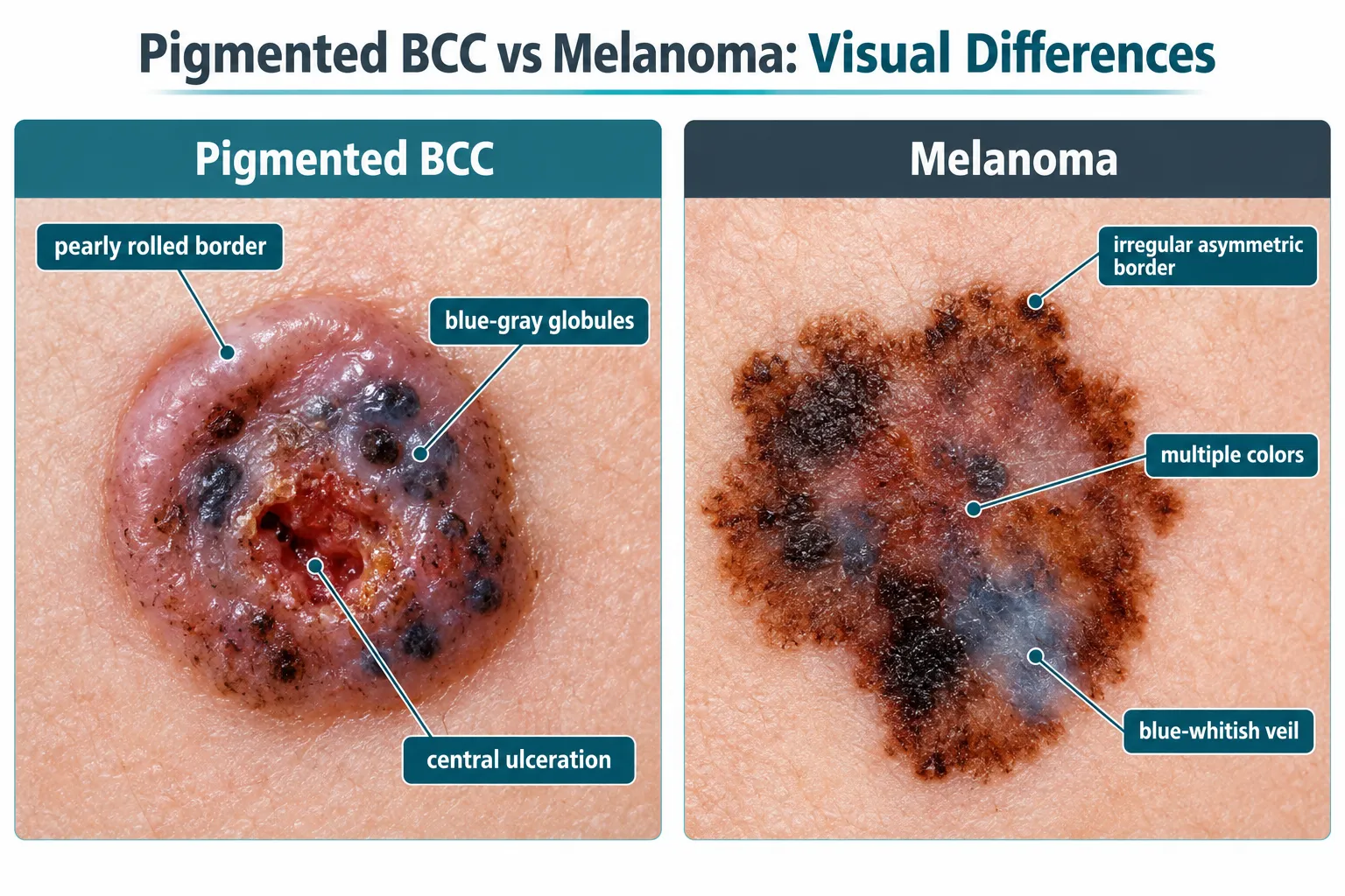
The confusion between these two conditions comes down to color, not biology. Basal cell carcinoma originates in the basal layer of the epidermis, the skin's structural cells, and pigmented BCC develops when melanocytes get pulled into the tumor and deposit pigment inside it. Melanoma, by contrast, is a cancer of the melanocytes themselves, meaning the pigment is not an incidental feature but the source of the disease. This distinction matters clinically because:
A 2026 case report described pigmented BCC of the nipple-areola complex in a 61-year-old woman that was clinically and dermoscopically indistinguishable from melanoma of the breast, a site where melanoma is already rare and easily missed. The authors noted that only dermoscopy combined with biopsy prevented the lesion from being mislabeled and undertreated[3]. This case illustrates a broader truth: location and appearance alone cannot settle a pigmented BCC vs melanoma question. For a deeper look at how melanoma itself is diagnosed and staged, see this overview of melanoma as a disease.
Pigmented BCC typically appears as a shiny, slightly raised nodule or flat plaque with a pearly or translucent edge and scattered dark pigment, while melanoma usually shows an irregular, asymmetric shape with multiple colors and a flat or unevenly raised surface. Despite these general patterns, both lesions can present atypically, which is where diagnostic errors happen.
Doctors often use the "ABCDE" rule for melanoma (asymmetry, border irregularity, color variation, diameter over 6mm, evolution) as a starting screening tool, but pigmented BCC can trigger several of these same red flags. Here is a side-by-side breakdown of typical features:
FeaturePigmented BCCMelanomaCommon colorBrown, blue-gray, or black with a translucent or pearly sheenBlack, brown, red, white, or blue in uneven patchesBorderOften rolled, well-defined edgeIrregular, notched, poorly definedSurfaceShiny, sometimes with visible small blood vesselsFlat, raised, or ulcerated, may bleed easilyGrowth patternSlow, over months to yearsCan grow rapidly, over weeks to monthsTypical locationFace, scalp, ears, sun-exposed skinTrunk, legs, back; also palms, soles, nail bedsUlcerationCentral ulcer common in nodular BCCPossible in advanced or nodular melanoma
A quick example: a 60-year-old man with a dark, pearly bump on his nose that has been present for a year and bleeds occasionally is more consistent with pigmented BCC. A 45-year-old woman with a rapidly changing, multicolored flat mole on her back that has doubled in size over three months looks more like melanoma. But these are patterns, not guarantees, and clinicians see exceptions to both often enough that visual impression is treated as a starting point, never a diagnosis[5][10].
Common mistake: Assuming a lesion cannot be cancerous because it looks "too shiny" or "too smooth" for melanoma. Nodular pigmented BCC frequently has a shiny surface, which can falsely reassure both patients and less experienced examiners.
Pigmented BCC gets mistaken for melanoma because both lesions can contain overlapping dermoscopic structures, including blue-gray coloring, irregular pigment networks, and blood vessel patterns that look similar under magnification. This overlap is a well-documented source of diagnostic error, not a rare fluke.
A 2025 dermoscopy-histopathology correlation study found that pigmented BCC most often shows blue-gray ovoid nests, leaf-like areas, and arborizing (tree-branch-shaped) vessels, while melanoma typically shows an atypical pigment network and a blue-whitish veil. Recognizing these specific patterns significantly improves diagnostic accuracy, but they are not always textbook-clean in real patients[4].
A 2025 review focused specifically on BCCs that were misdiagnosed as melanoma and quantified how often melanoma-mimicking features show up:
The authors concluded that overlapping dermoscopic signs are especially common on sun-damaged skin and the lower extremities, which are also common melanoma sites. Multiple case reports from 2023-2024 describe pigmented BCC mimicking nodular melanoma or lentigo maligna, particularly on the face and in patients with darker skin tones, where lesions presented as hyperpigmented nodules or flat macules with irregular pigmentation that were initially read as melanoma until biopsy proved otherwise[5]. Amelanotic melanoma, which paradoxically lacks the pigment most people associate with melanoma, adds a separate but related layer of diagnostic difficulty; you can read more in this guide to amelanotic melanoma.
A 2022 study identified 12 "trump" dermoscopic features that, when present, strongly favor a BCC diagnosis over melanoma even when the lesion otherwise looks melanoma-like. When those specific BCC signs are absent, diagnostic confidence and accuracy both drop noticeably, which is exactly the scenario that produces mislabeled cases[7].
Doctors distinguish pigmented BCC from melanoma using a combination of dermoscopy, clinical history, and biopsy with histopathology, and immunohistochemistry when the tissue findings are ambiguous. No single tool is considered sufficient on its own.
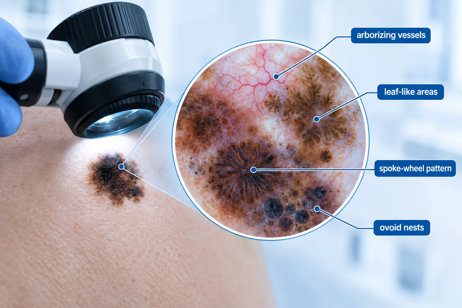
The typical diagnostic sequence looks like this:
Decision rule: if a lesion shows any dermoscopic feature typically associated with melanoma (atypical network, blue-whitish veil, irregular blotches) even alongside features that suggest BCC, biopsy should proceed as though melanoma is possible. The cost of a missed melanoma is far higher than the cost of biopsying a benign or low-risk lesion.
An emerging complication in this diagnostic process is artificial intelligence. Convolutional neural networks trained on pigmented lesion images frequently list melanoma among their top predictions for flat or macular pigmented lesions, including ones that turn out to be pigmented BCC, pigmented actinic keratosis, or solar lentigo. This means automated image-based screening tools can misclassify pigmented BCC as melanoma, reinforcing that expert dermoscopic review and histopathology remain necessary rather than optional, even as these technologies improve[8]. Patients relying on mole-mapping or AI screening tools should understand what these tools can and cannot detect; see this explainer on whether 3D mole mapping detects melanoma for more detail on the limits of imaging-based screening.
Pigmented BCC does not turn into melanoma. They are two biologically distinct cancers that arise from different cell types, and one does not transform into the other. It is possible, however, for a person to develop both conditions independently, including at the same time or on the same area of skin.
This is an important point because patients sometimes assume that a "less dangerous" pigmented BCC diagnosis rules out melanoma risk elsewhere on their body, or that a BCC left untreated could eventually "become" melanoma. Neither assumption is accurate.
Edge case: rarely, a biopsy of a single lesion reveals mixed or "collision" features, where pigmented BCC tissue sits immediately adjacent to a separate melanocytic lesion in the same piece of skin. This is not one cancer converting into another; it is two distinct growths that happen to occupy overlapping space. Pathologists identify this through careful histologic mapping and, when needed, immunohistochemistry to confirm two separate cell populations are present[1][2].
Because having one skin cancer type raises the statistical likelihood of having (or later developing) another, anyone diagnosed with pigmented BCC should still get a full skin check for other suspicious lesions, including ones that might be melanoma, rather than assuming the BCC diagnosis "explains" every dark spot on their body.
Pigmented BCC is not dangerous in the way melanoma is. Basal cell carcinoma, pigmented or not, almost never metastasizes and rarely causes death, while melanoma is an aggressive cancer with a meaningful risk of spreading to lymph nodes and distant organs if not caught early.
This prognostic gap is the single biggest reason clinicians insist on biopsy whenever melanoma is part of the differential diagnosis for a dark, changing lesion. Recent reviews and case series on pigmented BCC consistently emphasize that while it can look and behave visually like melanoma on the skin's surface, the two diseases are worlds apart biologically[5][10].
Key points on risk and outcomes:
Decision rule: treat any pigmented lesion suspicious for melanoma with the same urgency regardless of how "textbook BCC" it might otherwise look, because the downside of missing a melanoma (delayed treatment of an aggressive, potentially fatal cancer) vastly outweighs the downside of biopsying a lesion that turns out to be benign or low-risk BCC.
Pigmented BCC is more common in people with medium to darker skin tones and shows up most often on chronically sun-exposed areas like the face, ears, and scalp, while melanoma occurs across all skin tones but is diagnosed more frequently overall in fair-skinned populations, often on the trunk and legs. Basal cell carcinoma as a whole is far more common than melanoma, though pigmented BCC specifically is a less frequent subtype within that broader category.
Risk factor patterns worth knowing:
Example: a dermatology clinic seeing a large population of patients with darker skin tones is statistically more likely to encounter pigmented BCC as a diagnostic consideration for a dark facial nodule than a clinic serving a predominantly fair-skinned population, where the same lesion is more likely to prompt melanoma concern first. This does not mean either group is exempt from either disease; it means baseline suspicion should be adjusted, not eliminated.
Early pigmented BCC often shows up as a small, slow-growing, slightly raised bump with a pearly or translucent quality and visible dark pigment, sometimes with tiny surface blood vessels. It rarely causes pain in its early stages, which is part of why it can go unnoticed for months.
Warning signs to watch for include:
Quick example: a small, dark, shiny bump near the eyebrow that has slowly grown over eight months and occasionally bleeds after shaving is a classic early presentation worth having evaluated. In contrast, a flat, multicolored patch that appeared six weeks ago and has visibly changed shape leans more toward a melanoma workup, though again, only a biopsy settles the question definitively.
Common mistake: dismissing a persistent pigmented bump as an "age spot" or "mole" because it is small and not painful. Pigmented BCC's slow growth and lack of early symptoms are exactly what allow it to be mistaken for something benign, delaying diagnosis by months or longer.
Yes, pigmented BCC and melanoma require different treatment approaches because they behave differently biologically, even though both are typically managed with surgical removal as a first step. Melanoma treatment often involves more extensive margins, sentinel lymph node evaluation, and sometimes systemic therapy, while pigmented BCC treatment is usually more limited in scope.
A general comparison of treatment pathways:
Treatment considerationPigmented BCCMelanomaPrimary treatmentSurgical excision, Mohs micrographic surgery, or curettage and electrodesiccation for select casesWide local excision with margins based on tumor thicknessMargin requirementsTypically narrower (often 3-5mm for standard excision)Wider, determined by Breslow depth (often 1-2cm or more)Lymph node evaluationRarely neededSentinel lymph node biopsy often required for thicker lesionsSystemic therapyAlmost never neededImmunotherapy or targeted therapy for advanced or metastatic diseaseFollow-up intensityPeriodic skin checksMore frequent surveillance, often including imaging for higher-stage diseaseNon-surgical optionsTopical therapies or photodynamic therapy in select superficial casesNot a substitute for surgery in invasive melanoma
Because pigmented BCC and melanoma can look alike before biopsy, treatment planning always waits for a confirmed histopathologic diagnosis. Operating on cosmetically sensitive areas, such as the face, nipple-areola complex, or near the eyes, raises the stakes further, since excision margins may need to be limited for cosmetic or functional reasons even as the surgeon works to fully remove the tumor[11]. Patients navigating a melanoma diagnosis and looking for a qualified surgical team can review resources like the guide to finding a melanoma specialist near you or a dedicated melanoma surgeon in Toronto.
Decision rule: never proceed with a treatment plan based on visual or dermoscopic impression alone when melanoma is on the differential. Biopsy first, confirm the diagnosis, then match the treatment intensity to the confirmed pathology.
Untreated pigmented BCC continues to grow slowly and can eventually invade surrounding skin, cartilage, bone, or nerve tissue, causing disfigurement and functional problems, but it rarely spreads to distant organs or becomes fatal. This is different from untreated melanoma, which carries a substantial and growing risk of metastasis and death the longer it goes without treatment.
Consequences of delayed treatment for pigmented BCC can include:
By contrast, untreated melanoma can progress through local growth into regional lymph node involvement and then distant metastasis, a sequence that dramatically worsens prognosis at each stage. This is precisely why any lesion suspicious for melanoma, even one that might turn out to be pigmented BCC on biopsy, should not be watched and waited on. Getting a professional opinion promptly matters more than trying to guess the diagnosis from appearance. Individuals in the Toronto area seeking a prompt evaluation can look into options like Toronto's leading melanoma clinics or established melanoma specialists in Toronto.
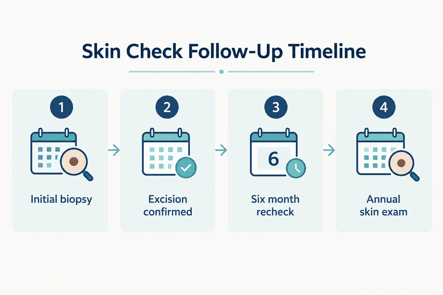
People previously diagnosed with pigmented BCC should have a full skin exam at least once a year, and more frequently, often every three to six months, if they have a history of multiple skin cancers, significant sun damage, or immunosuppression. This monitoring schedule should be set individually with a dermatologist based on personal risk factors.
General monitoring guidance:
Edge case: patients who have had pigmented BCC removed from a cosmetically sensitive site, such as the nipple-areola complex or near the eyelid, may need closer follow-up not just for recurrence but to monitor healing and scar changes that could otherwise be mistaken for new pigmented lesions.
Common mistake: assuming that because a prior lesion turned out to be "just BCC" rather than melanoma, future skin checks can be less frequent or less thorough. A BCC diagnosis is a marker of significant cumulative sun damage, which is itself a melanoma risk factor, so vigilance should not decrease after a BCC diagnosis.
Can a dermatologist tell pigmented BCC from melanoma just by looking at it?
A dermatologist can form a strong initial impression using dermoscopy, but visual and dermoscopic exams alone are not definitive. Biopsy with histopathology is required to confirm whether a lesion is pigmented BCC or melanoma[6].
Does pigmented BCC ever turn into melanoma over time?
No. Pigmented BCC and melanoma come from different cell types and one does not transform into the other. A person can, however, develop both cancers independently[1][2].
Is a black or very dark mole always melanoma?
No. Pigmented BCC can appear black or very dark because it contains melanocytes and melanophages within the tumor, producing a color that closely resembles melanoma despite being a completely different cancer[1].
Which is more dangerous, pigmented BCC or melanoma?
Melanoma is far more dangerous. It carries a meaningful risk of metastasis and death, especially if diagnosed late, while pigmented BCC is rarely fatal and mainly causes local tissue damage if untreated[5][10].
Why do AI skin-check apps sometimes flag pigmented BCC as melanoma?
Machine learning models trained on pigmented lesion images often list melanoma among the top predictions for flat, dark lesions, including ones that turn out to be pigmented BCC, pigmented actinic keratosis, or solar lentigo. Expert review and biopsy remain necessary because these tools can misclassify lesions[8].
Can you have pigmented BCC and melanoma at the same time?
Yes. The two are unrelated cancers, so a person can be diagnosed with both, sometimes on different areas of skin and occasionally at the same time during a single skin exam[2].
What does pigmented BCC look like on darker skin tones?
On darker skin, pigmented BCC often appears as a dark brown, blue-gray, or black nodule or plaque, sometimes with a shiny or slightly translucent quality, which can closely resemble nodular melanoma or lentigo maligna until biopsied[5].
How is pigmented BCC treated compared to melanoma?
Pigmented BCC is usually treated with surgical excision or Mohs surgery using narrower margins, while melanoma requires wider excision margins based on tumor thickness and sometimes sentinel lymph node biopsy or systemic therapy for more advanced disease.
Should every suspicious pigmented lesion be biopsied?
Yes, when melanoma is a realistic possibility. The cost of missing a melanoma diagnosis far outweighs the minor risk and cost of biopsying a lesion that turns out to be benign or low-risk pigmented BCC.
Is pigmented BCC rarer than regular basal cell carcinoma?
Yes. Standard, nonpigmented BCC is more common overall, especially in fair-skinned populations, while pigmented BCC is a specific subtype seen more frequently in people with medium to darker skin tones[9].
Pigmented BCC vs melanoma is not a distinction you can settle by staring at a mole in the mirror or running it through a phone app. Both conditions can produce dark, irregular, changing lesions, and both can trick even experienced clinicians using dermoscopy alone. The difference that actually matters is biological: pigmented BCC grows slowly and almost never kills, while melanoma can spread quickly and remains one of the more lethal skin cancers when caught late.
The practical takeaway is simple. Any new, changing, or unusual pigmented lesion deserves a professional evaluation, and any lesion where melanoma is a real possibility should go to biopsy rather than being watched. If you have a history of pigmented BCC, keep up with regular skin checks, since that history signals meaningful sun damage and a higher baseline risk for other skin cancers, including melanoma. If a lesion has been growing, bleeding, or changing color over the past few months, schedule an exam now rather than waiting for it to resolve on its own. Early, accurate diagnosis is what keeps a slow-growing BCC a minor procedure and catches a melanoma while it is still highly treatable.
[1] Pmc6551196 - https://pmc.ncbi.nlm.nih.gov/articles/PMC6551196/
[2] Pmc7250331 - https://pmc.ncbi.nlm.nih.gov/articles/PMC7250331/
[3] Pmc12968861 - https://pmc.ncbi.nlm.nih.gov/articles/PMC12968861/
[4] Full - https://www.frontiersin.org/journals/medicine/articles/10.3389/fmed.2025.1581601/full
[5] E261362 - https://casereports.bmj.com/content/17/9/e261362
[6] Gjct 9 151 - https://www.cancerresgroup.us/articles/GJCT-9-151.pdf
[7] Pmc9305787 - https://pmc.ncbi.nlm.nih.gov/articles/PMC9305787/
[8] dpcj - https://dpcj.org/index.php/dpc/article/download/2414/1906/47373
[9] jcp.bmj - https://jcp.bmj.com/content/62/2/120
[10] ijord - https://www.ijord.com/index.php/ijord/article/download/1872/1050/9807