
Last updated: October 6, 2026
Perineural invasion and other BCC/SCC pathology terms in plain English boil down to one idea: pathology reports describe how a skin cancer behaves under the microscope, and that behavior predicts how it might act in the body. Perineural invasion means cancer cells have grown into or around a nerve. It shows up in roughly 2 to 14 percent of squamous cell carcinomas and is less common in basal cell carcinomas, and it raises the risk of recurrence but does not mean a cure is impossible.[1][6][7][8]
Perineural invasion means a pathologist has seen cancer cells touching, surrounding, or growing inside the protective sheath of a nerve when looking at tissue under a microscope. It is a finding on a biopsy or excision report, not a symptom you would necessarily feel.[1][3][6][8]
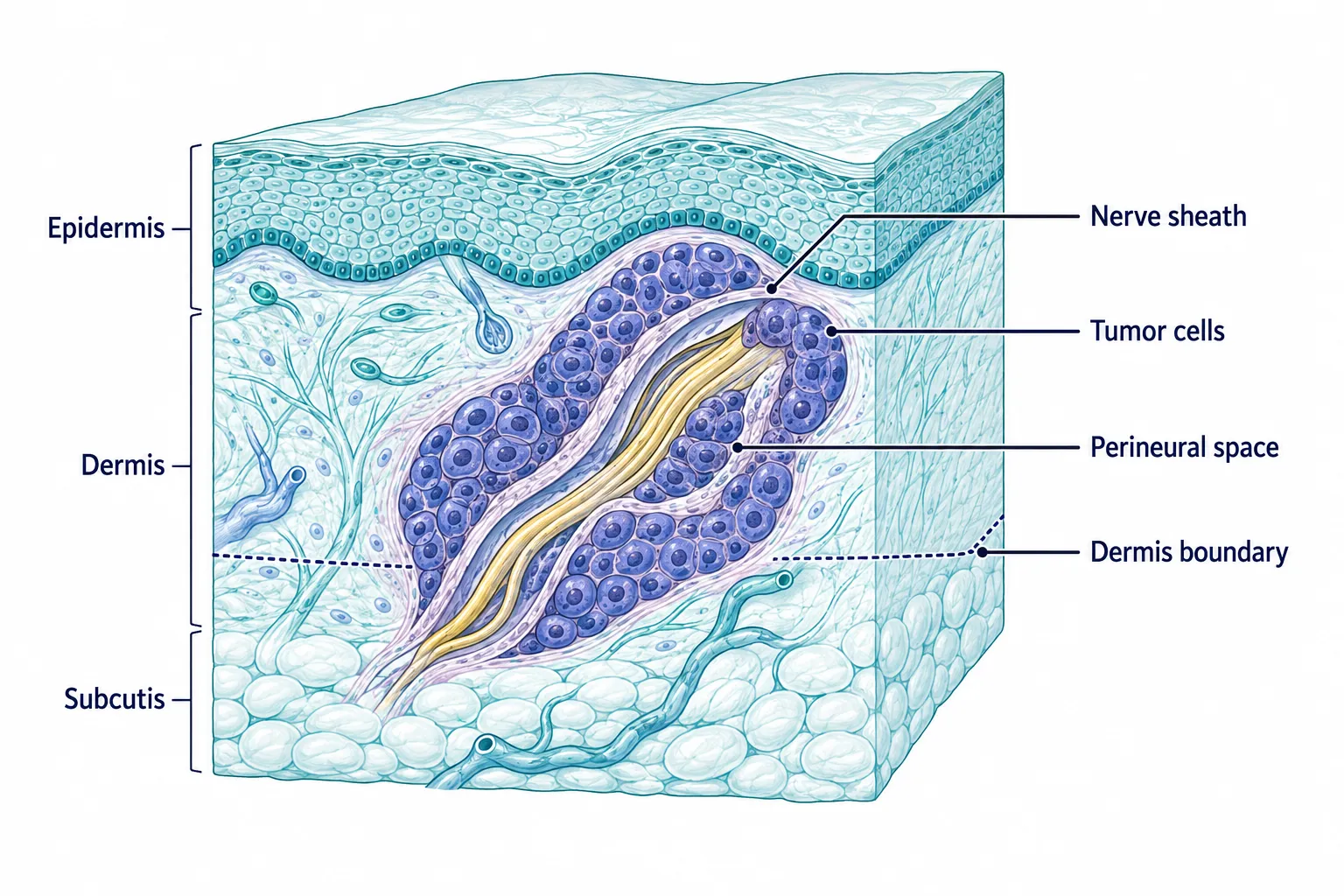
Nerves run through the skin like thin wires wrapped in insulation. When a skin cancer grows close to one of these nerves, cancer cells can sometimes track along the nerve's outer covering instead of staying in a tidy ball. Pathologists look for this pattern specifically because tumors that use nerves as a pathway can spread farther than the visible edge of the tumor suggests, which affects how much tissue a surgeon needs to remove.
What PNI is not:
Decision rule: If a pathology report mentions perineural invasion, the next question to ask is whether it is "focal" or "extensive," and whether it involves a small dermal nerve or a larger named nerve. That distinction, covered in detail later in this guide under incidental versus clinical PNI, matters more for prognosis than the word "perineural invasion" by itself.
Perineural invasion shows up in roughly 2 to 14 percent of cutaneous squamous cell carcinomas, making it notably more common in SCC than in basal cell carcinoma.[1][6][7][8] Across keratinocyte cancers as a group (BCC plus SCC combined), PNI is seen in about 2 to 6 percent of tumors overall.[7]
A few patterns are worth knowing:
Common mistake: Some patients assume that because PNI is "rare," a diagnosis of PNI must be unusual or alarming by definition. In reality, a 2 to 14 percent rate in SCC means it is uncommon but not exotic, and most dermatology and Mohs surgery practices see PNI cases regularly. The rarity of the finding does not equal rarity of a good outcome.
For general background on how skin cancers are classified and treated, see this overview of skin cancers explained.
Perineural invasion in basal cell carcinoma is taken seriously by dermatologists and surgeons, but it is generally less dangerous than PNI in squamous cell carcinoma because BCC rarely spreads to distant sites regardless of nerve involvement. The seriousness depends heavily on whether the nerve involved is small and incidental or large and symptomatic.
Why BCC with PNI is usually more manageable than SCC with PNI:
FactorBCC with PNISCC with PNIRisk of distant metastasisVery lowHigher, though still uncommon overallTypical nerve size involvedUsually small, dermal nervesCan involve larger nerves, especially in recurrent or facial tumorsMain concernLocal recurrence, tracking along tissue planesLocal recurrence plus lymph node or distant spreadUsual next stepWider excision or Mohs surgeryMohs surgery, often with specialist referral and sometimes imaging
Aggressive BCC subtypes, such as morpheaform (also called sclerosing) or infiltrative BCC, are more likely to show perineural invasion than nodular or superficial BCC. If a report lists both an aggressive subtype and PNI, that combination carries more weight than either finding alone.
Edge case: Periocular and periauricular BCC (tumors near the eye or ear) deserve special caution because nerves in these areas connect to structures that are harder to reconstruct and more sensitive to even minor spread. Clinical guidelines treat BCC near the eye with extra scrutiny when any high-risk feature, including PNI, is present.[9]
Doctors identify perineural invasion by examining tissue samples under a microscope after a biopsy or surgical excision; there is no blood test or standalone scan that detects it directly. A pathologist looks for tumor cells tracking along, inside, or around a nerve's protective covering within the removed tissue.
The process typically works like this:
Quick example: A patient has a scalp SCC removed. The pathology report notes "focal perineural invasion, single small-caliber nerve, no symptoms." No imaging is ordered because this fits the pattern of incidental PNI. A different patient has facial pain and jaw numbness alongside a similar tumor; this combination prompts an MRI to check whether the cancer is tracking along a named facial nerve.
For readers wanting to understand how a biopsy sample becomes a written report, this guide to mole biopsy testing and pathology explains the general process, even though that specific page focuses on pigmented lesions rather than BCC or SCC.
Perineural invasion and lymphovascular invasion both describe cancer cells found outside the main tumor mass, but PNI involves nerves while lymphovascular invasion (LVI) involves blood vessels or lymphatic channels. The practical difference matters because each pattern predicts a different kind of spread.
FeaturePerineural Invasion (PNI)Lymphovascular Invasion (LVI)Structure involvedNerve sheathBlood vessel or lymphatic vesselTypical spread riskLocal recurrence, nerve trackingLymph node involvement, distant metastasisHow it's foundMicroscopic exam of tissue near nervesMicroscopic exam of tissue near vesselsSymptom overlapNumbness, tingling, pain (in clinical PNI)Usually none directly, found incidentallyCommon next stepWider margins, Mohs surgery, imaging in severe casesLymph node evaluation, sometimes sentinel node biopsy or imaging
Both findings push a tumor into a higher-risk category on staging systems, but they are tracked separately on a pathology report because they point to different pathways of spread. A tumor can have one, both, or neither finding.
Decision rule: Choose to ask your doctor specifically which type of invasion was found if a report simply says "invasion present" without specifying nerve versus vessel. The treatment conversation changes meaningfully depending on which structure is involved.
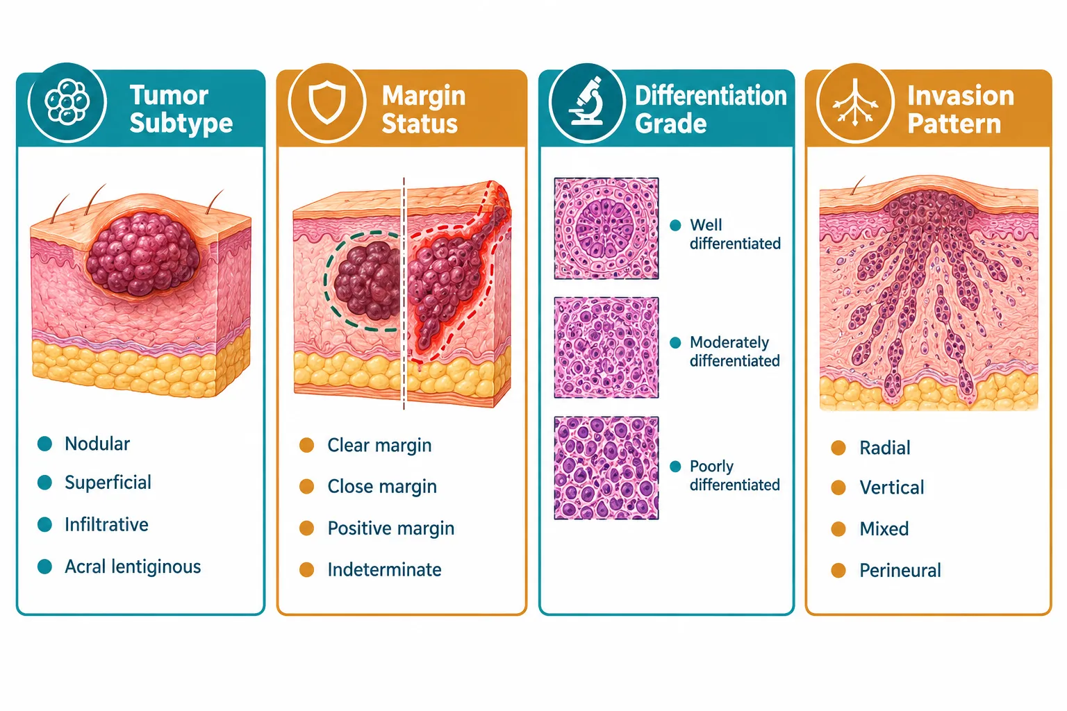
A handful of pathology terms appear on nearly every BCC and SCC report, and knowing them turns a confusing document into a readable one. Perineural invasion and other BCC/SCC pathology terms in plain English generally fall into five categories: margins, subtype, differentiation, depth, and invasion pattern.
Here is a working glossary for quick reference:
TermPlain-English MeaningMargins (clear/negative)No cancer cells found at the cut edge of the removed tissue; the tumor appears fully removedMargins (positive)Cancer cells are present at the cut edge, meaning more treatment is usually neededDifferentiationHow closely tumor cells resemble normal skin cells; poorly differentiated cells look less normal and tend to behave more aggressivelySubtype (BCC)The growth pattern: nodular and superficial are lower risk; infiltrative and morpheaform are higher riskSubtype (SCC)Keratinizing, non-keratinizing, or variants like spindle cell; some variants are more aggressive than classic SCCDepth of invasionHow far the tumor has grown below the skin surface, measured in millimetersPerineural invasionCancer cells found in or around a nerve sheathLymphovascular invasionCancer cells found in or around a blood or lymph vesselMitotic rateHow quickly tumor cells are dividing; a higher rate can suggest faster growthIn situCancer confined to the top layer of skin, not yet invasive
Common mistake: Treating every unfamiliar word on a pathology report as automatically alarming. Terms like "focal" (meaning limited to a small area) or "superficial" (meaning close to the surface) are often reassuring modifiers, not warning signs. Context changes the meaning of almost every term on this list.
For a broader look at how different skin growths are classified before they even reach a pathology report, this breakdown of 25 types of skin lesions explained is a useful companion resource.
Clark level and Breslow thickness are melanoma staging measurements, not BCC or SCC terms, and they measure how deep a melanoma has grown into the skin. Breslow thickness, measured in millimeters, is the more clinically important of the two today and directly influences melanoma treatment decisions.[4][5]
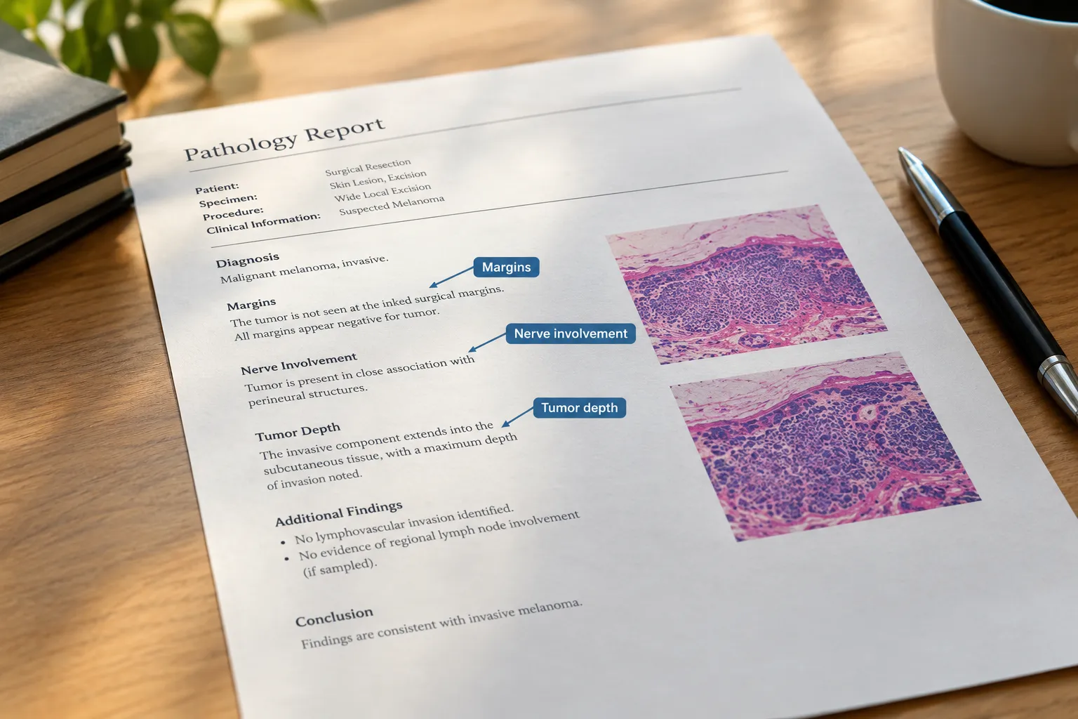
Why this section matters for BCC and SCC readers: Patients sometimes see "Clark level" or "Breslow thickness" mentioned in general skin cancer articles and wrongly assume these apply to their basal cell or squamous cell carcinoma report. They do not. BCC and SCC use their own depth and invasion measurements, including tumor thickness in some SCC staging systems and the perineural invasion status discussed throughout this guide, rather than Clark level or Breslow thickness.
If melanoma and its staging are relevant to your situation, this resource on neoadjuvant immunotherapy for stage III melanoma care covers how deeper or more advanced melanomas are being treated.
Certain tumor characteristics and anatomical locations raise the likelihood that a BCC or SCC will show perineural invasion. Knowing these risk factors helps explain why some tumors get closer monitoring even before a pathology result comes back.
Recognized risk factors include:
Edge case: A small, well-differentiated SCC on the trunk or limb found with incidental, focal PNI carries a very different risk profile than a large, poorly differentiated, recurrent SCC on the temple with PNI involving a named nerve and patient-reported numbness. Risk factors stack; a single factor rarely changes the whole treatment plan on its own.
Perineural invasion is one of the most common reasons dermatologists recommend Mohs micrographic surgery over standard excision, because Mohs allows the surgeon to examine 100 percent of the tumor margin in real time, including areas near nerves. This matters because nerve-tracking cancer cells can extend beyond what is visible or what a standard excision margin would capture.
Why Mohs surgery fits PNI cases well:
Decision rule: Choose Mohs surgery over standard excision if the pathology report shows PNI involving a larger or deeper nerve, if the tumor is recurrent, if it sits on the face or scalp, or if the subtype is aggressive. Choose standard excision with clear planned margins if PNI is incidental, focal, involves only a tiny dermal nerve, and the tumor is low-risk in every other category. The final decision should always come from the treating physician, since Mohs availability, tumor location, and overall health also factor in.
Common mistake: Assuming that finding PNI on an initial biopsy automatically requires immediate re-surgery with wider margins in every case. In reality, the full pathology picture (tumor type, location, size, differentiation, and whether the PNI is incidental or clinical) determines the next step, not PNI in isolation.
For practical information about minor surgical procedures and recovery expectations, this complete guide to minor surgery and mole removal outlines what patients can generally expect from in-office skin procedures.
Yes, most patients with perineural invasion, especially incidental PNI, are still cured with appropriate surgery and follow-up; PNI changes the treatment and monitoring plan far more than it changes the overall chance of cure for most cases. The outlook shifts meaningfully, though, when PNI is clinical rather than incidental.
A large cohort study comparing the two categories found:
These numbers show a real gap, but they also show that even clinical PNI does not guarantee a bad outcome; it means closer surveillance, often involving a head and neck or Mohs surgery specialist, additional imaging, and sometimes radiation therapy after surgery.
If PNI is found after the tumor has already been removed:
Quick example: A patient has a small SCC removed with clear margins, and the report later notes focal, incidental PNI in a single small nerve. The surgeon may simply recommend closer follow-up visits rather than additional surgery. Another patient with positive margins and PNI involving a larger nerve would likely need a second procedure and possibly radiation.
Edge case: Even with clinical PNI, long-term survival is still common when the disease is caught before it spreads beyond the local nerve pathway. The finding is a reason for a more careful plan, not a reason to assume the worst outcome.
Understanding a skin cancer pathology report comes down to locating five pieces of information: the diagnosis, the subtype, the margin status, the presence or absence of perineural or lymphovascular invasion, and any depth measurement. Reports can look dense, but they almost always follow a predictable structure.
A simple checklist for reading your report:
Common mistake: Focusing only on the word "carcinoma" and skipping past the modifiers around it. The difference between "superficial BCC, clear margins" and "infiltrative BCC, positive margins, perineural invasion" is enormous, even though both contain the word carcinoma.
If anything on the report is unclear, bring it to the follow-up appointment and ask the physician to walk through it line by line. For a step-by-step look at how pathology language is typically structured after a skin biopsy, see this guide to demystifying your pathology report after mole removal, which applies many of the same reading strategies even though it centers on pigmented lesions.
Does perineural invasion mean the cancer has already spread through the nervous system?
No. Perineural invasion means cancer cells were found touching or surrounding a nerve sheath in the removed tissue sample. It does not mean the cancer has traveled through the central nervous system or to distant organs; it is a local finding that raises the risk of local recurrence and, in more extensive cases, further spread.[1][3]
Is perineural invasion always extensive or dangerous?
No. Most perineural invasion found on skin cancer pathology reports is "incidental," meaning it involves a small nerve, was found only under the microscope, and comes with no symptoms. Extensive or "clinical" PNI, involving larger named nerves and symptoms like numbness or pain, is less common and carries higher risk.[2][3]
What is the difference between perineural invasion and lymphovascular invasion?
Perineural invasion involves cancer cells around a nerve sheath, while lymphovascular invasion involves cancer cells in a blood vessel or lymphatic channel. Both are risk markers tracked separately on pathology reports because they suggest different potential spread pathways.
Do Clark level and Breslow thickness apply to BCC and SCC?
No. Clark level and Breslow thickness are melanoma-specific measurements of how deep a melanoma has grown. BCC and SCC are evaluated with their own criteria, including subtype, differentiation, margin status, and perineural or lymphovascular invasion.[4][5]
How common is perineural invasion in squamous cell carcinoma specifically?
Perineural invasion appears in roughly 2 to 14 percent of cutaneous squamous cell carcinomas, depending on the study population and tumor characteristics such as size, location, and recurrence status.[1][6][7][8]
Can basal cell carcinoma with perineural invasion still be cured?
Yes, in most cases. BCC rarely metastasizes even with perineural invasion present, and treatment typically focuses on complete local removal, often with Mohs surgery, followed by closer monitoring.
What happens if perineural invasion is discovered after the tumor has already been removed?
The treatment team reviews the margin status and classifies the PNI as incidental or clinical. Depending on that review, the plan may involve observation with closer follow-up, a second surgical procedure, or referral for radiation or specialist evaluation.[2][7]
Should I automatically ask for Mohs surgery if my report mentions perineural invasion?
It is reasonable to ask whether Mohs surgery is appropriate, especially for facial, recurrent, or aggressive tumors with PNI. The final recommendation depends on the full pathology picture, not the PNI finding alone, so this decision should be made with the treating physician.
Why do pathology reports separate incidental PNI from clinical PNI?
Because the two categories carry meaningfully different risks. One study found local recurrence rates of about 17 percent for incidental PNI versus 37 percent for clinical PNI, and disease-specific death rates of about 6 percent versus 27 percent.[2] Separating the categories allows more accurate treatment planning.
What does "margins clear" mean on a BCC or SCC report?
It means no cancer cells were found at the outer edge of the tissue that was surgically removed, which generally suggests the tumor was fully excised. "Positive margins" means cancer cells reached the edge of the sample, usually prompting additional treatment.
Perineural invasion and other BCC/SCC pathology terms in plain English are not designed to confuse patients; they exist to give surgeons and oncologists a precise shared language for planning treatment. Perineural invasion specifically tells the care team whether a tumor might be tracking along a nerve pathway beyond what the eye can see, which shapes decisions about surgical technique, margin size, and follow-up frequency rather than determining a fixed outcome on its own.
The most useful next steps for anyone holding a BCC or SCC pathology report are straightforward. Read through the five core categories covered in this guide: diagnosis, subtype, margins, invasion findings, and depth. Write down any unfamiliar term and bring the list to the follow-up appointment. Ask directly whether any perineural invasion found is incidental or clinical, and whether the tumor's other features (location, size, differentiation, margin status) point toward Mohs surgery, standard excision, or additional imaging. A pathology report full of dense medical language becomes a manageable checklist once these core terms are translated into plain English, and that clarity is the foundation for making informed decisions about treatment and follow-up care.
[1] Pmc11545267 - https://pmc.ncbi.nlm.nih.gov/articles/PMC11545267/
[2] jamanetwork - https://jamanetwork.com/journals/jamadermatology/fullarticle/2635350
[3] Pmc11614312 - https://pmc.ncbi.nlm.nih.gov/articles/PMC11614312/
[4] jamanetwork - https://jamanetwork.com/journals/jamadermatology/fullarticle/2810838
[5] Pmc1874675 - https://pmc.ncbi.nlm.nih.gov/articles/PMC1874675/
[6] Pmc11416415 - https://pmc.ncbi.nlm.nih.gov/articles/PMC11416415/
[7] mdpi - https://www.mdpi.com/2072-6694/17/24/3921
[8] Pmc11965817 - https://pmc.ncbi.nlm.nih.gov/articles/PMC11965817/
[9] S41433 022 02306 W - https://www.nature.com/articles/s41433-022-02306-w
[10] pubmed.ncbi.nlm.nih.gov - https://pubmed.ncbi.nlm.nih.gov/37935106/