What is Angiokeratoma? Understanding This Common Skin Condition

You've noticed small, dark bumps on your skin—maybe on your feet, hands, or other reas—and you're wondering what they are. These tiny spots might be angiokeratomas, a surprisingly common skin condition that many people have but few understand. While they're usually harmless, knowing what you're dealing with can give you peace of mind and help you make informed decisions about treatment.

Angiokeratoma is a benign (non-cancerous) skin lesion that appears as small, dark red to black bumps on the skin's surface. Think of them as tiny blood vessels that have become dilated and covered by a thickened layer of skin. They're more common than you might think, affecting people of all ages, though they tend to appear more frequently as we get older.

At The Minor Surgery Center, we understand that any new skin growth can be concerning. That's why we're here to explain exactly what angiokeratomas are, why they develop, and what options you have if you'd like them removed.

Key Takeaways

Professional landscape medical infographic (1536x1024) for section "Understanding Angiokeratoma: The Basics". Type: Anatomical Diagram. Whit
  • Angiokeratomas are benign vascular lesions that appear as small, dark bumps on the skin caused by dilated blood vessels covered by thickened skin
  • Multiple types exist, including solitary angiokeratomas, angiokeratoma of Fordyce, and the rarer Fabry disease-related angiokeratomas
  • They're typically harmless but may bleed if injured or cause cosmetic concerns for some patients
  • Professional diagnosis is important to distinguish angiokeratomas from other skin lesions, including melanoma
  • Treatment options are available through minimally invasive solutions when removal is desired for medical or cosmetic reasons

Understanding Angiokeratoma: The Basics

Angiokeratoma literally means "blood vessel skin thickening"—and that's exactly what it is. These lesions develop when small blood vessels (capillaries) near the skin's surface become dilated and the overlying skin thickens.

The result? Small, raised bumps that range in color from bright red to dark purple or even black. They're usually between 1-5 millimeters in size—about the size of a pinhead to a small pea.

What Makes Angiokeratomas Different?

Unlike other skin lesions, angiokeratomas have some distinctive characteristics:

  • Vascular origin: They're made up of dilated blood vessels, which is why they're often dark red or purple
  • Warty surface: The overlying skin becomes thickened and may feel slightly rough or scaly
  • Tendency to bleed: Because they contain blood vessels, they may bleed easily if scratched or injured
  • Stable nature: They typically don't grow rapidly or change significantly once formed

Think of an angiokeratoma like a tiny varicose vein on the surface of your skin. Just as varicose veins are dilated blood vessels in the legs, angiokeratomas are dilated blood vessels in the skin—but on a much smaller scale.

The Medical Classification

Medically speaking, angiokeratomas are classified as benign vascular proliferations. They're part of a larger family of vascular lesions that includes conditions like cherry angiomas and pyogenic granulomas.

The key word here is benign. These lesions are not cancerous and won't become cancerous over time. However, proper diagnosis is essential because other skin conditions can sometimes look similar.

Types of Angiokeratoma: Not All Are the Same

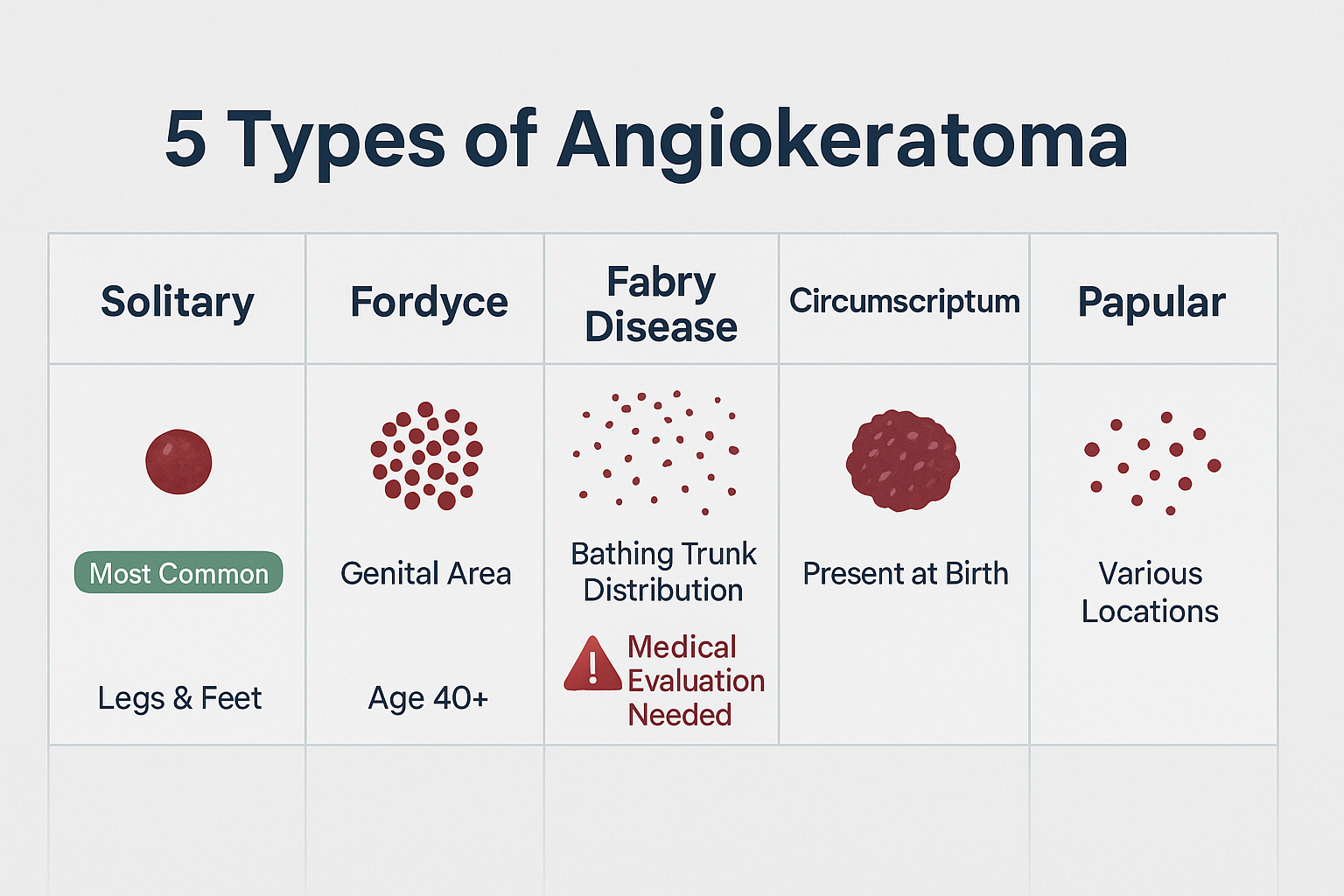
There are several distinct types of angiokeratomas, each with different characteristics and locations on the body. Understanding which type you have can help guide treatment decisions.

1. Solitary Angiokeratoma (Angiokeratoma of Mibelli)

This is the most common type—the kind most people encounter.

Characteristics:

  • Appears as single or few isolated lesions
  • Most commonly found on the legs, especially the feet and toes
  • Can also appear on fingers, hands, or other areas
  • More common in women than men
  • Often develops after minor trauma or injury to the area

These solitary lesions are completely benign and usually don't require treatment unless they're bothersome or frequently bleeding.

2. Angiokeratoma of Fordyce (Scrotal Angiokeratoma)

This type appears on the genital area and is more common than many people realize.

Characteristics:

  • Multiple small lesions on the scrotum in men or vulva in women
  • Typically develops in middle age or later
  • Becomes more common and numerous with age
  • Associated with increased venous pressure in the area
  • Completely benign despite the concerning location

Many people don't seek medical attention for these lesions due to embarrassment, but they're a normal variant that dermatologists see regularly.

3. Angiokeratoma Corporis Diffusum (Fabry Disease)

This is the rarest and most serious type, as it's associated with an underlying genetic condition called Fabry disease.

Characteristics:

  • Multiple lesions scattered across the body
  • Often appear in a "bathing trunk" distribution (lower trunk, buttocks, thighs)
  • Associated with Fabry disease, a genetic enzyme deficiency
  • Appears in childhood or adolescence
  • Requires medical evaluation and management of the underlying condition

Important distinction: While the first two types are simply cosmetic or minor nuisances, angiokeratomas associated with Fabry disease are a sign of a systemic condition that needs medical management.

4. Angiokeratoma Circumscriptum

This is a rare congenital (present from birth) form.

Characteristics:

  • Present at birth or appears in early childhood
  • Usually appears as a cluster of lesions in one area
  • Often on the legs or trunk
  • May be larger and more extensive than other types
  • Can sometimes be associated with underlying vascular malformations

5. Papular Angiokeratomas

These appear as multiple small papules (raised bumps) in various locations.

Characteristics:

  • Can occur anywhere on the body
  • Often appear in groups
  • May develop spontaneously without clear cause
  • Typically benign and stable

What Causes Angiokeratoma? Understanding the Risk Factors

The exact cause of angiokeratomas isn't always clear, but several factors can contribute to their development.

Primary Causes

Vascular Dilation and Damage

The fundamental cause is dilation of small blood vessels in the skin. This can happen due to:

  • Increased venous pressure: When pressure builds up in the veins, it can cause capillaries to dilate
  • Trauma: Minor injuries can damage blood vessels and trigger angiokeratoma formation
  • Aging: As we age, our blood vessels become more fragile and prone to dilation
  • Chronic venous insufficiency: Poor circulation can contribute to development

Genetic Factors

Some types of angiokeratomas run in families:

  • Fabry disease is inherited in an X-linked pattern
  • Some people may have a genetic predisposition to developing solitary angiokeratomas
  • Family history of vascular lesions may increase risk

Contributing Factors

Several conditions and circumstances can increase your likelihood of developing angiokeratomas:

Age-Related Changes

  • More common after age 40
  • Cumulative sun damage may play a role
  • Natural weakening of blood vessel walls over time

Pregnancy

  • Increased blood volume and hormonal changes
  • Higher venous pressure, especially in the pelvic area
  • Angiokeratomas may develop during pregnancy and persist afterward

Chronic Conditions

  • Varicose veins
  • Hemorrhoids (can be associated with scrotal angiokeratomas)
  • Hernias
  • Any condition that increases venous pressure

Occupational Factors

  • Jobs requiring prolonged standing
  • Heavy lifting
  • Activities that increase abdominal pressure

The Fabry Disease Connection

It's worth understanding this connection in more detail because it's the one type that requires medical attention beyond the lesions themselves.

What is Fabry Disease?

Fabry disease is a rare genetic disorder caused by a deficiency of the enzyme alpha-galactosidase A. This enzyme normally breaks down a fatty substance called globotriaosylceramide (GL-3). When the enzyme is deficient, GL-3 accumulates in cells throughout the body, including blood vessel walls.

Signs That Suggest Fabry Disease:

  • Multiple angiokeratomas appearing in childhood or adolescence
  • Distribution in the "bathing trunk" area
  • Family history of Fabry disease
  • Other symptoms like pain in hands and feet, kidney problems, heart issues, or stroke at a young age

If you have multiple angiokeratomas that appeared at a young age, especially with other unexplained symptoms, ask your doctor about Fabry disease screening.

Symptoms and Appearance: What to Look For

Recognizing an angiokeratoma involves understanding both how they look and how they feel.

Visual Characteristics

Color

  • Bright red when first developing
  • Dark red to purple as they mature
  • Blue-black in some cases
  • May appear similar to blood blisters or dark moles

Size and Shape

  • Usually 1-5 millimeters in diameter
  • Dome-shaped or slightly raised
  • Well-defined borders
  • May have a slightly warty or scaly surface

Texture

  • Firm to the touch
  • Slightly rough or scaly surface (hyperkeratosis)
  • May feel like a tiny bump or papule
  • Not typically painful unless injured

Physical Symptoms

Most angiokeratomas don't cause symptoms, but some people experience:

Bleeding

  • The most common symptom
  • Can occur spontaneously or after minor trauma
  • May be difficult to stop due to the vascular nature
  • More common with angiokeratomas in areas prone to friction

Itching

  • Occasional mild itching
  • More common when lesions are in areas that rub against clothing
  • Usually not severe

Cosmetic Concerns

  • Many people seek treatment primarily for appearance
  • Dark color can be mistaken for melanoma, causing anxiety
  • Visible lesions on hands, face, or other exposed areas may be bothersome

Rarely:

  • Mild tenderness
  • Burning sensation
  • Discomfort if located in areas of friction

Location Patterns

Where angiokeratomas appear can provide clues about their type:

Lower Extremities (Most Common)

  • Feet and toes
  • Ankles
  • Lower legs
  • Associated with venous pressure and minor trauma

Genital Area

  • Scrotum in men (Fordyce type)
  • Vulva in women
  • Increases in number with age

Trunk and Thighs

  • "Bathing trunk" distribution suggests possible Fabry disease
  • Requires medical evaluation

Hands and Fingers

  • Less common than lower extremities
  • May develop after injury

Other Areas

  • Can appear almost anywhere on the body
  • Face, arms, and other locations are less common

Diagnosis: How Doctors Identify Angiokeratoma

Professional landscape infographic (1536x1024) for section "Types of Angiokeratoma". Type: Visual Classification Chart. Light gray backgroun

Getting an accurate diagnosis is crucial because several other skin conditions can look similar to angiokeratomas—including some that require different treatment approaches.

Clinical Examination

The diagnostic process typically begins with a thorough visual examination by a healthcare provider.

What Your Doctor Will Look For:

  • Color and appearance: The characteristic dark red to black color
  • Size and shape: Typical dome-shaped appearance
  • Texture: Warty or scaly surface
  • Location: Where the lesions appear on your body
  • Number: Single lesion versus multiple lesions
  • Distribution pattern: Random versus specific patterns

Dermoscopy

Many dermatologists use a tool called a dermatoscope—a special magnifying device that allows them to see structures beneath the skin's surface.

Dermoscopic Features of Angiokeratoma:

  • Dark red to black lacunae (lake-like structures)
  • Well-defined borders
  • Absence of melanoma-specific patterns
  • Visible vascular structures

This non-invasive examination helps distinguish angiokeratomas from other lesions, particularly melanoma.

When a Biopsy is Needed

In some cases, your doctor may recommend a skin biopsy to confirm the diagnosis.

Reasons for Biopsy:

  • Uncertainty about the diagnosis
  • Lesion has unusual features
  • Rapid change in size or appearance
  • Need to rule out melanoma or other skin cancers
  • Patient anxiety requiring definitive diagnosis

The Biopsy Process:

At facilities like The Minor Surgery Center, the biopsy procedure is straightforward:

  1. Local anesthesia numbs the area
  2. Small sample of the lesion is removed
  3. Sent to pathology for microscopic examination
  4. Results typically available within a week

Our experienced surgical team performs these procedures with a comfort-first approach, ensuring you're at ease throughout the process.

Differential Diagnosis: What Else Could It Be?

Part of the diagnostic process involves ruling out other conditions that can look similar:

Melanoma

  • The most important condition to rule out
  • Can appear as dark, raised lesions
  • Key differences: melanoma typically shows irregular borders, color variation, and specific dermoscopic patterns
  • When in doubt, biopsy is recommended

Cherry Angioma

  • Also vascular lesions but typically brighter red
  • Usually don't have the warty surface of angiokeratomas
  • More common and generally smaller
  • Learn more about cherry angiomas

Pyogenic Granuloma

  • Rapidly growing vascular lesion
  • Often bleeds easily
  • Usually has a more lobulated appearance
  • Different treatment approach

Thrombosed Capillary or Venous Lake

  • Blue-black appearance
  • More common on lips and ears
  • Compressible (angiokeratomas are not)

Seborrheic Keratosis

  • Warty appearance but not vascular
  • Typically lighter in color
  • "Stuck on" appearance

Warts

  • Viral origin
  • Different surface appearance under dermoscopy
  • May have black dots (thrombosed capillaries)

Special Considerations for Fabry Disease Screening

If you have multiple angiokeratomas, especially if they appeared before age 30, your doctor may recommend screening for Fabry disease.

Screening Tests Include:

  • Enzyme assay: Measures alpha-galactosidase A activity
  • Genetic testing: Identifies mutations in the GLA gene
  • Family history: Assessment of relatives with similar symptoms
  • Additional symptoms: Evaluation for kidney, heart, or neurological involvement

Early diagnosis of Fabry disease is important because enzyme replacement therapy is available and can prevent serious complications.

Treatment Options: When and How to Remove Angiokeratoma

The good news? Most angiokeratomas don't require treatment. They're benign and won't harm your health. However, many people choose to have them removed for various reasons.

When to Consider Treatment

Medical Reasons:

  • Frequent bleeding that's difficult to control
  • Located in an area prone to trauma or irritation
  • Causing pain or discomfort
  • Interfering with daily activities

Cosmetic Reasons:

  • Visible location causing self-consciousness
  • Dark appearance causing anxiety about cancer
  • Personal preference for removal

Diagnostic Reasons:

  • Uncertainty about the diagnosis
  • Need to rule out melanoma or other conditions

Conservative Management (Watchful Waiting)

For many people, simply monitoring the angiokeratoma is the best approach.

When This Makes Sense:

  • Small, asymptomatic lesions
  • Located in non-visible areas
  • Not causing any problems
  • Confirmed diagnosis with no concern for malignancy

What to Watch For:

  • Rapid growth
  • Change in color or appearance
  • Increased bleeding
  • Development of new symptoms

Minimally Invasive Treatment Options

When treatment is desired, several effective options are available through expert outpatient care.

1. Laser Therapy

How It Works: Laser treatment targets the blood vessels within the angiokeratoma, causing them to coagulate and eventually be absorbed by the body.

Types of Lasers Used:

  • Pulsed dye laser (PDL): Most commonly used
  • Nd:YAG laser: Effective for deeper lesions
  • CO2 laser: For larger or more prominent lesions

Advantages:

  • Minimal scarring
  • Precise targeting
  • Quick procedure
  • Good cosmetic results
  • Can treat multiple lesions in one session

What to Expect:

  • Brief stinging sensation during treatment
  • Some redness and swelling afterward
  • Crusting for a few days
  • Healing within 1-2 weeks
  • May require multiple sessions for complete removal

2. Electrocautery (Electrodesiccation)

How It Works: An electric current is used to destroy the lesion through heat.

Advantages:

  • Effective for small to medium lesions
  • Quick procedure
  • Immediate results
  • Cost-effective

Considerations:

  • May leave a small scar
  • Some discomfort during healing
  • Risk of hyperpigmentation in darker skin types

3. Surgical Excision

How It Works: The angiokeratoma is surgically cut out and the wound is closed with sutures.

When This is Preferred:

  • Larger lesions
  • When biopsy is needed for diagnosis
  • Lesions that haven't responded to other treatments
  • Patient preference for complete removal

The Process at The Minor Surgery Center:

Our experienced surgical team specializes in safe and efficient surgery for skin lesions. Here's what you can expect:

  1. Consultation: We discuss your concerns and examine the lesion
  2. Personalized treatment plan: We explain the procedure and answer all questions
  3. Local anesthesia: The area is numbed for your comfort
  4. Precise excision: The lesion is carefully removed
  5. Closure: Sutures are placed for optimal healing
  6. Pathology: The tissue is sent for examination to confirm the diagnosis

Recovery:

  • Sutures typically removed in 7-14 days
  • Minimal downtime
  • Most people return to normal activities quickly
  • Follow-up to ensure proper healing

4. Cryotherapy

How It Works: Liquid nitrogen is applied to freeze and destroy the lesion.

Advantages:

  • No cutting required
  • Quick procedure
  • No need for anesthesia

Considerations:

  • May require multiple treatments
  • Can cause temporary hypopigmentation (lightening of skin)
  • Not ideal for all locations or skin types
  • Less predictable results than other methods

5. Shave Excision

How It Works: The raised portion of the angiokeratoma is shaved off at the skin level.

Advantages:

  • Quick and simple
  • Minimal scarring
  • Good for raised lesions

Considerations:

  • May not remove the entire lesion
  • Possibility of recurrence
  • Best for cosmetic improvement rather than complete removal

Choosing the Right Treatment

The best treatment depends on several factors:

Lesion Characteristics:

  • Size and depth
  • Location on the body
  • Number of lesions

Patient Factors:

  • Skin type
  • Healing ability
  • Cosmetic goals
  • Medical history

Practical Considerations:

  • Cost and insurance coverage
  • Recovery time available
  • Personal preferences

At The Minor Surgery Center, we work with you to develop a personalized treatment plan that addresses your specific needs and goals. Our comfort-first approach ensures you feel informed and confident about your decision.

Recovery and Aftercare: What to Expect

Proper aftercare is essential for optimal healing and the best cosmetic results, regardless of which treatment method you choose.

Immediate Post-Treatment Care

First 24-48 Hours:

Wound Care:

  • Keep the area clean and dry
  • Apply prescribed antibiotic ointment if recommended
  • Cover with a bandage as directed
  • Avoid getting the area wet (no swimming or prolonged water exposure)

Pain Management:

  • Mild discomfort is normal
  • Over-the-counter pain relievers (acetaminophen or ibuprofen) usually sufficient
  • Ice packs can reduce swelling (wrapped in cloth, not directly on skin)

Activity Restrictions:

  • Avoid strenuous exercise for 24-48 hours
  • No heavy lifting if lesion was on hand or arm
  • Elevate the area if possible to reduce swelling

Ongoing Care (Days 3-14)

Wound Monitoring:

  • Check daily for signs of infection (increased redness, warmth, pus, fever)
  • Some crusting is normal
  • Mild redness around the site is expected

Hygiene:

  • Gentle cleansing with mild soap and water
  • Pat dry, don't rub
  • Continue antibiotic ointment as directed

Sun Protection:

  • Keep the area covered or use high SPF sunscreen
  • UV exposure can cause hyperpigmentation
  • Especially important for the first 3-6 months

Suture Care (if applicable):

  • Keep sutures dry until removal
  • Don't pick at or pull on sutures
  • Return for removal as scheduled (typically 7-14 days)

Long-Term Healing

Weeks 2-8:

  • Scar will mature and fade
  • Redness gradually improves
  • Texture normalizes

Scar Management:

  • Silicone gel or sheets can improve scar appearance
  • Massage the area gently once fully healed
  • Continue sun protection
  • Consider vitamin E or other scar-reducing products

Signs of Complications

Contact your healthcare provider if you experience:

Infection Warning Signs:

  • Increasing pain after the first few days
  • Spreading redness
  • Warmth around the wound
  • Pus or foul-smelling drainage
  • Fever or chills
  • Red streaks extending from the wound

Healing Problems:

  • Wound edges separating
  • Excessive bleeding
  • Delayed healing beyond expected timeframe
  • Development of excessive scar tissue (keloid)

Other Concerns:

  • Allergic reaction to medications
  • Unexpected changes in the area
  • New lesions developing nearby

Expected Results and Realistic Expectations

Success Rates:

  • Most treatments are highly successful
  • Complete removal is usually achieved with surgical excision
  • Laser therapy may require 2-3 sessions for optimal results
  • Recurrence is uncommon but possible

Cosmetic Outcomes:

  • Small scars are common but usually fade well
  • Location affects scarring (areas of tension may have more visible scars)
  • Proper aftercare significantly improves cosmetic results
  • Most people are very satisfied with the outcome

Timeline:

  • Immediate improvement in appearance
  • Final cosmetic result visible in 3-6 months
  • Scars continue to mature for up to a year

Our team at The Minor Surgery Center provides detailed aftercare instructions and is always available to answer questions during your recovery. We want you to get back to your life quickly and confidently.

Prevention: Can You Avoid Angiokeratomas?

While you can't completely prevent angiokeratomas—especially if you have a genetic predisposition—certain strategies may reduce your risk or prevent new ones from forming.

General Prevention Strategies

Protect Your Skin:

  • Avoid trauma to areas prone to angiokeratomas
  • Wear protective footwear
  • Use padding in areas of friction
  • Be gentle when shaving or grooming

Manage Venous Health:

  • Exercise regularly to promote good circulation
  • Maintain a healthy weight
  • Avoid prolonged standing when possible
  • Elevate legs when resting
  • Consider compression stockings if you have venous insufficiency

Skin Care:

  • Keep skin well-moisturized
  • Use gentle cleansers
  • Avoid harsh scrubbing
  • Protect from sun damage with SPF

Specific Risk Reduction

For Lower Extremity Angiokeratomas:

  • Treat varicose veins if present
  • Manage chronic venous insufficiency
  • Avoid tight footwear that restricts circulation
  • Take breaks from standing if your job requires it

For Genital Angiokeratomas (Fordyce Type):

  • Manage hemorrhoids if present
  • Avoid chronic constipation and straining
  • Maintain healthy bowel habits
  • Address any conditions that increase pelvic venous pressure

Occupational Considerations:

  • Use ergonomic practices to reduce strain
  • Take regular breaks from prolonged standing
  • Use proper lifting techniques
  • Wear appropriate protective equipment

When Prevention Isn't Possible

Some angiokeratomas develop despite our best efforts, particularly:

  • Age-related changes
  • Genetic predisposition
  • Fabry disease-related angiokeratomas
  • Congenital forms

In these cases, early detection and appropriate management become the focus rather than prevention.

Monitoring for New Lesions

Regular Skin Checks:

  • Examine your skin monthly
  • Note any new dark spots or bumps
  • Take photos to track changes
  • Pay special attention to areas where you've had angiokeratomas before

Professional Evaluation:

  • Annual skin examination by a healthcare provider
  • More frequent checks if you have multiple lesions
  • Immediate evaluation of any rapidly changing lesions

When to Seek Medical Attention:

  • New dark lesions that bleed easily
  • Rapid growth of existing lesions
  • Multiple new lesions appearing simultaneously
  • Any lesion that looks different from your other angiokeratomas

Living with Angiokeratoma: Practical Advice

For many people, angiokeratomas are a long-term companion rather than something that needs immediate treatment. Here's how to manage them day-to-day.

Managing Bleeding Episodes

Bleeding is the most common issue people face with angiokeratomas.

Immediate Steps:

  1. Apply direct pressure with clean gauze or cloth
  2. Elevate the area above heart level if possible
  3. Hold pressure for 10-15 minutes without peeking
  4. Apply ice wrapped in cloth to help constrict blood vessels
  5. Use a hemostatic agent like styptic powder if available

Prevention:

  • Avoid scratching or picking at lesions
  • Be careful when shaving near angiokeratomas
  • Wear protective clothing over lesions in high-friction areas
  • Trim toenails carefully if you have foot angiokeratomas
  • Use electric razors instead of blades near lesions

When to Seek Help:

  • Bleeding doesn't stop after 20 minutes of pressure
  • Frequent bleeding episodes
  • Excessive blood loss
  • Signs of infection after bleeding

Clothing and Lifestyle Adjustments

Footwear Choices:

  • Wear well-fitting shoes that don't rub
  • Use padded socks if angiokeratomas are on feet
  • Avoid tight shoes that create pressure points
  • Consider custom orthotics if needed

Clothing Considerations:

  • Choose soft, non-irritating fabrics
  • Avoid tight clothing over affected areas
  • Use protective padding under clothing if needed
  • Consider seamless underwear for genital angiokeratomas

Activity Modifications:

  • Continue normal activities—angiokeratomas shouldn't limit you
  • Take precautions in contact sports
  • Use protective gear when appropriate
  • Be mindful during activities that could cause trauma

Psychological and Emotional Aspects

Dealing with Anxiety:

It's completely normal to worry when you first notice dark spots on your skin. Many people immediately think of skin cancer.

Coping Strategies:

  • Get a professional diagnosis for peace of mind
  • Educate yourself about the condition
  • Remember that angiokeratomas are benign
  • Join support groups if multiple lesions are affecting your quality of life
  • Consider treatment if anxiety is significant

Cosmetic Concerns:

Dark lesions in visible areas can affect self-confidence.

Options:

  • Makeup can effectively cover small lesions
  • Treatment is available and effective
  • Remember that most people don't notice them as much as you do
  • Focus on overall health and wellbeing

Communication:

  • Explain the condition to partners or family members who may be concerned
  • Educate them that angiokeratomas are not contagious
  • Share information from reliable sources
  • Be open about your treatment preferences

When Angiokeratomas Are Part of a Larger Condition

Living with Fabry Disease:

If your angiokeratomas are related to Fabry disease, management is more complex.

Comprehensive Care Includes:

  • Regular monitoring by specialists
  • Enzyme replacement therapy
  • Management of kidney, heart, and neurological symptoms
  • Genetic counseling for family planning
  • Support groups and patient advocacy organizations

Coordinated Treatment:

  • Dermatologist for skin manifestations
  • Nephrologist for kidney function
  • Cardiologist for heart involvement
  • Neurologist for pain and stroke prevention
  • Genetic counselor for family screening

Insurance and Financial Considerations

Coverage for Treatment:

  • Cosmetic removal may not be covered
  • Medical necessity (bleeding, pain) usually is covered
  • Get pre-authorization when required
  • Document symptoms thoroughly
  • Fabry disease treatment is typically covered

Cost Management:

  • Ask about payment plans
  • Compare costs between providers
  • Consider treating the most problematic lesions first
  • Inquire about package pricing for multiple lesions

At The Minor Surgery Center, we work with patients to understand their insurance coverage and provide transparent pricing for our minimally invasive solutions.

Angiokeratoma vs. Other Skin Conditions: Key Differences

Understanding how angiokeratomas differ from other skin lesions can help you recognize what you're dealing with and when to seek professional evaluation.

Angiokeratoma vs. Melanoma

This is the most important distinction because melanoma is a serious skin cancer.

FeatureAngiokeratomaMelanomaColorUniform dark red to blackOften multiple colors (brown, black, red, white, blue)BorderWell-defined, regularIrregular, poorly definedSymmetryUsually symmetricalOften asymmetricalSurfaceWarty, scalyVariable, may be smooth or irregularGrowthStable or slow growthOften rapid growth or changeBleedingCommon if traumatizedLess commonDermoscopyVascular lacunaeMelanoma-specific patterns

When in doubt, always get it checked. The peace of mind is worth it, and early detection of melanoma is crucial.

Learn more about identifying melanoma and other skin cancers.

Angiokeratoma vs. Cherry Angioma

Both are vascular lesions, but they have distinct differences.

Cherry Angioma:

  • Bright cherry-red color
  • Smooth, dome-shaped surface
  • Typically smaller (1-3mm)
  • More common (almost everyone over 30 has some)
  • Rarely bleed unless injured
  • Don't have the warty surface of angiokeratomas

Angiokeratoma:

  • Darker red to black color
  • Warty or scaly surface
  • Can be larger (up to 5mm or more)
  • Less common
  • Bleed more easily
  • Thickened overlying skin

Both are benign, but treatment approaches may differ. Read more about cherry angioma removal.

Angiokeratoma vs. Warts

Warts are caused by human papillomavirus (HPV) and are completely different in origin.

Warts:

  • Viral infection (contagious)
  • Rough, cauliflower-like surface
  • May have black dots (thrombosed capillaries)
  • Can spread to other areas
  • Often appear in clusters
  • Respond to antiviral treatments

Angiokeratoma:

  • Vascular malformation (not contagious)
  • Warty surface but different texture
  • Dark red to black throughout
  • Don't spread
  • Usually solitary or few in number
  • Don't respond to antiviral treatments

Angiokeratoma vs. Seborrheic Keratosis

Seborrheic keratoses are very common benign growths, especially in older adults.

Seborrheic Keratosis:

  • "Stuck on" appearance
  • Tan, brown, or black color (not vascular)
  • Waxy or scaly surface
  • Often multiple
  • Very common with age
  • No bleeding tendency

Angiokeratoma:

  • Appears more embedded in skin
  • Dark red to black (vascular origin)
  • Warty surface
  • May be solitary
  • Any age (though more common in adults)
  • Bleeds if traumatized

Angiokeratoma vs. Dermatofibroma

Dermatofibromas are benign fibrous nodules that can sometimes be confused with angiokeratomas.

Dermatofibroma:

  • Firm, button-like nodule
  • Brown to pink color
  • Dimples when pinched
  • Feels deeper in the skin
  • Doesn't bleed easily
  • Often on legs

Angiokeratoma:

  • Softer, more superficial
  • Dark red to black color
  • Doesn't dimple when pinched
  • More surface-level
  • Bleeds more easily
  • Various locations

Learn more about different types of skin lesions to better understand what you might be dealing with.

Angiokeratoma vs. Pyogenic Granuloma

Pyogenic granulomas are rapidly growing vascular lesions that can bleed profusely.

Pyogenic Granuloma:

  • Rapid growth (weeks to months)
  • Bright red color
  • Pedunculated (on a stalk) often
  • Bleeds very easily and profusely
  • Often appears after trauma
  • Requires treatment

Angiokeratoma:

  • Slow growth or stable
  • Darker red to black color
  • Sessile (flat-based) usually
  • Bleeds but usually controllable
  • May or may not have trauma history
  • Treatment optional in many cases

The Importance of Professional Diagnosis

While these comparisons can help you understand what you might have, only a healthcare professional can make a definitive diagnosis.

Why Professional Evaluation Matters:

  • Visual examination alone isn't always sufficient
  • Dermoscopy reveals features not visible to the naked eye
  • Biopsy provides definitive diagnosis when needed
  • Early detection of serious conditions like melanoma saves lives
  • Peace of mind knowing exactly what you're dealing with

At The Minor Surgery Center, our experienced surgical team has seen thousands of skin lesions and can quickly differentiate between various conditions. We use advanced diagnostic techniques and provide clear explanations so you understand exactly what's going on with your skin.

Frequently Asked Questions About Angiokeratoma

Professional landscape comparison infographic (1536x1024) for section "Diagnosis: How Doctors Identify Angiokeratoma". Type: Side-by-side Co

Are angiokeratomas dangerous?

No, angiokeratomas themselves are not dangerous. They're benign (non-cancerous) lesions that won't turn into cancer. However, the exception is when multiple angiokeratomas are a sign of Fabry disease—in that case, the underlying condition needs medical management, though the skin lesions themselves are still benign.

Can angiokeratomas spread or multiply?

Angiokeratomas don't spread like infections or warts. However, you may develop new ones over time, especially if you have risk factors like venous insufficiency or if they're related to Fabry disease. Existing angiokeratomas typically don't grow larger or multiply on their own.

Do I need to have my angiokeratoma removed?

Not necessarily. Many angiokeratomas don't require treatment. Consider removal if:

  • They bleed frequently
  • They're in a location that causes irritation
  • You're concerned about the appearance
  • There's uncertainty about the diagnosis
  • They're causing pain or discomfort

Will angiokeratoma removal leave a scar?

Most removal methods leave minimal scarring. The extent of scarring depends on:

  • Size and location of the lesion
  • Removal method used
  • Your individual healing characteristics
  • Proper aftercare

Laser therapy typically produces the least visible scarring, while surgical excision may leave a small linear scar. Our team at The Minor Surgery Center uses techniques designed to minimize scarring and optimize cosmetic outcomes.

Can angiokeratomas come back after removal?

Recurrence is uncommon but possible, especially if:

  • The lesion wasn't completely removed
  • Underlying risk factors (like venous insufficiency) persist
  • You have a genetic predisposition

Complete surgical excision has the lowest recurrence rate. If an angiokeratoma does recur, it can be treated again.

How can I tell if it's an angiokeratoma or melanoma?

While angiokeratomas and melanomas can both appear as dark lesions, there are key differences:

Angiokeratoma typically has:

  • Uniform dark red to black color
  • Regular, well-defined borders
  • Warty surface texture
  • Symmetrical shape

Melanoma often has:

  • Multiple colors
  • Irregular borders
  • Variable surface
  • Asymmetrical shape

However, only a professional can make this determination. If you have any doubt, get it checked. It's always better to be safe, and early melanoma detection is crucial. Learn more about different types of skin cancer.

Are angiokeratomas contagious?

No, angiokeratomas are not contagious. They're vascular malformations, not infections. You can't spread them to other people or to other parts of your own body.

Can children get angiokeratomas?

Yes, though they're less common in children. When angiokeratomas appear in childhood or adolescence, especially if there are multiple lesions, evaluation for Fabry disease may be recommended. The congenital form (angiokeratoma circumscriptum) is present from birth or appears in early childhood.

Does insurance cover angiokeratoma removal?

Coverage depends on the reason for removal:

  • Medical necessity (bleeding, pain, diagnostic uncertainty): Usually covered
  • Cosmetic reasons: Typically not covered
  • Fabry disease-related: Usually covered as part of disease management

Check with your insurance provider and get pre-authorization when required. We can help document medical necessity when appropriate.

How long does the removal procedure take?

Most angiokeratoma removal procedures are quick:

  • Laser therapy: 10-30 minutes depending on number of lesions
  • Electrocautery: 5-15 minutes
  • Surgical excision: 15-30 minutes including closure
  • Cryotherapy: 5-10 minutes

These times don't include preparation and aftercare instructions, but most patients are in and out within an hour for a straightforward removal.

What's the recovery time after angiokeratoma removal?

Recovery is typically quick:

  • Laser therapy: 1-2 weeks for complete healing
  • Surgical excision: 7-14 days until suture removal, then another week or two for complete healing
  • Electrocautery: 1-2 weeks
  • Cryotherapy: 2-4 weeks

Most people return to normal activities within a few days, with some restrictions on strenuous activity depending on the location.

Can angiokeratomas affect pregnancy?

Angiokeratomas don't affect fertility or pregnancy outcomes. However, pregnancy can sometimes trigger the development of new angiokeratomas due to increased blood volume and venous pressure. Genital angiokeratomas may become more prominent during pregnancy but typically don't cause complications with delivery.

When to See a Doctor: Red Flags and Recommendations

While angiokeratomas are benign, certain situations warrant professional medical evaluation.

Seek Immediate Medical Attention If:

🚨 Uncontrolled bleeding that doesn't stop with 20 minutes of direct pressure

🚨 Signs of infection including:

  • Increasing pain, redness, or swelling
  • Pus or foul-smelling drainage
  • Red streaks extending from the lesion
  • Fever or chills

🚨 Rapid change in size, color, or appearance over days to weeks

🚨 Severe pain associated with the lesion

Schedule a Consultation Soon If:

⚠️ New dark lesion appears that you haven't had evaluated

⚠️ Frequent bleeding episodes that interfere with daily life

⚠️ Multiple angiokeratomas appearing, especially in a pattern

⚠️ Lesions in childhood or adolescence, particularly if multiple

⚠️ Cosmetic concerns affecting your quality of life

⚠️ Uncertainty about diagnosis—when in doubt, get it checked

⚠️ Changes in existing lesions including:

  • Color changes
  • Size changes
  • Texture changes
  • New symptoms (pain, itching, bleeding)

Routine Evaluation Recommended For:

Annual skin checks if you have multiple angiokeratomas

Family history of Fabry disease or multiple angiokeratomas

New lesions that fit the typical appearance but you want confirmation

Treatment planning if you're considering removal

Follow-up after treatment to ensure proper healing

What to Expect at Your Appointment

When you visit The Minor Surgery Center or another healthcare provider, here's what typically happens:

Initial Consultation:

  1. Medical history: Discussion of your symptoms, when the lesion appeared, any changes
  2. Visual examination: Careful inspection of the lesion
  3. Dermoscopy: Magnified examination if available
  4. Discussion: Explanation of findings and recommendations
  5. Treatment planning: If treatment is desired or necessary

Our Approach:

We believe in clear communication and personalized care. You'll never feel rushed, and we'll answer all your questions. Our experienced surgical team will explain your options in plain language and help you make the decision that's right for you.

Questions to Ask Your Doctor:

  • What type of angiokeratoma do I have?
  • Do I need any additional testing?
  • What are my treatment options?
  • What are the risks and benefits of each option?
  • What will recovery be like?
  • What's the cost and will insurance cover it?
  • What happens if I choose not to treat it?
  • When should I come back for follow-up?

Don't hesitate to ask questions or express concerns. Your healthcare provider is there to help you understand your condition and make informed decisions.

The Minor Surgery Center Approach: Expert Care for Angiokeratoma

At The Minor Surgery Center, we specialize in providing expert outpatient care for skin lesions including angiokeratomas. Our approach combines medical expertise with a genuine understanding that you're a person, not just a case.

Why Choose The Minor Surgery Center?

Experienced Surgical Team

Our surgeons have performed thousands of minor surgical procedures, including angiokeratoma removal. We stay current with the latest techniques and technologies to provide you with the best possible outcomes.

Comfort-First Approach

We understand that any surgical procedure can be anxiety-inducing. That's why we:

  • Take time to explain everything clearly
  • Answer all your questions without rushing
  • Use effective local anesthesia for your comfort
  • Create a welcoming, reassuring environment
  • Provide detailed aftercare instructions

Minimally Invasive Solutions

We use the most appropriate technique for your specific situation:

  • Precision laser therapy when suitable
  • Careful surgical excision with optimal closure techniques
  • Advanced tools and methods to minimize scarring
  • Personalized treatment plans based on your goals

Efficient, Streamlined Care

We respect your time and your life outside the clinic:

  • Convenient appointment scheduling
  • Minimal wait times
  • Quick procedures with efficient recovery
  • Clear communication throughout the process
  • Follow-up care to ensure you're healing well

Our Treatment Philosophy

You're Not Just a Lesion

We see the whole person. That dark spot on your skin might seem small to others, but if it's causing you concern, we take that seriously. Whether you're worried about cancer, bothered by bleeding, or simply want it gone for cosmetic reasons—your concerns are valid and important.

Transparent Communication

We explain:

  • What we see and what we think it is
  • Why we recommend certain approaches
  • What the procedure will involve
  • What recovery will be like
  • What results you can expect
  • All costs upfront

No medical jargon without explanation. No surprises.

Safe and Efficient Surgery

Safety is paramount, but we also value efficiency. We use:

  • Proper sterile technique
  • Appropriate anesthesia
  • Proven surgical methods
  • Quality materials and equipment
  • Careful attention to cosmetic outcomes

What Sets Us Apart

Personalized Treatment Plans

No two patients are exactly alike. We consider:

  • Your specific lesion characteristics
  • Your medical history
  • Your cosmetic goals
  • Your lifestyle and schedule
  • Your preferences and concerns

Then we create a plan that's tailored to you.

Get Back to Your Life

We know you have things to do and places to be. Our goal is to:

  • Resolve your concern effectively
  • Minimize disruption to your life
  • Get you healed and back to normal quickly
  • Provide the support you need during recovery

Comprehensive Care

From initial consultation through complete healing:

  • Thorough evaluation and diagnosis
  • Clear treatment recommendations
  • Expert procedure performance
  • Detailed aftercare guidance
  • Available for questions during recovery
  • Follow-up to ensure optimal results

Our Commitment to You

When you choose The Minor Surgery Center for your angiokeratoma treatment, you're choosing:

Expertise: Board-certified surgeons with extensive experience

Quality: High standards for safety and outcomes

Compassion: Understanding that you're a person with concerns and goals

Transparency: Clear communication about everything

Efficiency: Respect for your time and schedule

Results: Commitment to the best possible outcome

Beyond Angiokeratomas

While we excel at angiokeratoma treatment, we also provide comprehensive care for many other conditions:

Whatever brings you to us, you'll receive the same high-quality, personalized care.

Ready to Take the Next Step?

If you have an angiokeratoma that's concerning you, we're here to help. Whether you need a diagnosis, want to discuss treatment options, or are ready to have it removed, our team is ready to provide the expert care you deserve.

Visit our blog for more information about skin conditions and minor surgical procedures, or contact us to schedule a consultation.

Conclusion: Understanding and Managing Angiokeratoma

Angiokeratomas are common, benign vascular lesions that affect many people. While they're typically harmless, they can cause bleeding, cosmetic concerns, or anxiety—all valid reasons to seek evaluation and treatment.

Key Points to Remember

Angiokeratomas are benign. They're not cancerous and won't become cancerous. This is perhaps the most important takeaway for anyone who's been worried about a dark spot on their skin.

Professional diagnosis is important. While angiokeratomas have characteristic features, other conditions can look similar. Getting a proper diagnosis gives you peace of mind and ensures you receive appropriate care.

Treatment is available but not always necessary. Many angiokeratomas don't require treatment. However, if yours is bleeding, bothersome, or causing concern, effective treatment options exist.

Multiple types exist. Understanding which type you have helps guide management. Most are simple solitary lesions, but multiple angiokeratomas—especially in young people—may warrant evaluation for Fabry disease.

Recovery is typically quick. Whether you choose laser therapy, surgical excision, or another method, most people heal quickly and get back to their normal activities with minimal disruption.

Taking Action

If you have an angiokeratoma or suspect you might:

Get it evaluated. A quick consultation can provide diagnosis and peace of mind. Don't let anxiety about a skin lesion affect your quality of life.

Consider your options. If treatment is recommended or desired, explore the different approaches. Ask questions until you feel comfortable with your decision.

Choose experienced providers. Angiokeratoma removal is a minor procedure, but expertise matters for optimal results and minimal scarring.

Follow aftercare instructions. Proper care during healing ensures the best cosmetic outcome and reduces the risk of complications.

Monitor your skin. Regular self-examinations help you notice new lesions or changes that warrant professional attention.

Moving Forward with Confidence

Living with angiokeratomas—or choosing to have them removed—doesn't have to be stressful. With proper understanding, professional care, and appropriate treatment when needed, you can address these lesions and move forward with confidence.

At The Minor Surgery Center, we're committed to helping you get back to your life, pain-free and worry-free. Our experienced surgical team provides personalized treatment plans with a comfort-first approach, ensuring you receive expert outpatient care that meets your specific needs.

Whether you're just beginning to learn about angiokeratomas or you're ready to discuss treatment options, we're here to help. Your skin health matters, your concerns are valid, and effective solutions are available.

Ready to take the next step? Contact The Minor Surgery Center to schedule a consultation. Let's work together to address your concerns and help you feel confident in your skin again.

Remember: knowledge is power. Now that you understand what angiokeratomas are, why they develop, and what options you have, you're equipped to make informed decisions about your care. And you don't have to navigate this alone—expert help is available when you need it.

References

[1] Schiller PI, Itin PH. Angiokeratomas: An update. Dermatology. 1996;193(4):275-282.

[2] Zaballos P, Daufí C, Puig S, et al. Dermoscopy of solitary angiokeratomas: A morphological study. Archives of Dermatology. 2007;143(3):318-325.

[3] Germain DP. Fabry disease. Orphanet Journal of Rare Diseases. 2010;5:30.

[4] Imperial R, Helwig EB. Angiokeratoma: A clinicopathological study. Archives of Dermatology. 1967;95(2):166-175.

[5] Ozdemir M, Baysal I, Engin B, Mevlitoglu I. Treatment of angiokeratoma of Fordyce with long-pulse neodymium-doped yttrium aluminum garnet laser. Dermatologic Surgery. 2009;35(1):92-97.

[6] Dohil MA, Baugh WP, Eichenfield LF. Vascular and pigmented birthmarks. Pediatric Clinics of North America. 2000;47(4):783-812.

[7] Eng CM, Germain DP, Banikazemi M, et al. Fabry disease: Guidelines for the evaluation and management of multi-organ system involvement. Genetics in Medicine. 2006;8(9):539-548.

[8] Borghi A, Corazza M, Minghetti S, Virgili A. Angiokeratoma circumscriptum: Successful treatment with argon laser. Dermatologic Therapy. 2010;23(3):308-311.

[9] Tsuji T, Sawabe M. Solitary angiokeratoma on the lower lip successfully treated with cryosurgery. Journal of Dermatology. 1994;21(7):499-501.

[10] Mehregan AH, Mehregan DA, Mehregan DR. Angiokeratomas: Clinical and histopathologic characteristics. Dermatology. 2002;204(1):31-37.

November 25, 2025
🇨🇦 Our clinic currently provides care to patients within Canada only. We apologize for any inconvenience this may cause.