Have you ever noticed a small, soft lump under your skin and wondered what it was? Or perhaps a doctor mentioned a "cyst" during a check-up, leaving you with questions. Cysts are incredibly common, affecting people of all ages and appearing in various parts of the body. While the word "cyst" might sound alarming, most are harmless and don't cause major problems. However, understanding what they are, what causes them, and when to seek medical attention is crucial for your peace of mind and health.
This comprehensive guide will take you on a deep dive into the world of cysts. We'll explore over 20 different types, from those you can see and feel on your skin to those that form deep inside your body. You'll learn about their unique characteristics, common symptoms, how they are diagnosed, and the various treatment options available. Our goal is to demystify these common growths, helping you feel more informed and confident about your health.
Key Takeaways
Cysts are common: They are typically sac-like pockets of tissue filled with fluid, air, pus, or other material, forming in various parts of the body.
Most are harmless: While many cysts are benign (non-cancerous) and don't require treatment, some can cause pain, become infected, or indicate an underlying condition.
Symptoms vary widely: Depending on the type and location, symptoms can range from no noticeable signs to pain, swelling, tenderness, or impaired function.
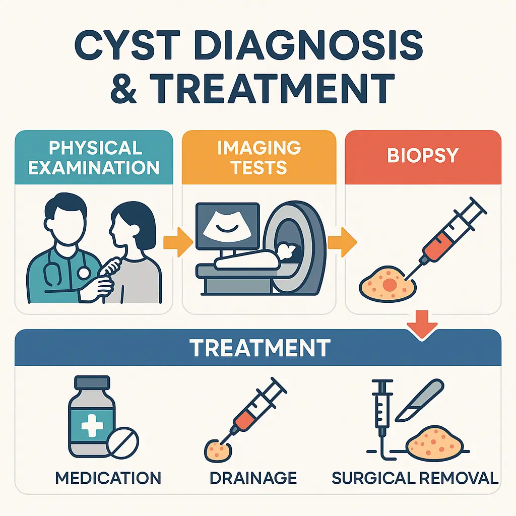
Diagnosis is key: A healthcare professional can diagnose a cyst through physical examination, imaging tests (like ultrasound or MRI), or biopsy.
Treatment options differ: Management can range from observation ("watchful waiting") to medication, drainage, or surgical removal, depending on the cyst's nature and symptoms.
What Exactly is a Cyst? 🤔
At its core, a cyst is a sac-like pocket of tissue that can form anywhere in the body. Think of it like a small balloon or a blister. This sac is usually filled with fluid, air, pus, or other materials. Cysts are different from tumors because they have a distinct wall or membrane that separates them from the surrounding tissue. They can vary greatly in size, from microscopic to as large as an orange or even bigger in rare cases.
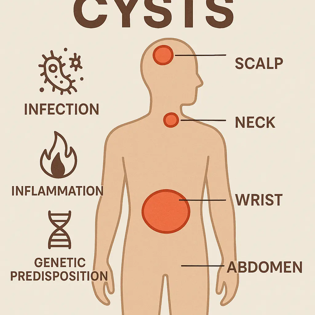
Cysts develop for many reasons. Sometimes, they form when a duct or tube gets blocked, causing fluid to build up. Other times, they might be the result of an infection, inflammation, a genetic condition, or even just normal body processes. For instance, some cysts are part of the body's natural healing process after an injury.
Most cysts are benign, meaning they are not cancerous and don't spread to other parts of the body. However, it's important to remember that some cysts can cause symptoms like pain, pressure, or affect how an organ works. In rare cases, a cyst might be a sign of a more serious underlying condition, or it might become infected. This is why getting a proper diagnosis from a healthcare professional is always the best course of action if you notice an unusual lump or experience new symptoms.
Common Characteristics of Cysts:
Feel: Often soft, smooth, and movable under the skin, but can also be firm.
Growth: Typically grow slowly over time.
Appearance: Can be flesh-colored, reddish, or yellowish, depending on the type and if it's inflamed.
Location: Can appear almost anywhere, including on the skin, in internal organs, or even in bones.
"Understanding the nature of a cyst is the first step towards effective management. Don't self-diagnose; always consult a medical professional."
When Should You Be Concerned About a Cyst? 🚨
While most cysts are harmless, there are certain signs and situations where it's wise to seek medical attention. Knowing when to worry can help you get timely care and ensure any potential issues are addressed.
You should consider seeing a doctor if a cyst:
Is painful or tender: This could indicate inflammation, infection, or pressure on nerves.
Grows rapidly in size: Quick growth can sometimes be a red flag.
Changes in appearance: Look for changes in color, shape, or if it becomes irregular.
Feels hard or fixed in place: Unlike typical cysts that are often movable, a hard, unmoving lump should be checked.
Causes functional problems: For example, a cyst near a joint that limits movement, or an internal cyst causing digestive issues or breathing difficulties.
Shows signs of infection: Redness, warmth, pus discharge, or fever.
Develops after an injury: Sometimes cysts can form after trauma, and a doctor can assess if it's related.
Is located in a sensitive area: Such as near the eye, in the breast, or in the groin.
You have any doubt or concern: It's always better to be safe and get a professional opinion.
A doctor can properly diagnose the cyst through a physical exam, possibly imaging tests (like ultrasound, MRI, or CT scan), or even a biopsy where a small sample of tissue is taken for examination under a microscope. This ensures you get an accurate diagnosis and the most appropriate advice or treatment. For a thorough check-up or to discuss any concerns, consider visiting a clinic that specializes in minor surgical procedures and diagnostics.
Diagnosing and Treating Cysts: A General Overview 🩺
When you visit a doctor because of a suspected cyst, they will follow a structured approach to figure out what it is and the best way to handle it.
How Cysts Are Diagnosed:
Physical Examination: The doctor will carefully look at and feel the lump. They'll note its size, shape, texture, whether it moves, and if it's tender.
Medical History: They'll ask about your symptoms, how long you've had the lump, if it's changed, and any other relevant health information.
Imaging Tests:
Ultrasound: Often the first choice for superficial cysts or those in soft tissues, as it can show if the lump is solid or fluid-filled.
MRI (Magnetic Resonance Imaging) or CT (Computed Tomography) Scan: Used for deeper cysts, especially in organs or the brain, to get detailed images.
X-ray: Less common for cysts unless it's suspected to be bone-related or to rule out other issues.
Biopsy: In some cases, especially if there's any concern about malignancy, a small sample of the cyst's fluid or wall is taken and examined under a microscope. This is the most definitive way to confirm the diagnosis.
Blood Tests: Rarely, blood tests might be used if an infection is suspected or to check for markers related to certain types of internal cysts.
General Treatment Approaches for Cysts:
Treatment for a cyst depends on several factors: its type, size, location, whether it's causing symptoms, and if it's benign or potentially problematic.
Watchful Waiting (Observation): Many small, harmless cysts that aren't causing any problems are simply monitored. The doctor might advise you to keep an eye on it and report any changes.
Medication:
Antibiotics: If the cyst is infected, antibiotics will be prescribed to clear the infection.
Anti-inflammatory drugs: Over-the-counter pain relievers or prescription anti-inflammatory medications can help manage pain and swelling.
Corticosteroid Injections: For some inflamed cysts (like certain acne cysts or ganglion cysts), a steroid injection can reduce inflammation and shrink the cyst.
Drainage (Aspiration): For fluid-filled cysts, the doctor can use a needle to drain the fluid. This is often done for ganglion cysts or large epidermal cysts. While it can relieve symptoms and reduce the size, the cyst wall remains, so it might fill up again.
Surgical Removal (Excision): This is the most definitive treatment, especially for cysts that:
Are symptomatic (painful, infected, interfere with function).
Are very large or growing rapidly.
Are cosmetically bothersome.
Are suspected to be pre-cancerous or cancerous.
Have recurred after drainage.
Surgical removal typically involves a minor procedure where the entire cyst wall is removed to prevent recurrence. This is often done as an outpatient procedure, meaning you can go home the same day. For more information on various conditions and their treatments, you can explore the conditions page of a specialized clinic.
Sclerotherapy: For some cysts, a substance is injected into the cyst after drainage to make the walls stick together, reducing the chance of recurrence.
The choice of treatment is always made in consultation with your doctor, considering your individual health and the specific characteristics of the cyst.
Exploring 20+ Types of Cysts: From Skin to Internal Organs 🗺️
Let's dive into the specifics of various cysts you might encounter. We'll categorize them for easier understanding.
I. Skin & Soft Tissue Cysts
These are some of the most common types of cysts, often visible or palpable just beneath the skin's surface.
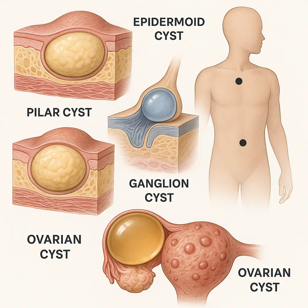
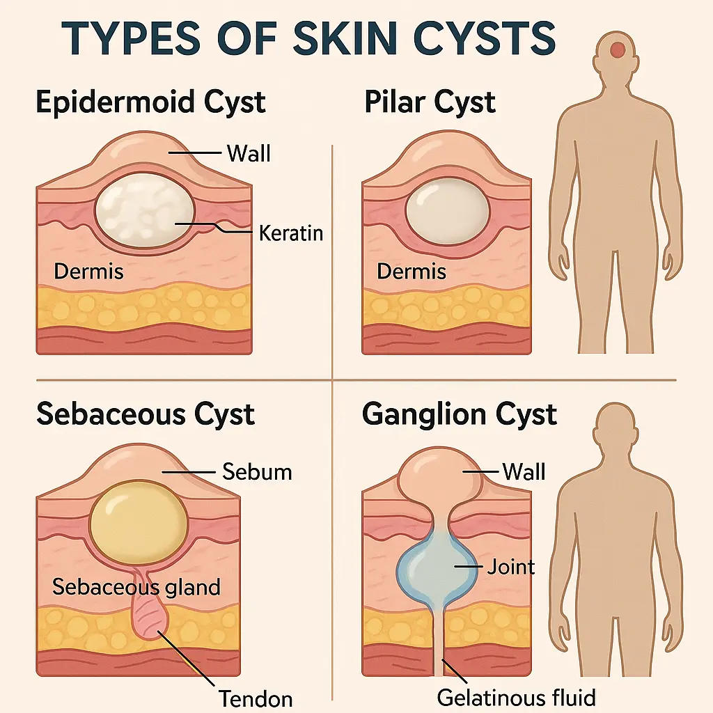
1. Epidermoid Cyst (Epidermal Inclusion Cyst)
What it is: An epidermoid cyst is a small, non-cancerous lump that forms just under the skin. It develops when skin cells that are normally shed on the surface get trapped deeper inside. These cells continue to multiply and form a sac, filling it with a soft, yellowish, cheesy substance called keratin.
Common Locations: Most often found on the face, neck, trunk, and sometimes the genitals.
Symptoms: Usually painless, firm to the touch, and can have a tiny blackhead-like pore in the center. If infected, it can become red, swollen, tender, and may drain pus.
Causes: Often results from blocked hair follicles, trauma to the skin, or sometimes occurs congenitally (from birth).
Diagnosis: Typically diagnosed by physical examination. If infected or unusual, a doctor might drain it or send a sample for lab analysis.
Treatment: Small, asymptomatic cysts often require no treatment. If large, inflamed, infected, or cosmetically bothersome, surgical removal is the most effective treatment to remove the entire cyst wall and prevent recurrence. Drainage is an option for infected cysts, but they may return.
2. Pilar Cyst (Trichilemmal Cyst)
What it is: Pilar cysts are also non-cancerous cysts that form from hair follicle cells. They are very similar to epidermoid cysts but have a different lining and are almost exclusively found on the scalp. Unlike epidermoid cysts, they contain a thick, pasty, white or yellowish substance, but it's not keratin.
Common Locations: Over 90% occur on the scalp.
Symptoms: Smooth, movable, firm lumps under the skin. They are usually painless unless they become infected or rupture. They can occur as single lumps or in clusters.
Causes: They are often hereditary, meaning they run in families.
Diagnosis: Clinical examination is usually sufficient.
Treatment: Surgical removal is the standard treatment, especially if they are large, numerous, or causing discomfort. They are typically easy to remove because they are well-defined.
3. Sebaceous Cyst (Misnomer)
What it is: The term "sebaceous cyst" is commonly used but is often a misnomer. Most lumps referred to as sebaceous cysts are actually epidermoid cysts or pilar cysts. A true sebaceous cyst, which originates from the sebaceous glands (oil-producing glands in the skin), is very rare. When they do occur, they contain oily, sebum-like material.
Common Locations: Can occur anywhere on the body where sebaceous glands are present, but rare.
Symptoms: Similar to epidermoid cysts – a small, movable lump under the skin.
Causes: Blockage of a sebaceous gland duct.
Diagnosis: A doctor can differentiate it from epidermoid or pilar cysts, usually after removal and lab analysis.
Treatment: Similar to epidermoid cysts, often surgical removal if symptomatic.
4. Ganglion Cyst
What it is: A ganglion cyst is a non-cancerous lump that usually forms along the tendons or joints of the wrists or hands. They are essentially sacs filled with a thick, jelly-like fluid (synovial fluid), which lubricates joints and tendons.
Common Locations: Most common on the back of the wrist, but can also occur on the palm side, ankles, feet, or knees.
Symptoms: A visible lump that can be soft or firm, fixed or movable. It may or may not be painful. Pain can worsen with joint movement. Size can fluctuate.
Causes: The exact cause isn't fully understood, but they may result from joint or tendon irritation or injury, causing the fluid to leak out and form a sac.
Diagnosis: Physical exam, transillumination (shining a light through it), and possibly ultrasound or MRI to confirm.
Treatment: Many disappear on their own. Options include observation, aspiration (draining the fluid with a needle), or surgical removal if persistent, painful, or affecting function. Recurrence is possible after aspiration.
5. Baker's Cyst (Popliteal Cyst)
What it is: A Baker's cyst is a fluid-filled sac that causes a bulge and a feeling of tightness behind the knee. It's not a true cyst in the sense of having its own distinct lining; rather, it's a bulge of the synovial capsule of the knee joint. It forms when excess joint fluid (synovial fluid) is pushed out of the joint and collects in a sac behind the knee.
Common Locations: Behind the knee (popliteal fossa).
Symptoms: A visible lump, stiffness, pain (especially with knee movement), and a feeling of pressure or fullness behind the knee. Symptoms often worsen with activity.
Causes: Almost always linked to an underlying knee joint problem, such as arthritis (especially osteoarthritis), meniscus tears, or other inflammatory conditions that cause excess fluid production.
Diagnosis: Physical exam. Ultrasound or MRI can confirm the diagnosis and help identify the underlying knee problem.
Treatment: Treating the underlying knee condition is key. Other treatments include rest, ice, compression, elevation (RICE), physical therapy, anti-inflammatory medications, fluid aspiration, or corticosteroid injections. Surgical removal is rare and only considered if other treatments fail and the cyst is very bothersome.
6. Ingrown Hair Cyst
What it is: An ingrown hair cyst, more accurately described as an inflamed ingrown hair or a pseudo-folliculitis, occurs when a hair curls back or grows sideways into the skin. This can lead to inflammation, forming a red, tender, pus-filled bump that resembles a cyst or boil.
Common Locations: Areas where hair is shaved, waxed, or plucked, such as the face, neck, armpits, bikini line, and legs.
Symptoms: Red, swollen, painful bump, sometimes with a visible hair trapped inside. It can be itchy and filled with pus.
Causes: Shaving, waxing, tight clothing, or naturally curly hair.
Diagnosis: Visual inspection.
Treatment: Often resolves on its own once the hair is released. Warm compresses can help. Avoid shaving the area. If infected, antibiotics may be needed. In some cases, a doctor may need to make a small incision to release the trapped hair or drain pus. Prevention involves proper shaving techniques and exfoliation.
7. Cystic Acne (Acne Cysts)
What it is: Cystic acne is the most severe form of acne. It develops when oil (sebum) and dead skin cells get trapped deep within hair follicles, leading to inflammation and infection. Unlike typical pimples, these lesions are large, painful, pus-filled lumps that form deep under the skin. They are not true cysts in the medical sense (they don't have an epithelial lining), but they are often referred to as "cysts" due to their size and appearance.
Common Locations: Face, chest, back, shoulders, and upper arms.
Symptoms: Large, painful, red, inflamed bumps that are soft and filled with pus. They can rupture, leading to scarring.
Causes: Hormonal fluctuations, genetics, certain medications, and excessive oil production.
Diagnosis: Clinical examination by a dermatologist.
Treatment: Requires professional medical treatment. Options include oral antibiotics, topical retinoids, isotretinoin (Accutane) for severe cases, steroid injections directly into the lesions to reduce inflammation, and drainage by a doctor. Self-treating can lead to scarring.
8. Chalazion
What it is: A chalazion is a small, usually painless lump or swelling that appears on the eyelid. It forms when a meibomian gland (an oil-producing gland in the eyelid) becomes blocked, causing oil to build up. It's often confused with a stye, but a stye is an acute infection of an eyelash follicle or eyelid gland, while a chalazion is a chronic, non-infectious inflammation.
Common Locations: Upper or lower eyelid.
Symptoms: A firm, round, painless lump on the eyelid. If large, it can cause blurry vision by pressing on the cornea. It may start as a red, tender area (like a stye) but then becomes painless.
Causes: Blocked meibomian gland.
Diagnosis: Visual inspection.
Treatment: Often resolves on its own within a few weeks to months. Warm compresses applied several times a day can help. If persistent or bothersome, an ophthalmologist may recommend steroid injections or surgical removal (incision and drainage).
9. Mucous Retention Cyst (Mucocele)
What it is: A mucocele is a common, harmless cyst that forms when a minor salivary gland duct gets blocked or injured, causing mucus to leak into the surrounding tissue and form a sac.
Common Locations: Most frequently found on the lower lip, but can also occur on the tongue, inner cheek, or floor of the mouth.
Symptoms: A soft, round, bluish or clear bump that is usually painless. It can fluctuate in size and sometimes rupture, releasing clear fluid, only to reform later.
Causes: Trauma (like biting the lip), blockage of a salivary gland duct.
Diagnosis: Visual inspection.
Treatment: Many mucoceles resolve on their own, especially if they are small. If persistent, bothersome, or frequently rupturing, surgical removal of the cyst and associated gland is usually performed to prevent recurrence.
10. Lipoma (Often Confused with Cysts)
What it is: While not a true cyst, a lipoma is a very common, non-cancerous growth of fatty tissue that often feels like a soft, rubbery lump under the skin. Because of its lump-like nature, it's frequently confused with a cyst. Unlike cysts, lipomas are solid masses of fat cells, not fluid-filled sacs.
Common Locations: Can appear anywhere on the body, but most common on the neck, shoulders, back, abdomen, arms, and thighs.
Symptoms: A soft, doughy, movable lump just under the skin. Usually painless unless it presses on a nerve or contains blood vessels.
Causes: Exact cause is unknown, but they tend to run in families.
Diagnosis: Physical exam. Ultrasound or biopsy might be used to confirm it's a lipoma and rule out other conditions.
Treatment: Usually no treatment is needed unless it's large, painful, growing rapidly, or cosmetically bothersome. Surgical removal is a simple procedure.
"Distinguishing between different types of lumps is crucial. If in doubt, always seek professional medical advice to ensure accurate diagnosis and appropriate care."
II. Internal Organ Cysts
These cysts form within internal organs and may not be visible externally. They are often discovered incidentally during imaging tests for other conditions.
11. Renal Cyst (Kidney Cyst)
What it is: A renal cyst is a fluid-filled sac that forms in the kidneys. Simple renal cysts are very common, especially as people age, and are almost always benign. They are distinct from cysts associated with polycystic kidney disease (PKD), which is a genetic disorder leading to numerous cysts and kidney failure.
Common Locations: Within the kidney.
Symptoms: Most simple renal cysts are asymptomatic and are discovered incidentally. Large cysts can cause pain in the back or side, fever (if infected), or high blood pressure (rarely, if pressing on vessels).
Causes: Unknown for simple cysts; likely due to weakening of kidney tubules. PKD is genetic.
Diagnosis: Often found during ultrasound, CT scan, or MRI of the abdomen.
Treatment: Simple cysts usually require no treatment, only observation. If a cyst is causing symptoms or looks suspicious, drainage (aspiration) or surgical removal may be considered.
12. Hepatic Cyst (Liver Cyst)
What it is: A hepatic cyst is a fluid-filled sac within the liver. Most are simple, benign cysts that cause no problems. Like renal cysts, they are distinct from cysts seen in polycystic liver disease (PCLD), a rare genetic condition causing multiple cysts.
Common Locations: Within the liver.
Symptoms: Most are asymptomatic. Large cysts can cause abdominal pain, bloating, or nausea by pressing on other organs.
Causes: Unknown for simple cysts. PCLD is genetic.
Diagnosis: Often found during abdominal ultrasound, CT scan, or MRI.
Treatment: Simple cysts usually require no treatment. If large and symptomatic, drainage or surgical removal (deroofing) may be performed.
13. Arachnoid Cyst
What it is: An arachnoid cyst is a fluid-filled sac that forms within the arachnoid membrane, one of the three membranes that cover the brain and spinal cord. These cysts contain cerebrospinal fluid (CSF) and are usually benign.
Common Locations: Most commonly found in the middle cranial fossa (side of the brain), but can be anywhere along the brain or spinal cord.
Symptoms: Many are asymptomatic and discovered incidentally. Symptoms depend on size and location, and can include headaches, seizures, hydrocephalus (fluid on the brain), weakness, or developmental delays in children.
Causes: Usually congenital (present at birth) due to abnormal development of the arachnoid membrane. Less commonly, they can result from head trauma, meningitis, or tumors.
Diagnosis: MRI or CT scan of the brain or spine.
Treatment: Asymptomatic cysts are usually monitored. If symptomatic, treatment involves surgical drainage or fenestration (creating an opening) to allow the fluid to drain into the CSF circulation.
14. Pancreatic Cyst
What it is: A pancreatic cyst is a fluid-filled sac in the pancreas. These cysts are diverse and can range from benign (like serous cystadenomas or pseudocysts) to potentially pre-cancerous (like mucinous cystic neoplasms or IPMNs) or cancerous. Pseudocysts are the most common type and form after inflammation or injury to the pancreas.
Common Locations: Within or near the pancreas.
Symptoms: Many are asymptomatic. Symptoms can include abdominal pain, nausea, vomiting, weight loss, or jaundice (yellowing of skin/eyes) if they press on bile ducts.
Causes: Pancreatitis (inflammation of the pancreas), abdominal trauma, or genetic factors.
Diagnosis: CT scan, MRI, or endoscopic ultrasound (EUS) with fine-needle aspiration (FNA) to analyze the fluid.
Treatment: Depends on the type, size, and symptoms. Benign cysts may be monitored. Pre-cancerous or symptomatic cysts often require surgical removal. Pseudocysts may be drained if large or symptomatic.
15. Splenic Cyst
What it is: A splenic cyst is a fluid-filled sac within the spleen. They can be true cysts (lined with cells) or pseudocysts (lacking a cellular lining, often due to trauma or infection).
Common Locations: Within the spleen.
Symptoms: Most are asymptomatic. Large cysts can cause abdominal pain, a feeling of fullness, or early satiety (feeling full after eating little).
Causes: True cysts are often congenital or parasitic (e.g., echinococcal). Pseudocysts result from trauma, infection, or inflammation.
Diagnosis: Ultrasound, CT scan, or MRI of the abdomen.
Treatment: Small, asymptomatic cysts are monitored. Large or symptomatic cysts may require surgical removal (splenectomy or partial splenectomy) or drainage.
16. Lung Cyst (Bronchogenic Cyst)
What it is: A bronchogenic cyst is a rare, non-cancerous congenital cyst that forms in the chest, usually near the airways or esophagus. It's a developmental anomaly that results from abnormal budding of the primitive foregut during fetal development.
Common Locations: Most often in the mediastinum (space between the lungs) or within the lung tissue itself.
Symptoms: Many are asymptomatic. Symptoms occur if the cyst grows large enough to compress airways or other structures, leading to coughing, shortness of breath, recurrent infections, or chest pain.
Causes: Congenital (developmental error during fetal growth).
Diagnosis: Chest X-ray, CT scan, or MRI.
Treatment: Surgical removal is usually recommended, even for asymptomatic cysts, due to the risk of complications like infection, rupture, or malignant transformation (though rare).
III. Gynecological Cysts
These cysts are specific to the female reproductive system.
17. Ovarian Cyst
What it is: Ovarian cysts are fluid-filled sacs that develop on or inside an ovary. They are very common, especially during a woman's reproductive years. Most are functional cysts, meaning they form as a normal part of the menstrual cycle and usually disappear on their own. Other types include dermoid cysts, endometriomas, and cystadenomas.
Common Locations: On or within the ovaries.
Symptoms: Often asymptomatic. Large or ruptured cysts can cause pelvic pain, bloating, pressure, abnormal bleeding, pain during intercourse, or frequent urination. A ruptured cyst or ovarian torsion (twisting of the ovary) can cause severe, sudden pain.
Diagnosis: Pelvic exam, ultrasound (transvaginal or abdominal), blood tests (like CA-125, especially if malignancy is suspected).
Treatment: Many functional cysts resolve on their own. Watchful waiting is common. Oral contraceptives can sometimes prevent new functional cysts. Surgical removal (laparoscopy or laparotomy) may be necessary for large cysts, those causing severe symptoms, those suspected to be cancerous, or those that persist.
18. Bartholin's Cyst
What it is: Bartholin's glands are two small glands located on each side of the vaginal opening, which produce fluid to lubricate the vagina. A Bartholin's cyst forms when the opening of one of these glands becomes blocked, causing fluid to back up and form a painless lump. If the cyst becomes infected, it can turn into a painful abscess.
Common Locations: On either side of the vaginal opening.
Symptoms: A small, soft, often painless lump. If infected (Bartholin's abscess), it becomes very painful, red, swollen, and tender, making sitting, walking, or intercourse difficult. Fever may be present.
Causes: Blockage of the Bartholin's gland duct, often due to infection (bacterial, including STIs) or inflammation.
Diagnosis: Physical examination.
Treatment: Small, asymptomatic cysts may not need treatment. Sitz baths (soaking in warm water) can help drainage. For symptomatic cysts or abscesses, treatment includes antibiotics (if infected), incision and drainage, or marsupialization (a surgical procedure to create a permanent opening for drainage and prevent recurrence).
19. Endometriomas (Chocolate Cysts)
What it is: Endometriomas are a specific type of ovarian cyst associated with endometriosis. Endometriosis is a condition where tissue similar to the lining of the uterus (endometrium) grows outside the uterus, often on the ovaries, fallopian tubes, and other pelvic organs. When this tissue grows on the ovary, it can form a blood-filled cyst, often dark brown, giving them the nickname "chocolate cysts."
Common Locations: On or within the ovaries.
Symptoms: Can cause chronic pelvic pain, painful periods (dysmenorrhea), pain during intercourse (dyspareunia), and infertility. The pain can be severe and debilitating.
Causes: Endometriosis.
Diagnosis: Pelvic exam, ultrasound, MRI. Laparoscopy (a surgical procedure) is often used for definitive diagnosis.
Treatment: Pain management (NSAIDs, hormonal therapy), and surgical removal. Surgery is often recommended for large endometriomas, those causing severe pain, or those contributing to infertility.
IV. Head & Neck Cysts
These cysts are found in the head and neck region and can be congenital or acquired.
20. Thyroglossal Duct Cyst
What it is: A thyroglossal duct cyst is a common type of neck lump that forms from remnants of the thyroglossal duct, a structure present during fetal development that helps the thyroid gland descend from the base of the tongue to its final position in the neck. If parts of this duct remain after birth, they can fill with fluid and form a cyst.
Common Locations: In the midline of the neck, usually just below the chin or near the Adam's apple. It characteristically moves upward when you stick out your tongue.
Symptoms: A soft, movable, painless lump in the front of the neck. It can become infected, leading to pain, redness, swelling, and sometimes drainage.
Causes: Congenital (failure of the thyroglossal duct to completely disappear after birth).
Diagnosis: Physical exam, ultrasound, CT scan, or MRI. Sometimes a fine-needle aspiration is done to confirm the fluid content.
Treatment: Surgical removal is recommended to prevent infection and recurrence. The "Sistrunk procedure" is a specific surgery that removes the cyst, the middle part of the hyoid bone (a bone in the neck), and the tract leading up to the tongue base to minimize recurrence.
21. Cystic Hygroma (Lymphatic Malformation)
What it is: A cystic hygroma is a congenital malformation of the lymphatic system, which is part of the immune system. It results from abnormal development of lymphatic vessels, leading to fluid-filled sacs, most commonly in the neck or armpit. They are benign but can grow large and cause problems due to compression.
Common Locations: Most often in the neck (especially on the left side) or armpit, but can occur elsewhere.
Symptoms: A soft, compressible, non-tender mass. Can cause breathing or swallowing difficulties if large and pressing on airways/esophagus. May become infected.
Causes: Congenital (developmental error during fetal growth).
Diagnosis: Often detected during prenatal ultrasound. After birth, physical exam, ultrasound, MRI, or CT scan.
Treatment: Surgical removal is the primary treatment. Sclerotherapy (injecting a substance to shrink the cyst) may also be used, especially for large or complex cysts.
22. Branchial Cleft Cyst
What it is: A branchial cleft cyst is a congenital abnormality that forms when tissues in the neck and collarbone area (branchial arches) that normally disappear during fetal development do not. They can form cysts, sinuses (small openings), or fistulas (tunnels) that drain to the skin.
Common Locations: Usually on one side of the neck, often near the front of the sternocleidomastoid muscle, or just below the ear.
Symptoms: A soft, movable lump in the neck, often noticed in childhood or early adulthood. Can become infected, leading to pain, redness, and pus drainage, sometimes through a small opening in the skin.
Causes: Congenital (failure of branchial arches to properly close during fetal development).
Diagnosis: Physical exam, ultrasound, CT scan, or MRI.
Treatment: Surgical removal is recommended to prevent recurrent infections and for cosmetic reasons. The entire tract, including any fistulas, must be removed to prevent recurrence.
V. Infection/Parasite Related Cysts
These cysts are often a result of infection or parasitic infestation.
23. Pilonidal Cyst
What it is: A pilonidal cyst is a sac-like structure that forms at the top of the crease between the buttocks, near the tailbone. It usually contains hair, skin debris, and can become infected, forming a painful abscess.
Common Locations: At the top of the natal cleft (buttock crease).
Symptoms: Often asymptomatic unless infected. When infected, it becomes a painful, red, swollen lump that may drain pus or blood. Fever can also occur.
Causes: Thought to be caused by ingrown hairs that puncture the skin and create a small tunnel or pit, leading to an inflammatory reaction and cyst formation. Friction and pressure in the area, along with excess hair, can contribute.
Diagnosis: Physical examination.
Treatment: If infected, incision and drainage of the abscess is the first step. For recurrent or chronic cysts, surgical removal of the cyst and its tracts is often necessary. This can involve various surgical techniques, including wide excision, marsupialization, or flap procedures. After treatment, maintaining good hygiene and hair removal in the area can help prevent recurrence.
24. Cystic Echinococcosis (Hydatid Cyst)
What it is: Cystic echinococcosis, also known as hydatid disease, is a parasitic infection caused by the tapeworm Echinococcus granulosus. It leads to the formation of slow-growing, fluid-filled cysts in various organs, most commonly the liver and lungs. These cysts can contain thousands of larval tapeworms.
Common Locations: Liver (most common), lungs, but can also occur in the brain, bones, kidneys, spleen, and other organs.
Symptoms: Often asymptomatic for years. Symptoms depend on the cyst's size and location. In the liver, it can cause abdominal pain, nausea, vomiting, or jaundice. In the lungs, cough, chest pain, or shortness of breath. Rupture of a cyst can lead to severe allergic reactions (anaphylaxis) or spread of the parasite.
Causes: Ingestion of eggs of the Echinococcus granulosus tapeworm, usually through contact with infected dog feces or contaminated food/water.
Diagnosis: Imaging (ultrasound, CT, MRI), blood tests (serology for antibodies), and sometimes biopsy (though this carries a risk of rupture and spread).
Treatment: Can involve anti-parasitic medications (albendazole), percutaneous aspiration and injection (PAIR) where the cyst is drained and injected with a scolicidal agent, or surgical removal. Treatment choice depends on cyst size, location, and type.
Understanding Your Symptoms: When to Consult a Doctor
It can be difficult to know when a lump or bump is something to worry about. This interactive guide can help you understand common scenarios and when it's best to seek professional medical advice. Remember, this is for informational purposes and not a substitute for a doctor's consultation.
It can be difficult to know when a lump or bump is something to worry about. This interactive guide can help you understand common scenarios and when it's best to seek professional medical advice. Remember, this is for informational purposes and not a substitute for a doctor's consultation.
Cyst Symptom Quick Guide
Answer a few questions to get a general idea of when to consult a doctor about a cyst or lump. This is not medical advice.
Is the lump or cyst painful, tender, red, or warm to the touch?
Yes
No
Has the lump grown rapidly in size, or changed in shape or color recently?
Yes
No
Is the lump causing problems with movement, breathing, eating, or other bodily functions?
Yes
No
Do you also have a fever, chills, or feel generally unwell?
Yes
No
Are you concerned about the lump for any reason, or is it cosmetically bothersome?
Yes
No
Get Advice
For professional medical advice or to schedule a consultation, please visit our Contact Us page or learn more about our services on our Conditions page.
Prevention and Living with Cysts 🌿
While not all cysts can be prevented, especially those that are congenital or form due to genetic predispositions, there are steps you can take to reduce the risk of certain types and manage existing ones.
General Prevention Tips:
Good Hygiene: Keeping your skin clean can help prevent blocked pores and follicles that lead to epidermoid cysts, pilar cysts, and acne cysts. Regular washing and gentle exfoliation can be beneficial.
Proper Shaving Techniques: For ingrown hair cysts and pilonidal cysts, shaving with the grain, using a sharp razor, and preparing the skin beforehand can reduce irritation. Laser hair removal can be a long-term solution for recurrent ingrown hairs.
Moisturize: Keeping skin hydrated can help maintain its barrier function and prevent dryness that can exacerbate certain skin conditions.
Manage Underlying Conditions: If you have conditions like acne, endometriosis, or arthritis, effectively managing them with your doctor's guidance can help prevent associated cysts (e.g., cystic acne, endometriomas, Baker's cysts).
Avoid Squeezing or Popping: Never try to squeeze or pop a cyst yourself. This can push the contents deeper, lead to infection, inflammation, and potentially scarring.
Protect from Trauma: While not always possible, avoiding repeated trauma to areas prone to cysts (like joints for ganglion cysts) can sometimes help.
Stay Hydrated: Drinking enough water supports overall body health, including skin health.
Healthy Diet: A balanced diet rich in fruits, vegetables, and whole grains can support overall skin health and reduce inflammation.
Living with a Cyst:
If you have a cyst that doesn't require immediate treatment, your doctor might recommend "watchful waiting." This means:
Monitor for Changes: Regularly check the cyst for any changes in size, shape, color, or if it becomes painful or inflamed. Keep a small log or take occasional photos to track its progress.
Keep it Clean: If it's a skin cyst, keep the area clean and dry.
Warm Compresses: For some skin cysts (like chalazions or small epidermal cysts), warm compresses can help reduce inflammation and encourage drainage.
Follow Doctor's Advice: Adhere to any specific instructions given by your healthcare provider.
Know When to Revisit: Be aware of the "red flag" symptoms discussed earlier (pain, rapid growth, infection signs) that warrant a follow-up visit.
Remember, even if a cyst is benign, it's always a good idea to discuss any concerns with a healthcare professional. They can provide accurate information, perform necessary diagnostics, and recommend the best course of action for your specific situation. For ongoing health information and insights, regularly check out the blog section of reputable medical sites.
Cysts are a common part of human anatomy, presenting in a remarkable variety of forms and locations. From the easily visible epidermoid cysts on our skin to the often-hidden ovarian or renal cysts deep within our bodies, each type has its unique characteristics, causes, and management strategies. While the sheer number of different cysts might seem overwhelming, the good news is that the vast majority are benign and pose no serious threat to your health.
However, understanding the distinctions between them and recognizing the signs that warrant medical attention is incredibly important. Whether a cyst is causing pain, growing rapidly, showing signs of infection, or simply causing you concern, consulting a healthcare professional is always the wisest step. They can provide an accurate diagnosis through physical examination and advanced imaging, and guide you towards the most appropriate treatment, which can range from simple observation to medication, drainage, or surgical removal.
Empowering yourself with knowledge about your body and knowing when to seek expert advice is key to maintaining your well-being. Don't hesitate to reach out to a trusted medical team for any lumps or bumps that worry you. You can find more information about various conditions and available treatments on our website.
December 9, 2025
🇨🇦
Our clinic currently provides care to patients within
Canada only.
We apologize for any inconvenience this may cause.