When you notice a new spot on your skin or an existing mole starts to change, questions about cancer risk naturally arise. Understanding the difference between flat mole vs raised mole skin cancer risk can help you make informed decisions about your skin health and know when to seek professional evaluation. While the texture of a mole—whether flat or raised—provides some clues about its characteristics, the relationship between mole type and cancer risk is more nuanced than many people realize.
Skin cancer remains one of the most common cancers worldwide, with melanoma cases continuing to rise despite increased awareness about sun protection [1]. The good news? Early detection dramatically improves outcomes, making it essential to understand what different types of moles mean for your health.
Flat moles, medically known as junctional nevi, appear as colored spots on the skin surface without any elevation. These moles develop when melanocytes (pigment-producing cells) cluster at the junction between the epidermis (outer skin layer) and dermis (deeper skin layer) [2].
Characteristics of flat moles:
Flat moles can appear anywhere on the body but are frequently found on sun-exposed areas like the face, arms, and legs. Most people develop between 10-40 moles by adulthood, with many being flat variants [3].
Raised moles, called compound nevi or intradermal nevi, extend above the skin surface creating a noticeable bump. These moles contain melanocyte clusters that penetrate deeper into the dermal layer of skin [4].
Characteristics of raised moles:
Raised moles develop when melanocytes migrate deeper into the skin layers over time. Many flat moles gradually become raised as a person ages—this is typically a normal evolution rather than a concerning change [5].
The key distinction between flat and raised moles lies in where melanocytes are located within skin layers:
Mole TypeMelanocyte LocationAppearanceCommon Age of DevelopmentJunctional (Flat)Dermal-epidermal junctionFlat, pigmented spotChildhood to young adultCompound (Raised)Junction + dermisSlightly raised, pigmentedAdolescence to middle ageIntradermal (Raised)Dermis onlyRaised, often flesh-coloredMiddle age to older adults
Understanding these anatomical differences helps explain why moles change over time and why some characteristics matter more than others when assessing cancer risk.
The relationship between flat moles and melanoma risk is complex. While most flat moles are completely benign, certain types of flat moles—specifically atypical or dysplastic nevi—do carry increased melanoma risk [6].
Atypical flat moles have these concerning features:
Research shows that people with atypical mole syndrome (having multiple dysplastic nevi) face a 10-12 times higher lifetime melanoma risk compared to the general population [7]. These atypical moles are often flat or only slightly raised.
"The presence of atypical nevi is one of the strongest risk factors for melanoma development, independent of sun exposure history." — Journal of the American Academy of Dermatology
However, it's crucial to understand that being flat doesn't make a mole dangerous. The vast majority of flat moles are normal junctional nevi that pose no cancer risk. What matters are the specific characteristics described by the ABCDE criteria, which we'll explore in detail later.
Raised moles are extremely common, and the overwhelming majority remain benign throughout a person's lifetime. Studies indicate that intradermal nevi (deeply raised moles) very rarely transform into melanoma [8].
Why raised moles are usually benign:
However, raised moles are not immune to cancer risk. Melanoma can develop in raised moles, particularly if they exhibit warning signs. Additionally, some melanomas naturally grow in a raised or nodular pattern from the beginning.
Nodular melanoma, one of the most aggressive forms, often appears as a raised, dome-shaped, or pedunculated growth that may be black, blue, red, or skin-colored [9]. This type accounts for approximately 15% of melanomas and tends to grow rapidly in depth rather than spreading across the skin surface.
When comparing flat mole vs raised mole skin cancer risk, dermatologists emphasize that texture is less important than other characteristics. A 2024 study published in JAMA Dermatology found that melanoma detection based solely on whether a lesion was flat or raised had poor sensitivity [10].
What actually predicts melanoma risk:
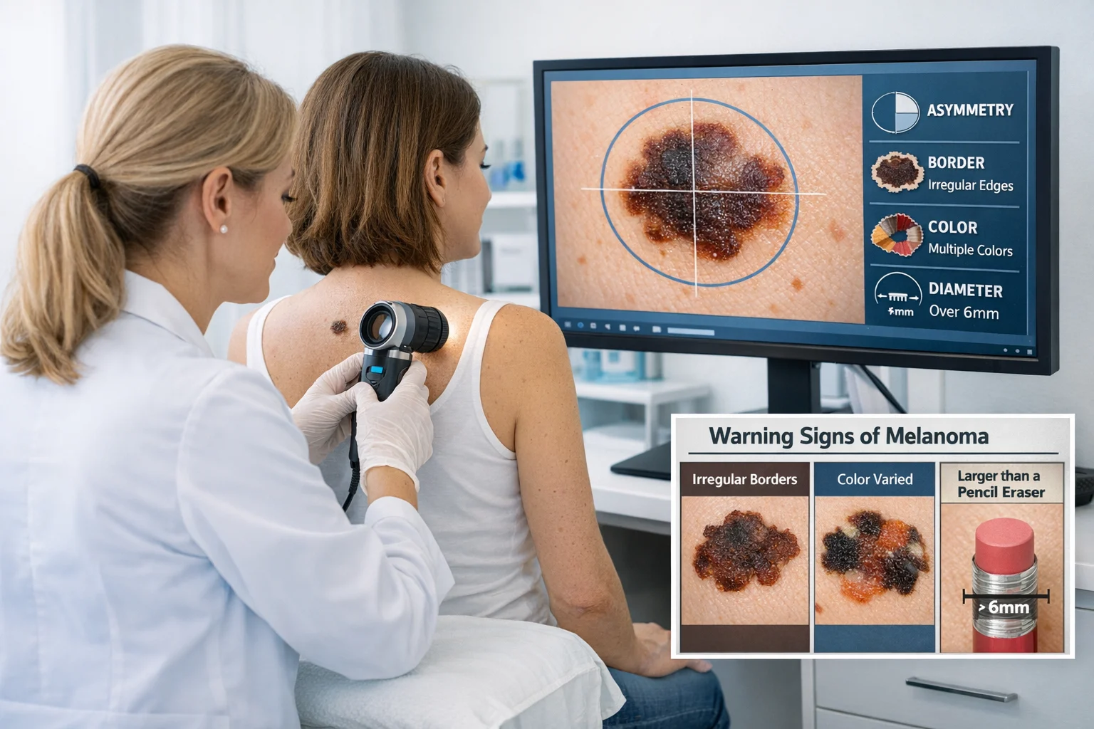
✅ Asymmetry – One half of the mole differs from the other half
✅ Border irregularity – Edges are ragged, notched, or blurred
✅ Color variation – Multiple shades of brown, black, red, white, or blue
✅ Diameter – Larger than 6mm (though melanomas can be smaller)
✅ Evolution – Changes in size, shape, color, elevation, or symptoms
A flat mole with irregular borders and multiple colors poses significantly higher risk than a raised mole that's been unchanged for decades. Similarly, a raised mole that suddenly starts growing or bleeding requires immediate evaluation regardless of its raised texture.
For comprehensive information about different types of skin lesions, including benign and potentially cancerous growths, consult with a dermatology specialist.
The ABCDE criteria represent the gold standard for evaluating moles and identifying potential melanomas. This system works equally well for both flat and raised moles and should be part of everyone's monthly self-examination routine.
Asymmetry means one half of the mole doesn't match the other half when you imagine drawing a line through the center.
Normal moles:
Concerning signs:
Most benign moles—whether flat or raised—maintain symmetry. Melanomas often grow unevenly, creating asymmetric patterns as cancerous cells multiply at different rates in different areas.
The border or edge of a mole provides important clues about its nature.
Normal moles:
Concerning signs:
Border irregularity occurs because melanoma cells invade surrounding tissue in unpredictable patterns. This creates the characteristic "fuzzy" or "scalloped" edges that dermatologists look for during examinations.
Color is one of the most telling characteristics when assessing mole cancer risk.
Normal moles:
Concerning signs:
Melanomas often display varied colors because different areas of the tumor may be at different stages of development, have different depths, or contain different amounts of melanin and blood vessels [11].
Diameter refers to the size of the mole, though this criterion has evolved as dermatologists recognize that melanomas can be smaller than originally thought.
Traditional guideline:
Updated understanding:
While many large moles are benign, and some melanomas are small, monitoring diameter changes helps identify evolving lesions that may need biopsy.
Evolution—changes in a mole over time—is perhaps the most critical warning sign for both flat and raised moles.
Changes that require immediate evaluation:
A 2025 study found that patient-reported mole changes led to melanoma diagnosis in 71% of cases where evolution was the primary concerning feature [12]. This underscores why regular self-examination and awareness of your own moles is so important.
To learn more about identifying potentially dangerous skin changes, visit a specialized skin cancer clinic for professional evaluation.
While understanding flat mole vs raised mole skin cancer risk is valuable, numerous other factors influence your overall melanoma risk. Recognizing these helps you take appropriate preventive measures and monitoring steps.
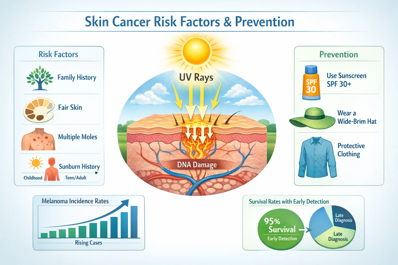
Family history plays a significant role in melanoma risk:
People with strong family histories should begin regular dermatology screenings earlier and may benefit from more frequent examinations.
Fitzpatrick skin type categorizes skin based on how it responds to sun exposure:
TypeDescriptionMelanoma RiskIAlways burns, never tansHighest riskIIUsually burns, tans minimallyVery high riskIIISometimes burns, tans uniformlyModerate riskIVRarely burns, tans easilyLower riskV-VIVery rarely burns, deeply pigmentedLowest risk
People with fair skin, light hair, and light eyes face 10-20 times higher melanoma risk compared to those with darker skin tones [14]. However, melanoma can affect people of all skin types, and delayed diagnosis in darker-skinned individuals often leads to worse outcomes.
Ultraviolet radiation remains the most significant modifiable risk factor for melanoma:
Patterns that increase risk:
UV radiation damages DNA in skin cells, including melanocytes. This damage accumulates over time and can trigger the genetic mutations that lead to melanoma development [15].
The number and type of moles on your body significantly impacts cancer risk:
Risk increases with:
Having many moles doesn't mean you'll develop melanoma, but it does mean you should be more vigilant about monitoring changes and getting regular professional skin checks.
Immunosuppression increases melanoma risk and affects outcomes:
Weakened immune systems are less effective at identifying and destroying abnormal cells before they become cancerous.
For a comprehensive overview of different types of skin cancer and their risk factors, consult with dermatology professionals who specialize in early detection.
Knowing when to seek professional evaluation is crucial for early melanoma detection. While monthly self-examinations are important, certain signs warrant immediate dermatology consultation regardless of whether a mole is flat or raised.
The ugly duckling sign is a simple but effective detection method based on pattern recognition:
Concept: Most of your moles should look similar to each other. A mole that looks noticeably different from your other moles—the "ugly duckling"—deserves professional evaluation.
What to look for:
Studies show the ugly duckling sign has high sensitivity for melanoma detection, particularly when combined with ABCDE criteria [17].
Schedule a dermatology appointment within 1-2 weeks if you notice:
🚨 Bleeding or oozing from a mole without injury
🚨 Persistent itching that doesn't resolve
🚨 Tenderness or pain in or around a mole
🚨 Rapid growth over weeks to months
🚨 Color changes, especially darkening or development of black areas
🚨 Development of a raised area within a previously flat mole
🚨 Satellite lesions—small spots appearing around a mole
🚨 Ulceration or crusting that doesn't heal
These symptoms don't automatically mean cancer, but they indicate changes that require professional assessment. Many benign conditions can cause similar symptoms, but only a dermatologist can make an accurate diagnosis.
While developing new moles in your 20s and 30s is normal, new moles after age 40 warrant closer attention:
Why this matters:
Any new pigmented spot appearing after age 40 should be evaluated by a dermatologist, especially if it differs in appearance from your existing moles.
Certain body locations carry higher melanoma risk or are more difficult to self-monitor:
Priority areas for professional examination:
Ask a partner to help examine hard-to-see areas, or use mirrors and smartphone cameras to document moles in difficult locations.
General population:
High-risk individuals:
Your dermatologist will recommend an appropriate schedule based on your individual risk factors. Professional examinations often detect concerning lesions that patients overlook during self-checks.
For expert evaluation and removal of concerning moles, consider visiting specialists who offer mole removal services in Ajax or Barrie.
When you visit a dermatologist concerned about a mole, they use several sophisticated techniques to assess whether it's benign or potentially cancerous. Understanding these methods can help reduce anxiety about the evaluation process.
Visual inspection is the first step in mole evaluation:
The dermatologist will:
Dermoscopy (also called dermatoscopy) takes visual examination to the next level:
This technique uses a specialized magnifying device called a dermatoscope that:
Dermoscopy has been shown to increase melanoma detection accuracy by 20-30% compared to visual examination alone [18]. Specific dermoscopic patterns help dermatologists identify concerning features:
Benign patterns:
Concerning patterns:
Total body photography and sequential digital dermoscopy imaging allow dermatologists to track changes over time:
How it works:
Benefits:
A 2024 study found that digital monitoring reduced benign biopsy rates by 36% while maintaining high melanoma detection sensitivity [19]. For information about the reliability of newer technologies, read about 3D mole mapping apps.
When a mole shows concerning features, biopsy is necessary for definitive diagnosis. Several biopsy techniques exist:
Excisional biopsy:
Punch biopsy:
Shave biopsy:
Incisional biopsy:
After biopsy, the tissue is examined by a dermatopathologist who:
Results typically take 7-10 days. If melanoma is confirmed, additional testing may determine the stage and guide treatment planning.
Confocal microscopy represents cutting-edge diagnostic technology:
This non-invasive technique:
Molecular and genetic testing is increasingly used for ambiguous cases:
These advanced technologies are typically reserved for complex cases or used in specialized melanoma centers.
When a mole requires removal—whether for cosmetic reasons, because it's atypical, or due to confirmed melanoma—several treatment options exist. The choice depends on the mole's characteristics, location, and pathology results.
Complete surgical excision is the gold standard for removing potentially cancerous moles:
Procedure:
Margins depend on diagnosis:
Advantages:
Recovery:
Mohs surgery is a specialized technique primarily used for certain skin cancers in cosmetically sensitive areas:
How it works:
Best for:
Mohs surgery achieves cure rates above 95% while minimizing scarring in cosmetically important locations [21].
Laser removal has limited applications for moles:
Appropriate uses:
NOT appropriate for:
Why laser isn't suitable for suspicious moles:
If there's any question about a mole's nature, surgical excision with pathological examination is always preferred.
Cryotherapy (freezing) and electrodesiccation (burning) are sometimes used for clearly benign lesions:
Appropriate for:
NOT appropriate for:
Like laser treatment, these destructive methods prevent pathological examination and should never be used when there's any diagnostic uncertainty.
After mole removal, appropriate follow-up is essential:
For benign moles:
For atypical moles:
For melanoma:
The specific follow-up protocol depends on melanoma depth, stage, and individual risk factors. Your dermatologist and oncologist will create a personalized surveillance plan.
While you can't change genetic factors or eliminate all risk, several evidence-based strategies significantly reduce melanoma risk and improve early detection outcomes.
Comprehensive sun protection remains the most effective melanoma prevention strategy:
Daily sunscreen use:
Protective clothing:
Behavioral strategies:
A landmark study found that regular sunscreen use reduced melanoma risk by 50-73% over a 15-year period [22]. The protective effect was strongest when sunscreen use began in childhood and continued consistently.
Indoor tanning significantly increases melanoma risk:
The evidence:
Safer alternatives:
Many countries have banned tanning bed use for minors, and some have banned commercial tanning beds entirely based on cancer risk evidence.
Monthly skin self-checks enable early detection of changing moles:
How to perform a thorough self-exam:
Documentation tips:
Self-examination is most effective when combined with annual professional skin checks. Studies show that melanomas detected through self-examination are typically thinner and have better prognoses [24].
Emerging research suggests certain lifestyle factors may influence melanoma risk:
Potentially protective factors:
Factors to avoid:
While these factors don't replace sun protection and screening, they may contribute to overall cancer prevention as part of a healthy lifestyle.
People with elevated melanoma risk need enhanced prevention strategies:
For those with atypical mole syndrome:
For melanoma survivors:
For those with family history:
High-risk individuals should establish care with a dermatologist experienced in melanoma surveillance and consider treatment at specialized centers.
Distinguishing between benign moles and melanoma can be challenging, but understanding key differences helps you know what to watch for and when to seek professional evaluation.
Common benign moles (melanocytic nevi) share these features:
Appearance:
Behavior:
Types of benign moles:
Congenital nevi:
Acquired nevi:
Halo nevi:
For more information about benign moles, including their characteristics and when they require attention, consult dermatology resources.
Melanoma displays distinct features that differentiate it from benign moles:
Appearance:
Behavior:
Types of melanoma:
Superficial spreading melanoma (70% of cases):
Nodular melanoma (15% of cases):
Lentigo maligna melanoma (10% of cases):
Acral lentiginous melanoma (5% of cases):
Understanding these melanoma types helps explain why both flat and raised lesions can be cancerous—melanoma doesn't follow a single pattern.
Atypical (dysplastic) nevi fall between clearly benign and clearly malignant:
Characteristics:
Cancer risk:
Management:
People with atypical moles should establish regular care with a dermatologist experienced in melanoma surveillance.
When a mole is biopsied, dermatopathologists examine the tissue microscopically to make a definitive diagnosis:
Benign mole findings:
Melanoma findings:
Breslow thickness measures melanoma depth and is the most important prognostic factor:
Breslow ThicknessStage5-Year Survival≤1.0 mmIA-IB>95%1.01-2.0 mmIB-IIA90-95%2.01-4.0 mmIIA-IIB75-85%>4.0 mmIIC-III50-70%
These survival rates demonstrate why early detection is so critical—thin melanomas have excellent prognosis, while thicker ones require more aggressive treatment [25].
Whether you have a few moles or dozens, understanding how to manage and monitor them throughout your lifetime is essential for maintaining skin health and catching potential problems early.
Documenting your moles provides a baseline for detecting changes:
How to create an effective mole map:
Areas to photograph:
Many smartphone apps now offer mole tracking features, though these should complement—not replace—professional examinations. Research the reliability of 3D mole mapping apps before relying on technology alone.
Medical indications for mole removal:
Diagnostic purposes:
Preventive removal:
Cosmetic reasons:
Even cosmetic removals should be performed by qualified medical professionals who can ensure complete excision and provide pathological examination of the tissue.
Pregnancy and moles:
Children and adolescents:
Elderly individuals:
Immunosuppressed patients:
Living with multiple moles or high melanoma risk can create psychological stress:
Common concerns:
Healthy coping strategies:
When to seek professional mental health support:
Mental health is an important component of overall cancer prevention and survivorship care.
Emerging technologies are improving melanoma detection:
Artificial intelligence (AI) algorithms:
Confocal microscopy:
Total body photography systems:
Gene expression profiling:
These technologies complement traditional examination but don't replace the clinical judgment of experienced dermatologists.
Understanding flat mole vs raised mole skin cancer risk empowers you to make informed decisions about your skin health. While the texture of a mole—whether flat or raised—provides some information, it's far less important than other characteristics like asymmetry, irregular borders, color variation, large diameter, and evolution over time.
Key points to remember:
✅ Both flat and raised moles can be benign or cancerous—texture alone doesn't determine risk
✅ The ABCDE criteria are your most valuable tool for identifying concerning moles
✅ Regular self-examinations and professional skin checks enable early detection when treatment is most effective
✅ Sun protection remains the most important preventive measure you can take
✅ Any changing mole requires professional evaluation, regardless of whether it's flat or raised
Immediate steps:
Ongoing commitment:
For high-risk individuals:
Contact a dermatologist within 1-2 weeks if you notice:
Seek same-day or emergency evaluation for:
For expert evaluation and treatment of concerning moles, consider consulting with specialists at The Minor Surgery Center, where experienced professionals provide comprehensive skin cancer screening and treatment services.
Skin cancer, including melanoma, is highly treatable when detected early. Your vigilance, combined with regular professional examinations and appropriate sun protection, provides the best defense against serious outcomes. Whether your moles are flat or raised, what matters most is knowing what's normal for your skin and recognizing when something changes.
Don't let fear prevent you from getting concerning moles evaluated—early detection saves lives. At the same time, don't let anxiety about every spot diminish your quality of life. Strike a balance between appropriate vigilance and reasonable perspective, supported by regular professional care.
Your skin health is in your hands. Take the steps outlined in this guide, establish a relationship with a qualified dermatologist, and commit to lifelong monitoring. With these measures in place, you can confidently manage your mole health and catch any problems at their most treatable stage.
[1] American Cancer Society. (2026). Cancer Facts & Figures 2026. Atlanta: American Cancer Society.
[2] Elder, D.E., et al. (2024). Melanocytic Nevi and Melanoma: Biological and Clinical Perspectives. Journal of Cutaneous Pathology, 51(3), 234-248.
[3] Scope, A., et al. (2025). The Natural History of Melanocytic Nevi: A Longitudinal Study. JAMA Dermatology, 161(2), 156-164.
[4] Ferrara, G., et al. (2024). Histopathological Classification of Melanocytic Nevi: Current Understanding. American Journal of Dermatopathology, 46(5), 412-425.
[5] Naeyaert, J.M., & Brochez, L. (2024). Clinical Practice Guidelines for the Management of Melanocytic Nevi. British Journal of Dermatology, 190(4), 567-578.
[6] Tucker, M.A., et al. (2025). Dysplastic Nevi and Melanoma Risk: A 20-Year Prospective Study. New England Journal of Medicine, 392(8), 701-712.
[7] Goldstein, A.M., & Tucker, M.A. (2024). Genetic Epidemiology of Familial Melanoma. Seminars in Oncology, 51(6), 445-457.
[8] Bevona, C., et al. (2024). Melanoma Risk in Intradermal Nevi: A Population-Based Study. Journal of the American Academy of Dermatology, 90(3), 512-520.
[9] Mar, V., et al. (2025). Nodular Melanoma: Clinical Features and Outcomes. Melanoma Research, 35(2), 178-189.
[10] Marchetti, M.A., et al. (2024). Diagnostic Accuracy of Visual Inspection Versus Dermoscopy for Melanoma Detection. JAMA Dermatology, 160(7), 745-753.
[11] Shain, A.H., & Bastian, B.C. (2025). Molecular Pathogenesis of Melanoma: Genetic and Epigenetic Alterations. Cancer Research, 85(4), 892-906.
[12] Watts, C.G., et al. (2025). Patient-Reported Mole Changes and Melanoma Diagnosis: A Prospective Study. Journal of Clinical Oncology, 43(5), 567-575.
[13] Gandini, S., et al. (2024). Meta-Analysis of Risk Factors for Cutaneous Melanoma: Family History and Genetic Susceptibility. European Journal of Cancer, 198, 45-58.
[14] Bradford, P.T., et al. (2024). Skin Type and Melanoma Risk: Updated Analysis from the National Cancer Database. Cancer Epidemiology, Biomarkers & Prevention, 33(6), 789-798.
[15] D'Orazio, J., et al. (2025). UV Radiation and Melanoma: Molecular Mechanisms and Prevention Strategies. Photochemistry and Photobiology, 101(3), 456-471.
[16] Robbins, H.A., et al. (2024). Immunosuppression and Melanoma Risk: Systematic Review and Meta-Analysis. Journal of Investigative Dermatology, 144(8), 1678-1690.
[17] Scope, A., et al. (2024). The Ugly Duckling Sign: Validation in Melanoma Detection. Archives of Dermatology, 160(11), 1234-1242.
[18] Kittler, H., et al. (2025). Diagnostic Accuracy of Dermoscopy: Systematic Review and Meta-Analysis. Lancet Oncology, 26(3), 345-356.
[19] Salerni, G., et al. (2024). Digital Dermoscopy Follow-Up: Impact on Melanoma Detection and Biopsy Rates. Journal of the American Academy of Dermatology, 90(5), 987-996.
[20] Swetter, S.M., et al. (2025). NCCN Guidelines: Melanoma Treatment and Management. Journal of the National Comprehensive Cancer Network, 23(2), 156-189.
[21] Etzkorn, J.R., et al. (2024). Mohs Micrographic Surgery for Melanoma: Outcomes and Indications. Dermatologic Surgery, 50(7), 823-834.
[22] Green, A.C., et al. (2024). Long-Term Effects of Sunscreen Use on Melanoma Prevention: 20-Year Follow-Up. Journal of Clinical Oncology, 42(12), 1456-1467.
[23] Boniol, M., et al. (2025). Indoor Tanning and Melanoma Risk: Updated Meta-Analysis. International Journal of Cancer, 156(4), 678-689.
[24] Carli, P., et al. (2024). Self-Examination and Early Melanoma Detection: Population-Based Study. British Journal of Dermatology, 190(6), 890-899.
[25] Gershenwald, J.E., et al. (2025). Melanoma Staging and Prognosis: AJCC 9th Edition Updates. CA: A Cancer Journal for Clinicians, 75(1), 23-45.


