

Last updated: September 29, 2026
Basal cell carcinoma (BCC) is not one disease with one look. Nodular BCC is the most common form and appears as a pearly bump; superficial BCC is a flat scaly patch usually on the trunk; infiltrative BCC grows as scar-like tissue that invades deeper and recurs more often; pigmented BCC is a color variant (usually of nodular or superficial BCC) that can look like melanoma. Understanding these BCC subtypes explained side by side helps explain why treatment, biopsy, and follow-up plans differ so much from one diagnosis to the next.
The fastest way to understand BCC subtypes explained together is to compare them on four points: appearance, common location, typical growth pattern, and risk level. Each subtype behaves differently under the microscope, which is why pathology reports matter as much as the visible lesion.
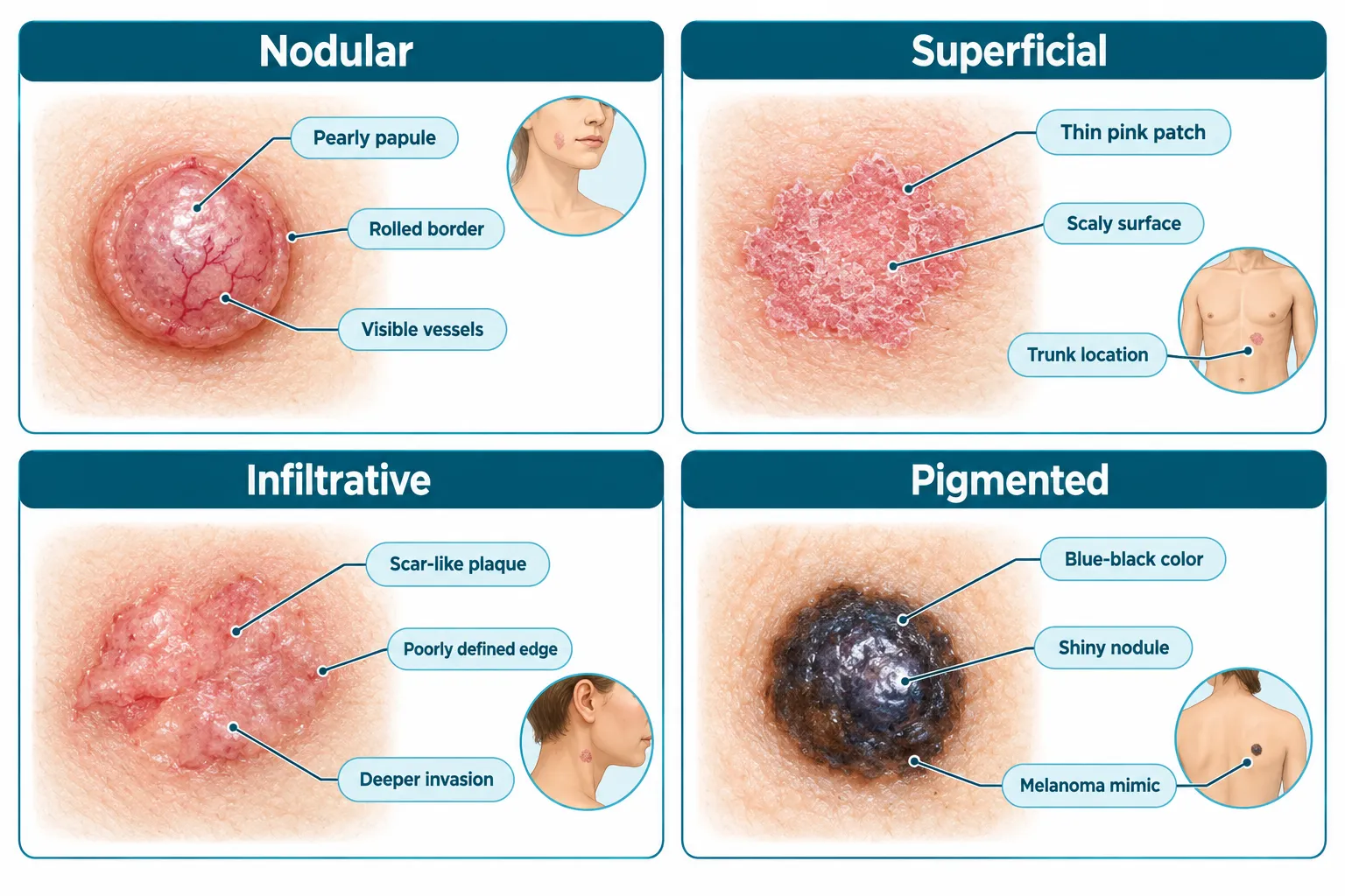
Recent histopathology reviews and a 2026-revised StatPearls chapter group BCC subtypes into two broad risk categories. Low-risk subtypes include nodular, superficial, pigmented, infundibulocystic, and fibroepithelial (Pinkus tumor). High-risk subtypes include infiltrative, micronodular, morpheaform (sclerosing), and basosquamous BCC.[1][4][13] The table below summarizes the four subtypes most patients ask about.
SubtypeTypical appearanceCommon locationGrowth patternRisk categoryNodularShiny, pearly papule or nodule with visible blood vessels and rolled edges; may ulcerateFace, sun-exposed skinSlow, outward, well-definedLow riskSuperficialThin, pink or red scaly patch or plaque; can look like eczemaTrunk, arms, legsSlow, spreads sideways in upper skin layerLow riskInfiltrativePoorly defined, scar-like or firm plaque; skin may look puckeredHead and neckDeep, finger-like invasion between collagen fibersHigh riskPigmentedBlue, brown, or black nodule or patch; often shinyFace or trunk, depending on underlying subtypeFollows the pattern of the underlying nodular or superficial tumorUsually low risk
Because appearance alone cannot confirm a subtype, a biopsy remains the standard step before finalizing a treatment plan. For readers building a broader picture of skin cancer categories, this comparison pairs well with an overview of 4 types of skin cancer.
Nodular BCC is a raised, pearly bump that grows outward as a solid mass, while superficial BCC is a flat, scaly patch that spreads sideways within the top layer of skin without forming a firm lump. The two subtypes also differ in typical body location and patient age at diagnosis.
Nodular BCC is described consistently across dermatology references as a shiny or pearly, firm papule or nodule with telangiectasia (small visible blood vessels), rolled borders, and sometimes a central crater or ulceration.[10][11][15] It tends to appear on chronically sun-exposed facial skin in older adults and is the single most common BCC subtype worldwide.[10][11][15]
Superficial BCC looks and behaves differently:
Decision rule: if a lesion is raised, shiny, and has visible blood vessels, suspect nodular BCC. If it is flat, scaly, and slow-growing on the chest or back, suspect superficial BCC. Either way, a shave or punch biopsy is the only reliable way to confirm which pattern is present, since both can occasionally mimic benign conditions covered in guides on benign mole features and other common skin marks.
A frequent mistake is assuming a flat, non-bleeding patch cannot be skin cancer. Superficial BCC often looks harmless for months or years before a biopsy is taken, which is why any persistent scaly patch that does not clear with standard eczema treatment deserves evaluation.
Nodular BCC is the most common subtype of basal cell carcinoma, making up an estimated 51-60% of all cases according to updated general dermatology references.[3][9][10] Superficial BCC ranks second at roughly 20-34%, and the aggressive infiltrative and morpheaform patterns together make up only about 5-10%.[3][9][10]
These proportions are consistent across primary care and oncology manuals published between 2020 and 2026, which means the ranking has held steady even as diagnostic tools have improved.[3][9][10] A few practical implications follow from this distribution:
Quick example: in a typical dermatology clinic reviewing 100 confirmed BCC biopsies, roughly 55 would be expected to show a nodular pattern, about 25 a superficial pattern, and only 5 to 10 an infiltrative or morpheaform pattern, based on the ranges reported in current literature.[3][9][10]
Infiltrative BCC looks like a flat, firm, scar-like patch with a poorly defined edge, while nodular BCC looks like a raised, shiny bump with a clear, rolled border. This visual difference reflects a real difference in how the two subtypes grow beneath the skin surface.
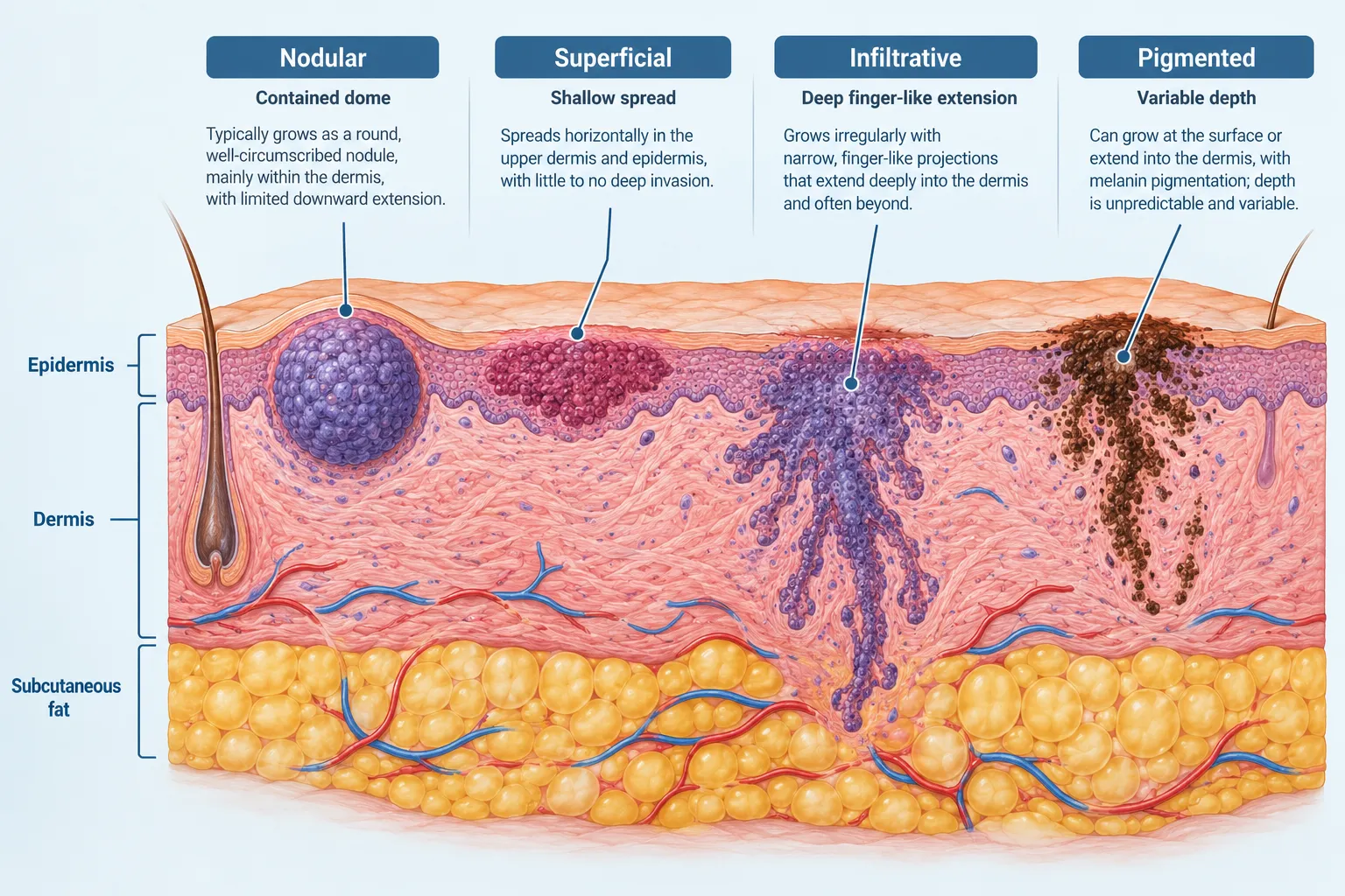
Under the microscope, infiltrative BCC (often grouped with morpheaform and micronodular patterns) is defined by narrow strands and cords of basaloid tumor cells that push between collagen bundles in the dermis, rather than forming one contained mass.[6][8][13] Clinically, this produces:
Nodular BCC, in contrast, tends to stay contained as a single dome-shaped growth with a border that is usually easy to trace by eye or with dermoscopy.
Infiltrative BCC means the tumor grows in thin, irregular strands that extend well past what is visible on the skin surface, rather than as one round mass. This subclinical spread is exactly why it is classified as high risk: current classifications emphasize its deeper invasion, wider subclinical spread, and higher local recurrence rates compared with nodular or superficial forms.[6][8][13]
In practical terms, a surgeon treating what looks like a small infiltrative lesion may find that the tumor extends several millimeters beyond the visible edge once tissue is examined layer by layer. This is the main reason infiltrative BCC is usually treated with Mohs micrographic surgery rather than a standard excision with a fixed margin.
Depth of invasion varies clearly by subtype: superficial BCC generally stays confined to the upper dermis, nodular BCC grows as a rounded mass that can extend into the mid-to-deep dermis, and infiltrative or morpheaform BCC can penetrate into deep dermis and occasionally subcutaneous fat while still looking deceptively small on the surface.[1][10][13]
Common mistake: judging depth by the size of the visible lesion. A one-centimeter infiltrative BCC can have roots extending farther under the skin than a two-centimeter nodular BCC, which is why pre-operative planning for high-risk subtypes often includes wider surgical margins or Mohs technique.
Yes, pigmented BCC can closely resemble melanoma because both can appear as blue, brown, or black skin lesions with irregular pigment. Dermoscopy and biopsy are typically needed to tell the two apart with confidence.
Pigmented BCC is not considered a separate histologic subtype in recent dermoscopy and pathology reviews. Instead, it is recognized as a pigmented variant of nodular or superficial BCC that contains melanin within tumor cell nests and surrounding melanophages (pigment-carrying immune cells).[4][9][12] Clinically, pigmented BCC:
Because of this overlap, any dark, irregular, or changing lesion should be evaluated the same way a suspected melanoma would be, with dermoscopy and, in most cases, a biopsy. Readers comparing pigmented lesions more broadly may also find it useful to review differences between benign mole characteristics and more concerning pigmented growths, or check a wider list of lesion types in 25 types of skin lesions explained.
Decision rule: if a dark lesion is shiny, has visible surface blood vessels, or shows rolled edges alongside the pigment, suspect pigmented BCC and biopsy accordingly. If it has an irregular, multicolored, asymmetric pattern without those classic BCC features, melanoma must be ruled out first.
Yes, pigmented BCC can and does appear on darker skin tones, and some clinical reviews note that BCC in patients with more pigmented skin is more likely to show the pigmented variant than in lighter-skinned patients. Increased baseline melanin activity in darker skin can make pigmented presentations more common, though BCC overall is less frequent in deeply pigmented skin than in fair skin. A 2025 dermoscopy-focused review confirms that pigmented features can occur across all BCC types, with pigmentation more often tied to the non-aggressive subtypes.[12][14] This makes routine skin checks and prompt evaluation of new dark spots important for all skin tones, not just fair-skinned patients.
Infiltrative BCC, along with the closely related micronodular and morpheaform subtypes, spreads and grows faster than nodular or superficial BCC. Contemporary cancer-organization summaries updated through September 2026 specifically note that infiltrative and micronodular BCCs "grow and spread more quickly than nodular and superficial BCC," which reinforces their high-risk classification and the need for closer follow-up.[2][11][15]
Growth speed differences by subtype:
Edge case: a long-neglected nodular BCC can eventually become locally destructive even though it is technically "low risk," because size and depth increase over years without treatment. Subtype risk describes typical behavior, not a guarantee, which is why any suspicious lesion should be evaluated rather than watched indefinitely.
Dermatologists diagnose BCC subtypes using a combination of visual exam, dermoscopy (a handheld magnification tool), and biopsy with histopathology, which remains the gold standard for confirming subtype. Dermoscopic patterns give strong clues before a single cell is examined under a microscope.
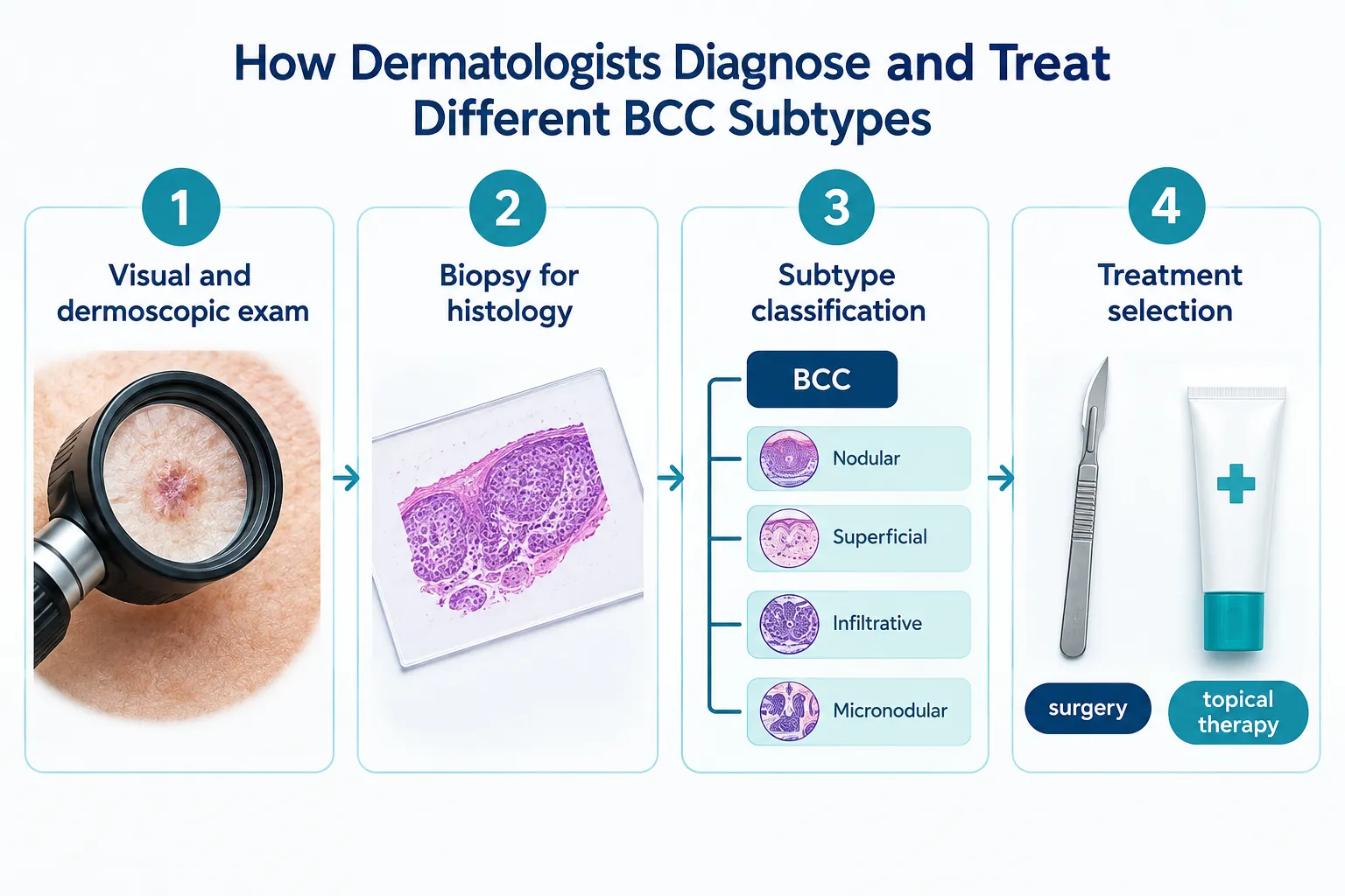
Refined dermoscopic criteria help distinguish the main subtypes before biopsy results return:
These dermoscopic patterns support pre-operative risk stratification, meaning a clinician can often predict whether a lesion is likely low-risk or high-risk before the biopsy confirms it, which helps guide how the biopsy itself is performed (shave versus punch versus incisional).
Biopsy remains necessary because dermoscopy alone cannot always distinguish BCC from other lesions with overlapping features, including some forms of actinic keratosis or early Bowen's disease, both of which can present as scaly patches similar to superficial BCC. A pathologist examining the biopsy sample identifies the specific growth pattern (nodular, superficial, infiltrative, or a mixed pattern) under the microscope, which then guides the treatment plan.
Common mistake: relying on a single small biopsy to rule out an aggressive component. Because BCCs can be mixed-pattern tumors, a superficial-only biopsy of a larger lesion can miss an infiltrative component growing deeper in the same tumor.
Yes, a single BCC lesion can contain more than one histologic subtype, commonly referred to as a mixed-pattern tumor. This is one of the most important reasons biopsy sampling and full excision pathology matter more than a quick visual diagnosis.
Mixed-pattern BCCs most often combine a low-risk pattern (such as nodular or superficial) with a higher-risk pattern (such as infiltrative or micronodular) in different areas of the same tumor.[1][13] Practical implications include:
Example: a facial lesion initially biopsied and read as nodular BCC may, after full excision, show a small area of infiltrative growth at the deep margin. In that scenario, the surgical plan and follow-up schedule typically shift to match the higher-risk infiltrative component rather than the original nodular reading.
The best treatment for each BCC subtype depends on its risk category: low-risk subtypes (nodular, superficial, pigmented variants of these) often respond well to topical therapy, photodynamic therapy, or standard excision, while high-risk subtypes (infiltrative, morpheaform, micronodular) are usually managed with Mohs micrographic surgery or wide excision.[1][10][13]
Recent clinical guidance breaks treatment choice down by subtype and lesion characteristics:
Pull quote: "Treatment decisions in BCC are made by subtype and risk category, not by the size of the lesion alone. A small infiltrative tumor can require a more aggressive surgical approach than a larger nodular one."
In general, superficial BCC is at least as easy to treat as nodular BCC, and it offers more nonsurgical options because it stays confined to the upper skin layer. Superficial BCC responds well to topical treatments and photodynamic therapy in many cases, options that are less commonly first-line for nodular BCC because nodular tumors form a deeper, more solid mass.[1][10][13]
That said, "easier" depends on location and patient preference:
Choose topical or light-based therapy if: the lesion is confirmed superficial BCC, is located away from the face, and the patient prefers to avoid a surgical scar. Choose surgical excision or Mohs if: the lesion is nodular, infiltrative, located on the face or ears, or has recurred after previous treatment.
Superficial and nodular BCC generally have the best prognosis and the lowest recurrence rates when treated appropriately, because both are classified as low-risk subtypes with well-defined growth patterns. Infiltrative, morpheaform, micronodular, and basosquamous subtypes carry the highest recurrence risk and require closer long-term follow-up.[1][13]
A simplified comparison of expected outcomes by risk category:
Risk categorySubtypes includedGeneral prognosisRecurrence risk after appropriate treatmentLow riskNodular, superficial, pigmented (nodular/superficial-based), fibroepithelial, infundibulocysticExcellentLowHigh riskInfiltrative, micronodular, morpheaform, basosquamousGood with prompt, appropriate surgeryHigher, especially if margins are narrow
Pigmented BCC is consistently categorized as low risk across recent histopathology and clinical reviews, provided the underlying architecture is nodular or superficial.[4][9][12] Its melanoma-like appearance, however, means it still demands careful dermoscopic evaluation and usually biopsy to avoid a missed or delayed diagnosis, with management guided by the underlying subtype rather than the pigment itself.[4][9][12]
Edge case: even a "low risk" subtype can recur if the initial excision margins were too narrow or if part of the tumor was missed. Recurrence risk is tied to complete removal as much as it is tied to subtype, which is why follow-up skin checks matter regardless of which subtype was originally diagnosed.
Misreading a BCC subtype, or misunderstanding what the pathology report means, can lead to under-treatment or unnecessary anxiety. A few recurring mistakes are worth flagging directly:
What are the four main BCC subtypes discussed in this guide?
The four subtypes most often discussed are nodular, superficial, infiltrative, and pigmented BCC. Nodular and superficial are the two most common; infiltrative is a high-risk pattern; pigmented is a color variant that can occur with either nodular or superficial BCC.
Is nodular or superficial BCC more dangerous?
Neither is considered dangerous compared with infiltrative or morpheaform BCC; both nodular and superficial are classified as low-risk subtypes.[1][4][13] Nodular BCC can become locally destructive if left untreated for years, but it does not carry the same invasive growth pattern as infiltrative BCC.
Does pigmented BCC always mean a worse prognosis?
No. Pigmented BCC generally shares the prognosis of its underlying subtype, most often nodular or superficial, both of which are low risk.[4][9][12] The pigment itself is not a marker of aggressiveness.
Can BCC subtypes change over time?
A BCC does not typically transform from one subtype to another, but an untreated tumor can develop additional patterns within the same lesion over time, which is why mixed-pattern tumors occur.
What is the safest way to tell BCC apart from melanoma at home?
There is no reliable way to distinguish BCC, including pigmented BCC, from melanoma without professional evaluation. Any new, changing, or irregular pigmented spot should be checked by a clinician using dermoscopy and biopsy if needed.
Which BCC subtype is most likely to be treated with Mohs surgery?
Infiltrative, morpheaform, and micronodular BCC are most often treated with Mohs micrographic surgery because of their tendency to extend beyond visible margins.[1][10][13] Nodular BCC on the face is also frequently treated with Mohs for cosmetic and margin-control reasons.
How often should someone be checked after BCC treatment?
Follow-up schedules vary by clinician and risk category, but periodic skin checks after any BCC diagnosis are standard practice, with high-risk subtypes typically monitored more closely than low-risk ones.
Can superficial BCC be treated without surgery?
Yes, superficial BCC frequently responds to topical treatments such as imiquimod or 5-fluorouracil, or to photodynamic therapy, particularly when located on the trunk or limbs.[1][10][13] Surgical options remain available and are often preferred on the face.
BCC subtypes explained side by side come down to one core idea: appearance predicts risk, but only a biopsy confirms it. Nodular BCC is the common pearly bump on sun-exposed skin. Superficial BCC is the flat scaly patch that mimics a rash. Infiltrative BCC is the scar-like, deceptively aggressive pattern that needs more careful surgery. Pigmented BCC is a color variant that can look alarming but usually behaves like the nodular or superficial tumor underneath it.
The practical next step is simple. Any new or changing skin lesion, whether it is shiny, scaly, or dark, should be examined by a clinician who can use dermoscopy and biopsy to identify the exact subtype. From there, treatment can be matched to actual risk rather than to how the lesion happens to look on the surface. Patients who have already been treated for one BCC subtype should keep up with routine skin checks, since new lesions and, less commonly, mixed patterns within a treated area can still develop.
[1] Nbk482439 - https://www.ncbi.nlm.nih.gov/books/NBK482439/
[2] Pmc9681969 - https://pmc.ncbi.nlm.nih.gov/articles/PMC9681969/
[3] P161 - https://www.aafp.org/afp/2012/0715/p161
[4] Pmc8630439 - https://pmc.ncbi.nlm.nih.gov/articles/PMC8630439/
[6] Nbk470301 - https://www.ncbi.nlm.nih.gov/books/NBK470301/?report=printable
[8] Basal Cell Carcinoma Pathology - https://dermnetnz.org/topics/basal-cell-carcinoma-pathology
[9] P339 - https://www.aafp.org/afp/2020/0915/p339
[10] Basal Cell Carcinoma - https://www.msdmanuals.com/professional/oncology/cancers-of-the-skin/basal-cell-carcinoma