Last updated: February 17, 2026
Finding a small, round bump beneath your skin can trigger immediate concern. While most skin cysts are benign, understanding the specific type you're dealing with determines the best treatment approach and helps predict whether it will recur after removal. Epidermoid, sebaceous, and pilar cysts may look similar on the surface, but they originate from different skin structures, behave differently, and require distinct management strategies.
Understanding epidermoid vs sebaceous vs pilar cyst: how they differ (and why it matters) empowers patients and healthcare providers to make informed decisions about observation versus intervention. The differences extend beyond academic classification—they influence rupture risk, infection likelihood, preferred removal techniques, and recurrence rates. Misidentifying the cyst type can lead to incomplete removal and frustrating regrowth.

The primary distinction lies in their origin within the skin. Epidermoid cysts form when surface skin cells become trapped deeper in the skin, usually after follicle damage. Pilar cysts develop specifically from the outer root sheath of hair follicles, which explains their scalp predominance. Sebaceous cysts originate from the oil-producing sebaceous glands themselves—though this term is technically outdated since true sebaceous cysts are extremely rare[5].
Each type contains different material: epidermoid cysts hold keratin (a protein found in skin cells) that appears thick and cheese-like, pilar cysts contain keratin from hair follicle cells that's often firmer, and genuine sebaceous cysts would contain oily sebum. Location, appearance, and texture provide the first clues to identification, but definitive diagnosis often requires examination of the cyst contents or wall during removal.
Epidermoid cysts are the most common type of skin cyst, forming when cells from the outer layer of skin (epidermis) become trapped beneath the surface[2][3]. This trapping usually occurs after trauma to a hair follicle, skin injury, or blockage of a skin pore. The trapped cells continue producing keratin—the same protein that makes up hair and the outer layer of skin—which accumulates inside a sac-like structure.
Common characteristics of epidermoid cysts include:
The central punctum serves as a key identifying feature. This tiny opening represents the original follicle or pore where the cyst originated. When squeezed (which dermatologists strongly advise against), this opening may release foul-smelling keratin debris.
Epidermoid cysts carry a higher risk of rupture compared to other cyst types[1][4][6]. When the cyst wall breaks—whether from trauma, pressure, or spontaneous rupture—the keratin contents leak into surrounding tissue. The body recognizes this material as foreign, triggering an inflammatory response that causes redness, swelling, pain, and sometimes infection.
Choose immediate medical evaluation if:
For more information about managing ruptured cysts, see our guide on deep-seated cysts.
Pilar cysts (also called trichilemmal cysts) originate from the outer root sheath of hair follicles rather than from surface skin cells[2][3][7]. This fundamental difference in origin creates distinct characteristics that help differentiate them from epidermoid cysts. Approximately 90% of pilar cysts occur on the scalp, though they can occasionally appear on the face or arms[1][4][7].
Distinctive features of pilar cysts:
The absence of a central opening makes pilar cysts less prone to spontaneous drainage but doesn't eliminate rupture risk entirely. The cyst wall in pilar cysts tends to be thicker and more durable than in epidermoid cysts, contributing to their typically stable, asymptomatic nature.
Unlike epidermoid cysts, which often appear as solitary lesions, pilar cysts frequently occur in multiples[7]. It's not uncommon for patients to develop 5-10 or more pilar cysts on the scalp over time. This pattern reflects the hereditary nature of pilar cysts and their origin from hair follicle structures.
Common mistake: Assuming all scalp bumps are pilar cysts. While pilar cysts dominate scalp locations, epidermoid cysts, lipomas, and other growths can also appear on the scalp. Proper identification matters because treatment approaches differ.
The term "sebaceous cyst" creates significant confusion in both medical literature and everyday conversation. Historically, all three cyst types were called sebaceous cysts because experts believed they originated from sebaceous glands (the oil-producing glands attached to hair follicles)[5]. Modern understanding reveals this classification was incorrect—most cysts labeled "sebaceous" are actually epidermoid or pilar cysts.
True sebaceous cysts:
When healthcare providers use the term "sebaceous cyst" in 2026, they're often referring to epidermoid cysts rather than true sebaceous origin cysts. This outdated terminology persists despite being technically inaccurate[5].
Understanding whether you have a true sebaceous cyst versus an epidermoid or pilar cyst affects treatment planning. True sebaceous cysts require complete removal of the sebaceous gland to prevent recurrence[6]. Epidermoid and pilar cysts need removal of the entire cyst wall (sac) but don't require gland excision since they don't originate from glands.
Treatment implications:
For detailed information about surgical approaches, visit our epidermoid cyst removal surgery guide.
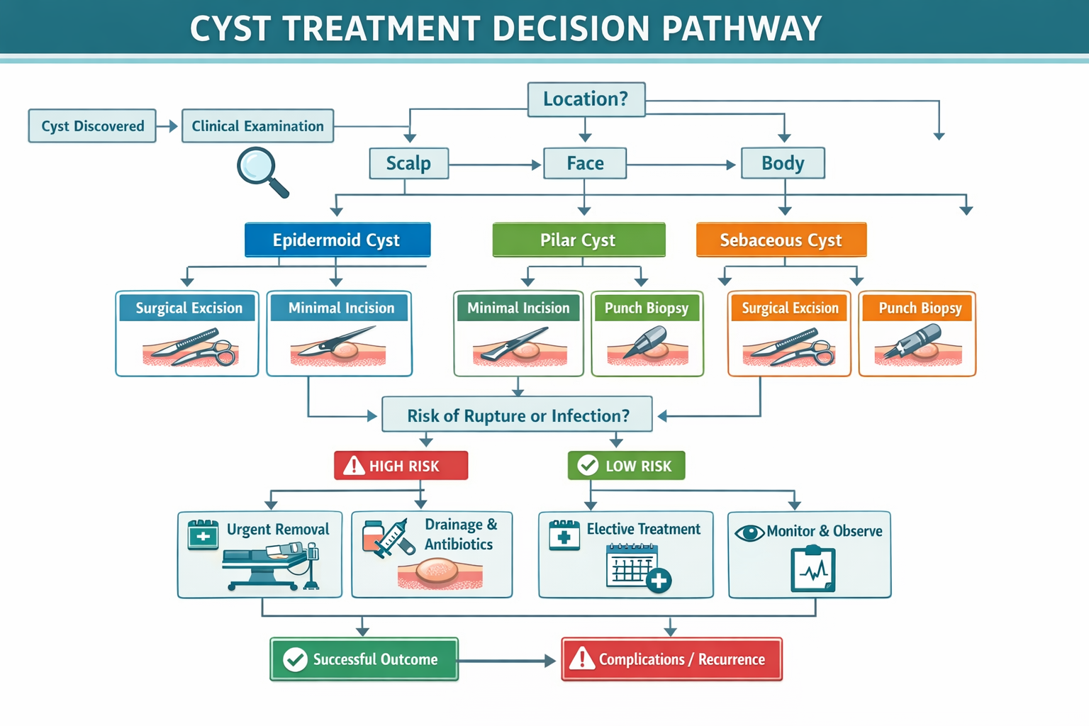
While definitive diagnosis requires professional examination and sometimes pathological analysis, several visual and tactile clues help distinguish between epidermoid, sebaceous, and pilar cysts. Healthcare providers use a combination of location, appearance, and physical characteristics to make initial assessments.
FeatureEpidermoid CystPilar CystSebaceous CystMost Common LocationFace, neck, chest, backScalp (90% of cases)Variable, anywhere with sebaceous glandsCentral OpeningUsually present (punctum)Typically absentMay show enlarged poreTextureRubbery, moderately firmFirm, well-definedDome-shaped, smoothMobilityModerateHigh (moves easily)VariableContentsThick, cheese-like keratinFirmer keratinOily, waxy sebumSmell When RupturedFoul, distinctive odorLess odorousMinimal odorMultiple CystsUsually solitaryOften multiple on scalpVariableGenetic PatternNot typically hereditaryOften runs in familiesRare overall
Examination steps healthcare providers use:
Location alone often narrows the diagnosis significantly. A smooth, firm bump on the scalp without a central opening is most likely a pilar cyst. A bump on the face or upper back with a visible dark punctum strongly suggests an epidermoid cyst. However, exceptions exist—epidermoid cysts can appear on the scalp, and pilar cysts occasionally develop on the face.
Edge case: Some cysts show features of both types. Hybrid cysts or cysts that have been traumatized may not fit neatly into one category. In these situations, removal and pathological examination provide definitive classification.
Understanding epidermoid vs sebaceous vs pilar cyst: how they differ (and why it matters) extends beyond satisfying curiosity—it has practical implications for treatment decisions, outcome expectations, and long-term management. The cyst type influences everything from rupture risk to the surgical technique that offers the best cosmetic outcome.
Different cyst types respond better to specific removal techniques. Epidermoid cysts, with their higher rupture and inflammation risk, often benefit from complete excision using minimal-incision or punch-laser techniques that remove the entire cyst wall while minimizing scarring[6]. Pilar cysts, being firmer and more defined, can often be removed through very small incisions with excellent cosmetic results.
Treatment matching:
Recurrence rates differ by cyst type and removal completeness. Epidermoid cysts that rupture before removal have higher recurrence rates because inflammation makes complete sac removal more difficult[6]. Pilar cysts, when completely excised, rarely recur. True sebaceous cysts require complete gland removal—if any gland tissue remains, a new cyst can form[6].
Recurrence prevention factors:
For information about ensuring complete removal, see our article on cyst sac removal.
Epidermoid cysts carry the highest risk of rupture, inflammation, and secondary infection[1][4][6]. The keratin contents are particularly irritating to surrounding tissue when released. Pilar cysts tend to remain stable and asymptomatic for years, though they can still rupture with trauma. Understanding your specific risk profile helps with monitoring decisions.
Risk-based monitoring approach:
Treatment approaches for epidermoid, pilar, and sebaceous cysts range from observation to surgical excision. The optimal strategy depends on cyst type, size, location, symptoms, and patient preferences. Not all cysts require removal—many can be safely observed if they remain small and asymptomatic.
For small, asymptomatic cysts that aren't growing or causing cosmetic concerns, observation is a reasonable approach. This is particularly appropriate for pilar cysts, which tend to remain stable for years. Patients choosing observation should monitor for changes in size, tenderness, or appearance.
Observation is appropriate when:
Seek treatment if:
Complete surgical excision remains the most effective treatment for permanent cyst removal. The procedure involves removing the entire cyst, including its wall (sac), through an incision. When performed correctly, excision provides the lowest recurrence rates across all cyst types.
Standard excision procedure:
For epidermoid cysts, minimal-incision techniques using smaller cuts can achieve excellent results with less scarring[6]. Pilar cysts, being firmer and more defined, often "pop out" easily through small incisions. Sebaceous cysts require careful removal of all gland tissue to prevent recurrence[6].
Learn more about the step-by-step process in our cyst removal surgery guide.
Simple drainage (incision and drainage) provides quick relief for painful, inflamed cysts but doesn't prevent recurrence. Since the cyst wall remains in place, the cyst almost always refills. Drainage is typically reserved for infected cysts that need immediate decompression before definitive excision can be performed.
Drainage considerations:
Common mistake: Attempting home drainage or squeezing cysts. This increases infection risk, causes scarring, and can trigger rupture into surrounding tissue, making subsequent surgical removal more difficult. For information on why home treatment is problematic, see our article on drawing out a sebaceous cyst at home.
For inflamed cysts that aren't infected, intralesional corticosteroid injection can reduce inflammation and size. This approach doesn't remove the cyst but can shrink it and alleviate symptoms. It's sometimes used as a bridge therapy before surgical excision or for patients who cannot undergo surgery.
Injection therapy:
Recovery time varies based on cyst type, size, location, and removal technique. Most patients experience minimal downtime after uncomplicated cyst excision, though healing continues for several weeks beneath the skin surface.
Days 1-3:
Days 4-7:
Weeks 2-4:
Months 1-6:
For detailed recovery information, visit our guide on cyst removal recovery time.
Faster healing occurs with:
Slower healing may occur with:
Scarring depends more on location and technique than cyst type. Facial cysts typically receive the most meticulous closure techniques to minimize scarring. Scalp cysts often heal with scars hidden by hair. Back and chest cysts may develop more visible scars due to skin tension in these areas.
Scar minimization strategies:
For more information about scarring, see our article on whether cyst removal leaves a scar.
Epidermoid, pilar, and true sebaceous cysts are benign (non-cancerous) growths. The vast majority remain benign throughout a person's lifetime. However, extremely rare cases of malignant transformation have been reported in medical literature, almost exclusively in long-standing, large cysts that have been present for many years.
The risk of cancer developing in or from these cysts is extraordinarily small. When it does occur, it's typically squamous cell carcinoma arising in a long-standing epidermoid cyst. The overall incidence is estimated at less than 0.1% of all epidermoid cysts, and most cases involve cysts present for decades.
Cancer warning signs (seek immediate evaluation):
Reassuring features:
Most removed cysts are sent for pathological examination, which involves a pathologist examining the tissue under a microscope. This serves two purposes: confirming the diagnosis and ruling out any unexpected findings. While cancer is rare, pathology provides definitive diagnosis and peace of mind.
Pathology is especially important when:
While most skin cysts remain harmless, several complications can develop, particularly with epidermoid cysts. Understanding potential problems helps patients recognize when medical attention is needed and why some cysts warrant earlier removal rather than continued observation.
Cyst rupture—when the cyst wall breaks and contents leak into surrounding tissue—is the most common complication, especially for epidermoid cysts[1][4][6]. The body recognizes the released keratin as foreign material, triggering an inflammatory response. This creates a tender, red, swollen area that can be quite painful.
Rupture characteristics:
Management of ruptured cysts:
Secondary bacterial infection can occur in any cyst type but is most common after rupture or attempted home drainage. Infected cysts become acutely painful, with surrounding redness, warmth, and sometimes pus drainage. Fever may develop with more severe infections.
Signs of infection:
Treatment for infected cysts:
Even without complications, cysts can cause cosmetic distress, particularly on visible areas like the face, neck, or arms. The psychological impact of a visible bump shouldn't be minimized—cosmetic concerns are valid reasons for seeking removal.
Cosmetic considerations:
Rarely, cysts develop near nerves or blood vessels, creating potential complications during removal. This is more common with deeper cysts or those in anatomically complex areas. Experienced surgeons can usually navigate these situations safely, but patients should be aware of the possibility.
Potential nerve-related complications:
Not every skin cyst requires immediate medical attention, but certain situations warrant prompt evaluation. Understanding when to seek care helps prevent complications and ensures appropriate treatment timing.
Seek medical care within 24-48 hours if:
These symptoms suggest infection, rupture, or acute inflammation requiring treatment. Delaying care can lead to worsening infection or abscess formation.
Schedule a regular appointment if:
You can monitor at home if:
Common mistake: Waiting until a cyst becomes inflamed or infected before seeking care. Elective removal of a non-inflamed cyst is easier, has better cosmetic outcomes, and lower complication rates than emergency treatment of an infected cyst.
The cost of cyst removal varies significantly based on cyst type, size, location, complexity, and whether the procedure is considered medically necessary or cosmetic. Insurance coverage also plays a major role in out-of-pocket expenses.
Cost variables include:
Most insurance plans cover cyst removal when medically necessary—meaning the cyst is symptomatic, infected, rapidly growing, or interfering with function. Purely cosmetic removal of small, asymptomatic cysts may not be covered.
Generally covered situations:
May not be covered:
For detailed cost information, visit our guide on how much it costs to remove a cyst.
For procedures not covered by insurance or for patients without insurance, many practices offer:
Cost-saving strategies:
Location provides the strongest clue—pilar cysts occur on the scalp 90% of the time, while epidermoid cysts favor the face, neck, chest, and back. Epidermoid cysts typically show a central punctum (small dark opening), while pilar cysts have smooth surfaces without openings. However, definitive diagnosis often requires examination of the cyst contents or wall during removal.
No, many cysts can be safely observed if they remain small, stable, and asymptomatic. Removal is recommended when cysts are painful, infected, rapidly growing, cosmetically concerning, or located in areas prone to trauma. The decision should balance the cyst's characteristics, symptoms, and patient preferences.
Recurrence risk depends on removal completeness. When the entire cyst wall (sac) is removed, recurrence rates are very low (under 5%). Incomplete removal—especially of inflamed or ruptured cysts—increases recurrence risk significantly. Simple drainage without wall removal almost always leads to recurrence.
Healthcare providers strongly advise against home drainage or squeezing. This increases infection risk, can trigger rupture into surrounding tissue (making surgical removal more difficult), causes scarring, and rarely provides lasting relief since the cyst wall remains. Professional treatment offers safer, more effective results.
Most straightforward cyst removals take 15-45 minutes, depending on cyst size, location, and complexity. Larger cysts, inflamed cysts, or those in anatomically complex areas may take longer. The procedure is typically performed under local anesthesia in an office setting.
No, epidermoid, pilar, and sebaceous cysts are not contagious. They result from trapped skin cells or blocked glands, not from infections that can spread between people. You cannot "catch" a cyst from someone else or transmit one to another person.
The foul odor from ruptured or drained epidermoid cysts comes from keratin breakdown by bacteria. Keratin itself is odorless, but when exposed to bacteria on the skin surface or when trapped for long periods, it develops a characteristic unpleasant smell often described as "cheesy" or putrid.
While rare, small cysts occasionally resolve spontaneously, particularly after rupturing and completely draining. However, most cysts persist indefinitely unless removed. Temporary size fluctuations can occur, especially with inflammation or partial drainage, but complete spontaneous resolution is uncommon.
Cysts are non-infected, fluid-filled or keratin-filled sacs that develop slowly over time. Boils (furuncles) are infected hair follicles filled with pus that develop rapidly, are painful from the start, and show signs of infection (redness, warmth, tenderness). Boils require different treatment than cysts.
Earlier removal often offers advantages: smaller incisions, better cosmetic results, easier surgery, and lower complication risk. However, not all cysts require removal. Discuss the specific cyst's characteristics, growth pattern, and your concerns with a healthcare provider to make an informed decision.
No, cyst type is determined by origin and doesn't change. An epidermoid cyst remains epidermoid, and a pilar cyst remains pilar. However, trauma, inflammation, or incomplete removal can alter a cyst's appearance, sometimes making classification more difficult.
There's no guaranteed prevention method since cysts result from skin cell trapping or gland blockage, often after minor trauma you might not notice. However, protecting skin from injury, treating acne appropriately, and avoiding manipulation of existing bumps may reduce risk. Pilar cysts have a genetic component that can't be prevented.
Understanding epidermoid vs sebaceous vs pilar cyst: how they differ (and why it matters) empowers patients to make informed decisions about their care. While these three cyst types may appear similar superficially, their distinct origins, behaviors, and treatment requirements make accurate identification valuable. Epidermoid cysts, with their central punctum and higher rupture risk, require different management considerations than the smooth, scalp-predominant pilar cysts or the rare true sebaceous cysts.
The good news is that all three types are benign, and excellent treatment options exist when removal becomes necessary or desired. Whether you choose observation for a small, stable cyst or pursue surgical excision for symptom relief or cosmetic improvement, working with an experienced healthcare provider ensures the best outcomes.
If you have a skin cyst:
For cyst removal:
The differences between epidermoid, sebaceous, and pilar cysts extend beyond academic classification—they influence practical decisions about treatment timing, technique selection, and outcome expectations. Armed with this knowledge, you can engage in meaningful conversations with healthcare providers and make choices aligned with your health goals and personal values.
For expert evaluation and treatment of skin cysts in the Greater Toronto Area, The Minor Surgery Center offers specialized care with experienced surgeons and state-of-the-art techniques designed to achieve optimal cosmetic and medical outcomes.
[1] The Difference Between Epidermoid Pilar Sebaceous Cysts - https://beflawlessmn.com/2022/02/13/the-difference-between-epidermoid-pilar-sebaceous-cysts/
[2] Epidermoid Cysts - https://www.beaconhealthsystem.org/library/diseases-and-conditions/epidermoid-cysts?content_id=CON-20305522
[3] Drc 20352706 - https://www.mayoclinic.org/diseases-conditions/epidermoid-cysts/diagnosis-treatment/drc-20352706
[4] Epidermoid Cyst Vs Pilar Cyst Treatment - https://www.worldsfamousdermatologist.com/epidermoid-cyst-vs-pilar-cyst-treatment/
[5] Epidermoid And Pilar Cysts Sebaceous Cysts Leaflet - https://patient.info/skin-conditions/epidermoid-and-pilar-cysts-sebaceous-cysts-leaflet
[6] Sebaceous Cyst Vs Epidermoid Cyst Spot The Difference Best Treatments - https://www.mountcastleplasticsurgery.com/our-blog/sebaceous-cyst-vs-epidermoid-cyst-spot-the-difference-best-treatments/
[7] 23092 Pilar Trichilemmal Cyst - https://my.clevelandclinic.org/health/diseases/23092-pilar-trichilemmal-cyst
[8] 1395420 Types Of Cysts You Might Have Sebaceous Epidermoid And More - https://www.northbranchdermatology.com/blog/1395420-types-of-cysts-you-might-have-sebaceous-epidermoid-and-more/


