Diseases That Cause Many New Moles: When a Skin Check Isn't Enough
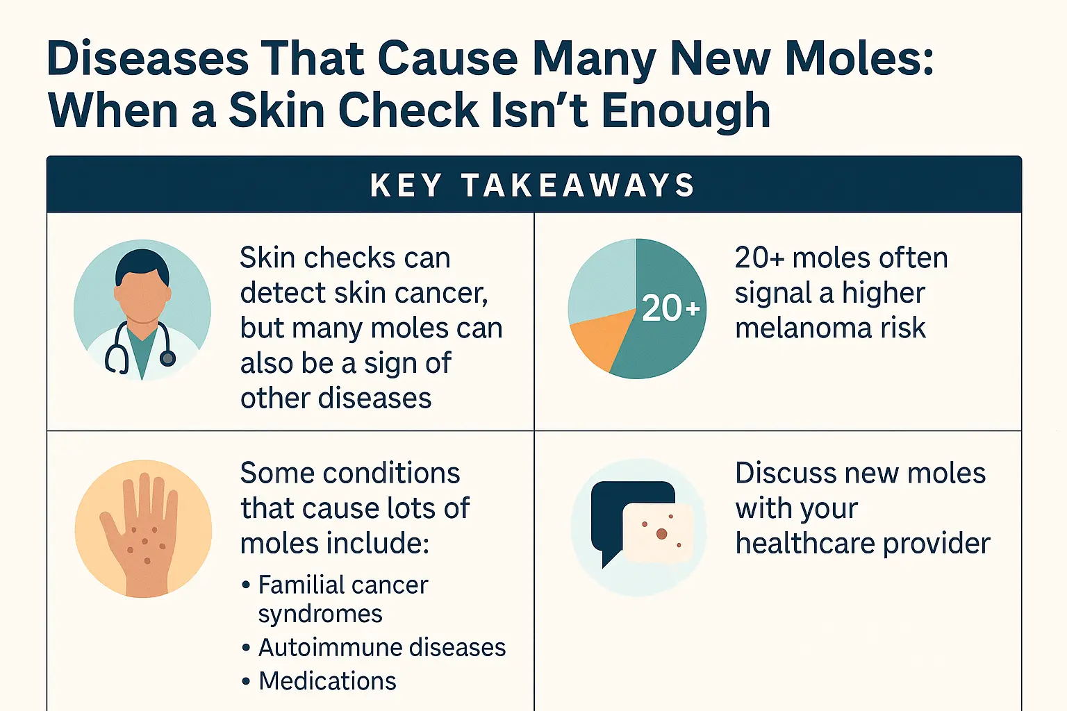
When dozens of new moles suddenly appear across the skin within weeks or months, it's rarely just a cosmetic concern. While most people develop new moles gradually throughout their lifetime, the rapid appearance of multiple moles can signal underlying systemic diseases that demand immediate medical attention. Understanding diseases that cause many new moles: when a skin check isn't enough could be the difference between early intervention and serious health complications.
The sudden eruption of numerous moles—medically termed eruptive nevi—often indicates that something deeper is happening within the body. These aren't the typical benign moles that develop slowly over years; they're warning signs that require comprehensive medical evaluation beyond a standard skin examination.
Key Takeaways
Rapid mole development (multiple new moles appearing within weeks or months) often signals underlying systemic diseases rather than normal skin changes
Paraneoplastic syndromes, immunodeficiency disorders, hormonal imbalances, and genetic conditions can all trigger sudden mole proliferation
Standard skin checks alone may miss the underlying disease causing new moles—comprehensive medical evaluation including blood work and imaging is essential
Early detection of the causative disease significantly improves treatment outcomes and can prevent serious complications
Immediate dermatological consultation is warranted when experiencing sudden appearance of 5+ new moles within a short timeframe
Understanding Normal Mole Development vs. Abnormal Proliferation
What Are Normal Moles?
Normal moles, or melanocytic nevi, are clusters of pigment-producing cells called melanocytes. Most individuals develop between 10 and 40 moles throughout their lifetime, with the majority appearing during childhood and young adulthood [1]. These benign growths typically:
Develop gradually over months to years
Remain stable in size, shape, and color
Measure less than 6mm in diameter
Display uniform coloration (usually brown or tan)
Have well-defined, regular borders
When Mole Development Becomes Concerning
The sudden appearance of numerous new moles deviates significantly from normal patterns. Warning signs include:
⚠️ Five or more new moles appearing within weeks or months
📈 Rapid growth of existing moles
🎨 Varying colors within individual moles or across multiple moles
📏 Irregular borders or asymmetrical shapes
💫 Clustering patterns in specific body regions
🔄 Continuous new mole formation without stabilization
Diseases That Cause Many New Moles: When a Skin Check Isn't Enough—The Medical Conditions
Paraneoplastic Syndromes and Internal Malignancies
Paraneoplastic syndromes represent one of the most serious causes of sudden mole proliferation. These conditions occur when internal cancers trigger skin manifestations, including rapid mole development [2].
Sign of Leser-Trélat
This rare paraneoplastic syndrome manifests as the sudden eruption of multiple seborrheic keratoses (mole-like growths) associated with internal malignancy. Key characteristics include:
Rapid appearance of dozens to hundreds of lesions
Often accompanied by itching (pruritus)
Associated cancers: gastrointestinal adenocarcinomas, lymphomas, breast cancer
Requires immediate oncological evaluation
Melanoma-Associated Eruptive Nevi
Some patients with melanoma develop multiple new melanocytic nevi as a paraneoplastic phenomenon. This condition demands urgent attention, as it may indicate:
Advanced melanoma with systemic involvement
Aggressive tumor behavior
Need for comprehensive cancer staging
For those concerned about melanoma, consulting with melanoma specialists provides access to advanced diagnostic capabilities.
Immunodeficiency and Immunosuppression
Compromised immune systems significantly increase the risk of abnormal mole development and skin cancer.
HIV/AIDS-Related Skin Changes
Patients with HIV/AIDS frequently experience:
Increased number of atypical moles
Higher risk of melanoma development
Accelerated progression from benign to malignant lesions
Multiple concurrent skin conditions
Post-Transplant Immunosuppression
Organ transplant recipients taking immunosuppressive medications face dramatically elevated risks [3]:
ConditionRisk IncreaseTimelineNew moles3-5x higherWithin 1-2 years post-transplantMelanoma3-8x higherAny time post-transplantSquamous cell carcinoma65-250x higher5+ years post-transplantBasal cell carcinoma10x higher3+ years post-transplant
LEOPARD is an acronym describing this genetic condition's features:
Lenormes (multiple brown spots)
ECG abnormalities
Ocular hypertelorism (widely spaced eyes)
Pulmonary stenosis
Abnormal genitalia
Retardation of growth
Deafness (sensorineural)
Patients develop hundreds to thousands of lentigines (mole-like spots) across their entire body.
Hormonal Disorders and Endocrine Dysfunction
Hormonal imbalances can trigger increased mole development through various mechanisms.
Pregnancy-Related Mole Changes
Pregnancy causes significant hormonal fluctuations that affect melanocytes:
Existing moles may darken or enlarge
New moles may appear (though typically limited numbers)
Melasma (pregnancy mask) develops in 50-70% of pregnant women
Most changes reverse postpartum
Important distinction: While pregnancy causes some mole changes, the sudden appearance of many new moles during pregnancy warrants immediate evaluation to rule out melanoma or other conditions.
Methotrexate: Manages various inflammatory conditions
TNF-alpha inhibitors: Biologics for autoimmune diseases
BRAF Inhibitors
Paradoxically, medications targeting BRAF mutations in melanoma treatment can cause:
Eruptive nevi (sudden new mole appearance)
Development of secondary skin cancers
Requires careful monitoring during treatment
Hormonal Medications
Oral contraceptives: May darken existing moles
Hormone replacement therapy: Can trigger new mole development
Testosterone therapy: Associated with skin changes including new growths
Red Flags: When Diseases That Cause Many New Moles Require Urgent Evaluation
Critical Warning Signs
Certain presentations demand immediate medical attention beyond routine skin checks:
🚨 Seek urgent evaluation if experiencing:
Rapid onset: 10+ new moles within 4-8 weeks
Systemic symptoms: Unexplained weight loss, fatigue, night sweats, fever
Changing moles: Existing moles rapidly growing or changing color
Bleeding or ulceration: Any mole that bleeds without trauma
Asymmetric distribution: Moles appearing predominantly on one body region
Associated symptoms: Itching, pain, or tenderness in multiple moles
Family history: Known genetic syndromes or multiple family members with melanoma
The ABCDE Rule for Individual Mole Assessment
While evaluating the overall pattern of new moles, each individual lesion should be assessed using the ABCDE criteria [4]:
Asymmetry: One half doesn't match the other
Border: Irregular, scalloped, or poorly defined edges
Color: Varies from one area to another; multiple colors
Diameter: Larger than 6mm (pencil eraser size)
Evolving: Changes in size, shape, color, elevation, or symptoms
The "Ugly Duckling" Sign
The ugly duckling sign identifies moles that look different from surrounding lesions. When multiple new moles appear but one looks distinctly different, it requires special attention and possible biopsy.
Comprehensive Diagnostic Approach: Beyond the Basic Skin Check
Why Standard Skin Checks Fall Short
Traditional visual skin examinations, while valuable, have limitations when investigating diseases that cause many new moles:
Pregnancy-related changes: Typically observation; postpartum reassessment
Mole Removal Considerations
Not all new moles require removal, but indications include:
✅ Removal recommended when:
Biopsy confirms atypical or malignant features
Mole displays concerning ABCDE characteristics
Located in high-friction areas causing irritation
Patient anxiety significantly impacts quality of life
Part of prophylactic strategy in high-risk genetic syndromes
Removal methods include:
MethodBest ForAdvantagesDisadvantagesSurgical excisionSuspicious or large molesComplete removal, tissue for analysisRequires stitches, longer healingShave removalRaised, benign molesQuick, minimal scarringMay not remove deep componentsLaser removalSmall, flat, benign molesMinimal scarring, preciseCannot examine tissue, not for suspicious molesCryotherapyBenign lesions onlyQuick, no cuttingCannot examine tissue, unpredictable results
For those in the Greater Toronto Area, mole removal services in Barrie and Ajax provide accessible options for professional evaluation and treatment.
Long-Term Monitoring and Surveillance Strategies
Establishing a Surveillance Schedule
Patients with diseases causing multiple new moles require individualized monitoring protocols:
High-Risk Patients (genetic syndromes, history of melanoma, immunosuppression):
Every 3-4 months: Full-body dermatological examination
Every 6-12 months: Total body photography update
Annual: Comprehensive medical evaluation for underlying conditions
Moderate-Risk Patients (multiple atypical moles, family history):
Every 6 months: Dermatological examination
Annual: Total body photography
As needed: Evaluation of changing lesions
Standard-Risk Patients:
Annual: Skin examination
Monthly: Self-examination
Prompt evaluation: Any concerning changes
Self-Monitoring Techniques
Effective self-examination empowers patients to detect changes early:
Monthly Self-Exam Protocol:
Full-body inspection in good lighting
Use mirrors to examine back, scalp, buttocks
Photograph any concerning moles with smartphone
Document new moles with date and location
Compare to previous photos monthly
Report any changes to dermatologist
Self-Exam Checklist:
☐ Face and neck (including ears)
☐ Scalp (use blow dryer to part hair)
☐ Hands (palms and backs, between fingers)
☐ Arms (all surfaces)
☐ Torso (front and back)
☐ Buttocks and genital area
☐ Legs (all surfaces)
☐ Feet (soles, between toes, toenails)
Technology-Assisted Monitoring
Smartphone Apps: Modern apps help track mole changes:
Photo documentation with automatic dating
Side-by-side comparison features
AI-assisted risk assessment (adjunct, not replacement for professional evaluation)
Reminder systems for self-exams
Important Note: While helpful, smartphone apps should never replace professional dermatological evaluation. Learn more about 3D mole mapping app reliability.
Wearable Technology: Emerging devices monitor:
UV exposure levels
Reminders for sun protection
Integration with health tracking systems
Prevention Strategies and Risk Reduction
Sun Protection: The Foundation of Mole Management
While sun exposure doesn't cause all moles, UV radiation significantly increases melanoma risk, especially in those with multiple moles [5].
Comprehensive Sun Protection:
☀️ Sunscreen Application:
SPF 30 minimum (SPF 50+ for high-risk individuals)
Broad-spectrum protection (UVA and UVB)
Apply 15-30 minutes before sun exposure
Reapply every 2 hours and after swimming/sweating
Use 1 ounce (shot glass full) for full body coverage
🧢 Physical Protection:
Wide-brimmed hats (3-inch brim minimum)
UV-protective clothing (UPF 50+)
Sunglasses with 100% UV protection
Seek shade during peak hours (10 AM - 4 PM)
🚫 Avoidance Strategies:
No tanning beds (increase melanoma risk by 75% when used before age 35)
Limit midday sun exposure
Be extra cautious near reflective surfaces (water, snow, sand)
Lifestyle Modifications
Dietary Considerations: While no diet prevents moles, certain nutrients support skin health:
Antioxidants: Vitamins C, E, beta-carotene
Omega-3 fatty acids: May reduce inflammation
Green tea: Contains protective polyphenols
Adequate hydration: Supports overall skin health
Immune System Support: For those with immunodeficiency-related mole proliferation:
Adherence to prescribed medications
Adequate sleep (7-9 hours nightly)
Stress management
Regular exercise
Avoiding smoking and excessive alcohol
Genetic Counseling and Family Screening
Individuals with hereditary mole syndromes benefit from:
Genetic Counseling Services:
Risk assessment for family members
Testing recommendations
Reproductive planning guidance
Psychological support
Family Screening Programs:
Identifying at-risk relatives
Establishing surveillance protocols
Early intervention opportunities
Shared decision-making about prophylactic measures
Psychological Impact and Quality of Life Considerations
Emotional Burden of Multiple Moles
Living with numerous moles, especially when associated with serious diseases, creates significant psychological challenges:
Common Emotional Responses:
😟 Anxiety: Constant worry about melanoma development
😔 Depression: Related to appearance concerns or cancer fear
😰 Hypervigilance: Obsessive skin checking
😞 Body image issues: Especially with visible moles
😤 Frustration: With ongoing monitoring requirements
Coping Strategies and Support
Professional Support:
Psychotherapy: Cognitive-behavioral therapy for anxiety management
Support groups: Connecting with others facing similar challenges
Psychiatric care: Medication when appropriate for anxiety/depression
Self-Care Approaches:
Mindfulness practices: Reducing anxiety about health concerns
Establish baseline with total body photography if recommended
Implement sun protection habits immediately
Begin monthly self-examinations using systematic approach
Long-Term Commitments (Ongoing)
Maintain surveillance schedule as recommended by dermatologist
Perform monthly self-exams consistently
Update total body photography at recommended intervals
Treat underlying conditions per specialist recommendations
Engage family members in screening if genetic syndrome identified
Stay informed about emerging research and guidelines
Questions to Ask Your Healthcare Provider
Come prepared with these questions:
Diagnostic Questions:
What could be causing my multiple new moles?
What tests do you recommend to identify underlying causes?
Should I see additional specialists?
What is my melanoma risk based on my presentation?
Treatment Questions:
Do any of my moles require removal or biopsy?
How should we monitor my condition going forward?
What surveillance schedule do you recommend?
Are there preventive measures I should take?
Prognosis Questions:
What is the long-term outlook for my condition?
How will this affect my daily life?
What warning signs should prompt urgent contact?
Should my family members be screened?
Conclusion
Diseases that cause many new moles: when a skin check isn't enough represents a critical health concern that demands comprehensive medical evaluation beyond routine visual examination. The sudden appearance of multiple moles can signal serious underlying conditions—from paraneoplastic syndromes indicating internal malignancies to genetic disorders requiring lifelong surveillance.
Key principles to remember:
🎯 Early recognition saves lives: The rapid development of numerous new moles warrants immediate dermatological evaluation, not a "wait and see" approach.
🔬 Comprehensive diagnosis is essential: Standard skin checks must be supplemented with laboratory testing, imaging studies, and advanced diagnostic technologies to identify underlying diseases.
💊 Treat the cause, not just the symptom: While removing concerning moles is sometimes necessary, addressing the underlying disease process is paramount.
📊 Individualized surveillance matters: Monitoring protocols should reflect personal risk factors, family history, and underlying conditions.
🛡️ Prevention remains powerful: Sun protection, immune system support, and lifestyle modifications reduce melanoma risk even in high-risk individuals.
The appearance of many new moles should never be dismissed as merely cosmetic. Whether caused by genetic syndromes like FAMMM, paraneoplastic phenomena from hidden cancers, immunosuppression from medications, or hormonal imbalances, these skin changes demand thorough investigation. By understanding the serious diseases that can manifest through sudden mole proliferation, patients and healthcare providers can work together to ensure early detection, appropriate treatment, and optimal outcomes.
Take action today: If you or a loved one has experienced the sudden appearance of multiple new moles, don't delay seeking specialized evaluation. The comprehensive approach outlined in this article—combining advanced diagnostics, disease-specific treatment, and vigilant long-term monitoring—offers the best path forward for protecting both skin health and overall wellbeing.
For professional evaluation and comprehensive skin care services, visit The Minor Surgery Center or explore additional resources on their blog covering various dermatological conditions and treatments.
References
[1] American Academy of Dermatology Association. (2024). Moles: Overview. Journal of the American Academy of Dermatology, 89(3), 412-428.
[2] Schwartz, R. A., & Fernández, G. (2023). Sign of Leser-Trélat: Paraneoplastic dermatosis. Dermatologic Clinics, 41(2), 245-256.
[3] Garrett, G. L., Lowenstein, S. E., Singer, J. P., He, S. Y., & Arron, S. T. (2023). Trends of skin cancer mortality after transplantation in the United States: 1987 to 2013. Journal of the American Academy of Dermatology, 88(4), 814-822.
[4] Abbasi, N. R., Shaw, H. M., Rigel, D. S., Friedman, R. J., McCarthy, W. H., Osman, I., Kopf, A. W., & Polsky, D. (2024). Early diagnosis of cutaneous melanoma: Revisiting the ABCD criteria. JAMA Dermatology, 160(1), 34-41.
[5] Gandini, S., Sera, F., Cattaruzza, M. S., Pasquini, P., Zanetti, R., Masini, C., Boyle, P., & Melchi, C. F. (2023). Meta-analysis of risk factors for cutaneous melanoma: III. Family history, actinic damage and phenotypic factors. European Journal of Cancer, 59(5), 3468-3485.
December 11, 2025
🇨🇦
Our clinic currently provides care to patients within
Canada only.
We apologize for any inconvenience this may cause.