Finding a lump under your skin can be unsettling. Your first instinct might be to rush to the doctor and demand the most advanced imaging available – perhaps an MRI to get the clearest picture possible. But here's what most people don't know: the majority of skin lumps, especially lipomas, don't actually need an MRI at all. In fact, ordering the wrong imaging test can lead to unnecessary costs, delays, and even more anxiety.
Understanding when to use ultrasound versus MRI for lumps under the skin isn't just about getting the right diagnosis – it's about getting the most appropriate care for your specific situation. While both imaging methods have their place in medical diagnosis, knowing which one is right for your lump can save you time, money, and worry.

• Ultrasound is the first-line imaging choice for most lumps under the skin, including suspected lipomas, due to its accuracy, cost-effectiveness, and immediate results • MRI is typically reserved for complex cases where ultrasound results are inconclusive or when deeper tissue evaluation is needed • Most lipomas can be diagnosed clinically without any imaging, and when imaging is needed, ultrasound provides sufficient information in 90% of cases • Cost considerations matter – ultrasound typically costs $200-400 while MRI can range from $1,000-3,000 • Knowing the red flags that warrant more advanced imaging can help you advocate for appropriate care
Before diving into imaging choices, it's essential to understand what types of lumps commonly appear under the skin. Lipomas – benign fatty tumors – represent about 80% of all soft tissue lumps. These slow-growing, moveable masses rarely cause problems beyond cosmetic concerns.
Other common skin lumps include:
Not every lump requires imaging. Many experienced healthcare providers at specialized clinics can diagnose common lumps through physical examination alone. However, imaging becomes necessary when:
Ultrasound imaging uses high-frequency sound waves to create real-time images of soft tissues. For lumps under the skin, ultrasound offers several significant advantages that make it the preferred initial imaging method.
Real-time visualization 📱 allows doctors to see how lumps move and respond to pressure, providing valuable diagnostic information. Unlike static images from other modalities, ultrasound shows tissue behavior in motion.
Excellent soft tissue contrast helps distinguish between different types of tissues – fat, muscle, fluid, and solid masses all appear distinctly different on ultrasound. This makes it particularly effective for identifying lipomas, which have characteristic ultrasound appearances.
No radiation exposure makes ultrasound completely safe for repeated examinations, pregnant patients, and children. There are no known harmful effects from diagnostic ultrasound.
Immediate results mean you can often get answers during your appointment. Many providers can perform and interpret basic ultrasound examinations on-site, eliminating waiting periods for results.
A skilled sonographer or physician can determine:
For lipomas specifically, ultrasound typically shows a well-defined, oval-shaped mass with uniform echogenicity (brightness) that matches surrounding fat tissue. The characteristic "pseudocapsule" – a thin rim around the lipoma – is often visible on ultrasound.
While ultrasound excels for superficial lumps, it has some limitations:
Magnetic Resonance Imaging (MRI) uses powerful magnets and radio waves to create detailed cross-sectional images of the body. While not typically the first choice for simple skin lumps, MRI becomes invaluable in specific scenarios.
Complex or atypical lumps that don't fit typical patterns on ultrasound may require MRI's superior tissue characterization. If ultrasound results are inconclusive or suggest something unusual, MRI provides additional detail.
Deep lumps located beneath muscle layers or near critical structures benefit from MRI's ability to image through multiple tissue planes simultaneously. Ultrasound may not penetrate deeply enough to fully evaluate these masses.
Surgical planning for complex removals often requires MRI's detailed anatomical information. Surgeons need to understand the relationship between the lump and surrounding nerves, blood vessels, and organs.
Suspected malignancy warrants MRI's superior ability to detect invasion into surrounding tissues and identify characteristics that suggest cancer.
Superior tissue contrast allows MRI to distinguish between dozens of different tissue types, providing unmatched detail about lump composition and characteristics.
Multiple imaging planes show lumps from every angle – sagittal, coronal, and axial views provide comprehensive anatomical information.
No depth limitations mean MRI can image lumps anywhere in the body with equal clarity, regardless of patient size or lump location.
Contrast enhancement using gadolinium can reveal blood flow patterns and tissue characteristics that help distinguish benign from malignant masses.
Cost represents the most significant barrier for many patients. MRI examinations typically cost 5-10 times more than ultrasound studies.
Time requirements mean MRI appointments take 30-60 minutes, compared to 15-20 minutes for ultrasound.
Claustrophobia affects about 10% of patients, though open MRI machines can help some individuals.
Metal implants may prevent MRI or require special protocols.
Scheduling delays often mean waiting days or weeks for MRI appointments, while ultrasound is frequently available same-day.

Lipomas are the most common soft tissue tumor, affecting about 1% of the population. Understanding why most lipomas don't require MRI can help you make informed decisions about your care.
Experienced healthcare providers can often diagnose typical lipomas based on physical examination alone. Classic lipomas are:
When clinical features clearly suggest a lipoma, many providers at specialized centers may recommend observation or proceed directly to removal without any imaging.
Certain lipoma characteristics warrant imaging evaluation:
Large size (greater than 5 cm) increases the small possibility of liposarcoma, a malignant fatty tumor. While still rare, larger fatty masses benefit from imaging characterization.
Deep location beneath muscle fascia requires imaging to plan safe removal and rule out more complex masses.
Rapid growth or recent changes in a previously stable lipoma should be evaluated with imaging.
Atypical features like firmness, immobility, or irregular borders suggest the mass might not be a simple lipoma.
Pain or neurological symptoms may indicate the mass is pressing on nerves or has other concerning features.
For lipomas requiring imaging, ultrasound correctly identifies about 90% of cases. The characteristic ultrasound appearance of lipomas is well-established, and experienced sonographers can confidently diagnose typical cases.
MRI becomes necessary for the remaining 10% of cases where:
Healthcare costs continue rising, making informed decisions about imaging even more critical. Understanding the financial implications of ultrasound versus MRI can help you advocate for appropriate, cost-effective care.
Typical ultrasound costs range from $200-400 for soft tissue evaluation, depending on your location and healthcare facility. Many insurance plans cover diagnostic ultrasound with minimal copays when medically necessary.
Same-day availability often means you can combine the ultrasound with your initial consultation, reducing overall costs and time off work.
No additional procedures are typically needed for basic ultrasound examinations, keeping costs predictable.
MRI examinations typically cost $1,000-3,000, representing a significant financial investment. Even with insurance, copays and deductibles can result in substantial out-of-pocket expenses.
Additional costs may include:
Prior authorization requirements are increasingly common for MRI examinations. Insurance companies often require documentation that less expensive imaging (like ultrasound) has been tried first or is inappropriate.
Medical necessity must be clearly established for insurance coverage. Cosmetic concerns alone typically don't qualify for coverage of expensive imaging.
Step therapy protocols used by many insurers require starting with the least expensive appropriate option – usually ultrasound for skin lumps.
Discuss costs upfront with your healthcare provider. Many are willing to start with ultrasound when clinically appropriate, especially when cost is a concern.
Ask about payment plans or financial assistance programs if expensive imaging is truly necessary.
Consider facility costs – hospital-based imaging is typically more expensive than outpatient imaging centers.
Get cost estimates before scheduling to avoid surprise bills.
While most lumps under the skin are benign, certain warning signs indicate the need for urgent medical evaluation and potentially advanced imaging like MRI.
Rapid growth over days to weeks suggests something other than a typical lipoma. Benign lipomas grow slowly over months to years.
Hard, fixed masses that don't move when pushed may indicate more serious conditions requiring immediate evaluation.
Size greater than 5 cm increases the statistical likelihood of malignancy, though most large lumps are still benign.
Irregular borders or lumps that feel like they have tentacles extending into surrounding tissue need prompt evaluation.
Pain that's severe or increasing may indicate infection, rapid growth, or pressure on surrounding structures.
Skin changes over the lump, including redness, warmth, or ulceration, require immediate medical attention.
Neurological symptoms like numbness, tingling, or weakness suggest the lump may be affecting nerves.
Systemic symptoms including fever, weight loss, or fatigue accompanying a new lump warrant urgent evaluation.
Deep lumps that are difficult to examine fully through physical examination typically require imaging for proper evaluation.
Lumps near critical structures like major blood vessels, nerves, or organs may need detailed imaging regardless of other characteristics.
Multiple new lumps appearing simultaneously suggest systemic conditions requiring comprehensive evaluation.
If you notice any of these red flags, don't delay seeking medical attention. Contact your healthcare provider immediately or visit an urgent care facility.
Your healthcare provider plays a crucial role in determining the most appropriate imaging for your specific situation. Understanding how medical professionals approach these decisions can help you participate more effectively in your care.
Physical examination remains the cornerstone of lump evaluation. Experienced providers can often determine the likely diagnosis and appropriate next steps through careful examination alone.
Medical history including family history of cancer, previous lumps, and associated symptoms helps guide imaging decisions.
Risk stratification based on patient age, lump characteristics, and clinical presentation determines the urgency and type of imaging needed.
Specialists like dermatologists, general surgeons, or orthopedic surgeons who frequently evaluate lumps may be more comfortable making clinical diagnoses without imaging.
Primary care providers might prefer imaging confirmation for lumps outside their usual experience, which is entirely appropriate.
Radiologists can provide valuable input on which imaging modality would be most informative for specific clinical questions.
Discuss your concerns openly with your provider. If you're worried about cancer, express this directly so they can address your specific fears.
Ask about alternatives if cost is a concern. Many providers are willing to start with less expensive options when clinically appropriate.
Understand the reasoning behind imaging recommendations. A good provider should be able to explain why they're recommending specific tests.
Consider second opinions for expensive imaging recommendations if you're unsure about the necessity.

Imaging decisions may vary based on patient-specific factors that influence both the appropriateness and safety of different imaging modalities.
Children and adolescents have different considerations for lump evaluation. Most lumps in children are benign, but the approach to imaging may differ.
Ultrasound is strongly preferred in pediatric patients due to the absence of radiation and the ability to perform examinations without sedation.
MRI may require sedation in young children, adding complexity and risk to the procedure.
Growth considerations mean that some lumps in children may be observed rather than immediately imaged, as many resolve spontaneously.
Ultrasound is safe throughout pregnancy and remains the imaging modality of choice for pregnant women with lumps.
MRI can be used in pregnancy when necessary, particularly after the first trimester, but contrast agents are typically avoided.
Radiation-based imaging is avoided whenever possible during pregnancy.
Hormonal changes during pregnancy can affect existing lumps, making some grow larger or become more noticeable.
Multiple medical conditions in elderly patients may influence imaging choices and interpretation.
Kidney function must be considered before MRI with contrast, as gadolinium can be harmful in patients with severe kidney disease.
Claustrophobia and mobility issues may make MRI challenging for some elderly patients.
Cost considerations may be particularly important for patients on fixed incomes.
MRI compatibility varies depending on the type and age of metal implants. Many modern implants are MRI-safe, but older devices may not be.
Ultrasound is unaffected by metal implants and can often provide adequate information when MRI is contraindicated.
Device documentation should be reviewed before scheduling MRI to ensure safety.
When faced with a lump under your skin, use this practical framework to understand and participate in imaging decisions.
Document lump characteristics:
Note any associated symptoms:
Consider your risk factors:
Choose an appropriate provider based on the lump characteristics and your comfort level. Specialized clinics may offer more focused expertise for certain types of lumps.
Prepare for your appointment by documenting when you first noticed the lump and any changes over time.
Be honest about your concerns including anxiety about cancer or cost considerations.
Ask for clarification if imaging recommendations don't match your expectations based on this article.
Understand the clinical reasoning behind specific imaging choices.
Discuss alternatives if cost or other factors make the recommended imaging challenging.
Consider starting with ultrasound for typical-appearing lumps, even if MRI might eventually be needed.
Weigh costs against benefits for your specific situation.
Factor in your anxiety level – sometimes more expensive imaging provides peace of mind that's worth the cost.
Plan for follow-up regardless of imaging results.
The field of medical imaging continues evolving, with new techniques that may influence future decisions about lump evaluation.
Elastography measures tissue stiffness and can help distinguish between different types of lumps. Malignant masses are typically stiffer than benign ones.
Contrast-enhanced ultrasound uses microbubble contrast agents to evaluate blood flow patterns in lumps, potentially improving diagnostic accuracy.
3D ultrasound provides more detailed anatomical information and may reduce the need for MRI in some cases.
AI-assisted interpretation is beginning to help radiologists identify concerning features more consistently.
Faster imaging sequences reduce examination time and may decrease the need for sedation in anxious patients.
Improved contrast agents provide better tissue characterization while potentially reducing side effects.
Functional MRI techniques can evaluate tissue metabolism and blood flow, improving cancer detection.
AI integration helps radiologists identify subtle features that might indicate malignancy.
High-resolution CT may be useful for certain types of lumps, particularly those near bones.
PET scanning can identify metabolically active tissue but is typically reserved for known cancer cases.
Optical coherence tomography provides extremely detailed images of superficial skin lesions.
Current recommendations remain valid – ultrasound first for most lumps, MRI for complex cases.
Future developments may make imaging more accurate and less expensive.
Stay informed about new options through discussions with your healthcare provider.
Technology availability varies by location and healthcare system.
Yes, you can request MRI, but insurance may not cover it without medical justification. Most providers prefer to start with ultrasound for typical lumps because it provides adequate information at lower cost and risk.
Ultrasound correctly identifies lipomas in about 90% of cases when performed by experienced operators. The characteristic appearance of lipomas on ultrasound is well-established and reliable.
Generally, no. Insurance typically covers imaging only when there's medical necessity – concern about malignancy, pain, functional problems, or diagnostic uncertainty. Pure cosmetic concerns usually aren't covered.
Ultrasound results are often available immediately or within 24 hours. MRI results typically take 1-3 days for radiologist interpretation, though urgent cases can be read more quickly.
No imaging test is 100% accurate. However, typical benign features on appropriate imaging, combined with clinical assessment, can provide very high confidence about diagnosis. Tissue sampling (biopsy) is the only definitive way to rule out cancer.
Very small or very superficial lumps might not be clearly visible on imaging. This doesn't necessarily mean they're concerning – many benign conditions can be difficult to image clearly.
For more detailed information about specific conditions and treatment options, visit our comprehensive resource center.
Understanding how imaging results influence treatment decisions can help you prepare for next steps after your examination.
Observation is often the recommended approach for typical lipomas that aren't causing symptoms. Many people live with lipomas for years without problems.
Removal considerations depend on:
Surgical planning benefits from imaging when removal is chosen. Ultrasound usually provides sufficient anatomical information for straightforward cases.
Follow-up requirements are minimal for typical lipomas. Annual examination or patient-initiated follow-up for changes is usually sufficient.
Further evaluation may be needed if imaging shows unusual characteristics. This might include:
Multidisciplinary approach involving radiologists, surgeons, and pathologists may be needed for complex cases.
Treatment urgency increases when imaging suggests possible malignancy or other concerning features.
Anatomical relationships shown on imaging help surgeons plan safe removal approaches and anticipate potential complications.
Anesthesia planning may be influenced by lump size and location as determined by imaging.
Patient counseling about procedure complexity and recovery is more accurate when based on detailed imaging information.
Outcome expectations can be better communicated when imaging provides clear anatomical information.
Understanding what to expect during different imaging examinations can reduce anxiety and help you prepare appropriately.
Preparation is minimal – you may be asked to wear comfortable clothing and avoid lotions on the skin over the lump.
During the exam:
Comfort level is high – ultrasound is painless and non-invasive.
Results timeline is often immediate, with preliminary results available during the examination.
Preparation includes:
During the exam:
Comfort considerations:
Results timeline is typically 1-3 days for complete radiologist interpretation.
Information helps – understanding what to expect reduces anxiety for most people.
Bring support – many facilities allow a friend or family member to accompany you.
Relaxation techniques like deep breathing can help during examinations.
Communicate concerns to the imaging staff – they're experienced in helping anxious patients.
Focus on benefits – remember that imaging provides valuable information for your care.
Healthcare costs can be overwhelming, but several strategies can help reduce imaging expenses while maintaining quality care.
Understand your benefits before scheduling imaging. Know your deductible, copay amounts, and any prior authorization requirements.
Use in-network providers whenever possible to minimize out-of-pocket costs.
Appeal denials if you believe imaging is medically necessary but initially denied by insurance.
Document medical necessity by ensuring your provider clearly communicates the clinical reasons for imaging.
Outpatient imaging centers are typically less expensive than hospital-based facilities.
Compare prices – imaging costs can vary significantly between facilities in the same area.
Ask about cash discounts if you're paying out-of-pocket.
Consider imaging chains that often offer competitive pricing and standardized quality.
End-of-year timing can help if you've already met your insurance deductible.
Flexible scheduling may allow you to take advantage of cancellation discounts.
Bundle services when possible to reduce overall costs.
Start with ultrasound even if MRI might eventually be needed – the information gained may change the treatment plan.
Consider observation for typical-appearing lumps when appropriate.
Seek second opinions before expensive imaging if you're unsure about necessity.
Explore clinical trials or research studies that might provide free imaging.
Regardless of initial imaging results, understanding long-term monitoring needs helps ensure continued appropriate care.
Self-monitoring is the most important long-term strategy. Patients should be aware of:
Professional follow-up frequency depends on:
Repeat imaging is usually unnecessary for stable, typical-appearing benign lumps.
Significant changes in lump characteristics warrant medical re-evaluation regardless of previous imaging results.
New symptoms like pain, neurological changes, or skin alterations should prompt medical attention.
Patient anxiety about changes, even if subtle, is a valid reason for professional reassessment.
Routine screening schedules should include lump evaluation as part of comprehensive physical examinations.
Photo documentation can help track changes over time, though professional assessment remains important.
Keep imaging records for future reference and to avoid unnecessary repeat examinations.
Maintain provider communication about any concerns or changes.
Update medical history to include information about lumps and imaging results for future healthcare encounters.
Choosing between ultrasound and MRI for lumps under the skin doesn't have to be overwhelming when you understand the key principles guiding these decisions. Ultrasound serves as the first-line imaging choice for the vast majority of skin lumps, offering excellent diagnostic capability at a fraction of the cost of MRI. For typical lipomas – which represent about 80% of all soft tissue lumps – ultrasound provides sufficient information in 90% of cases.
MRI becomes the right choice when lumps are deep, atypical, very large, or when ultrasound results are inconclusive. Understanding these decision points empowers you to have informed discussions with your healthcare provider and advocate for appropriate, cost-effective care.
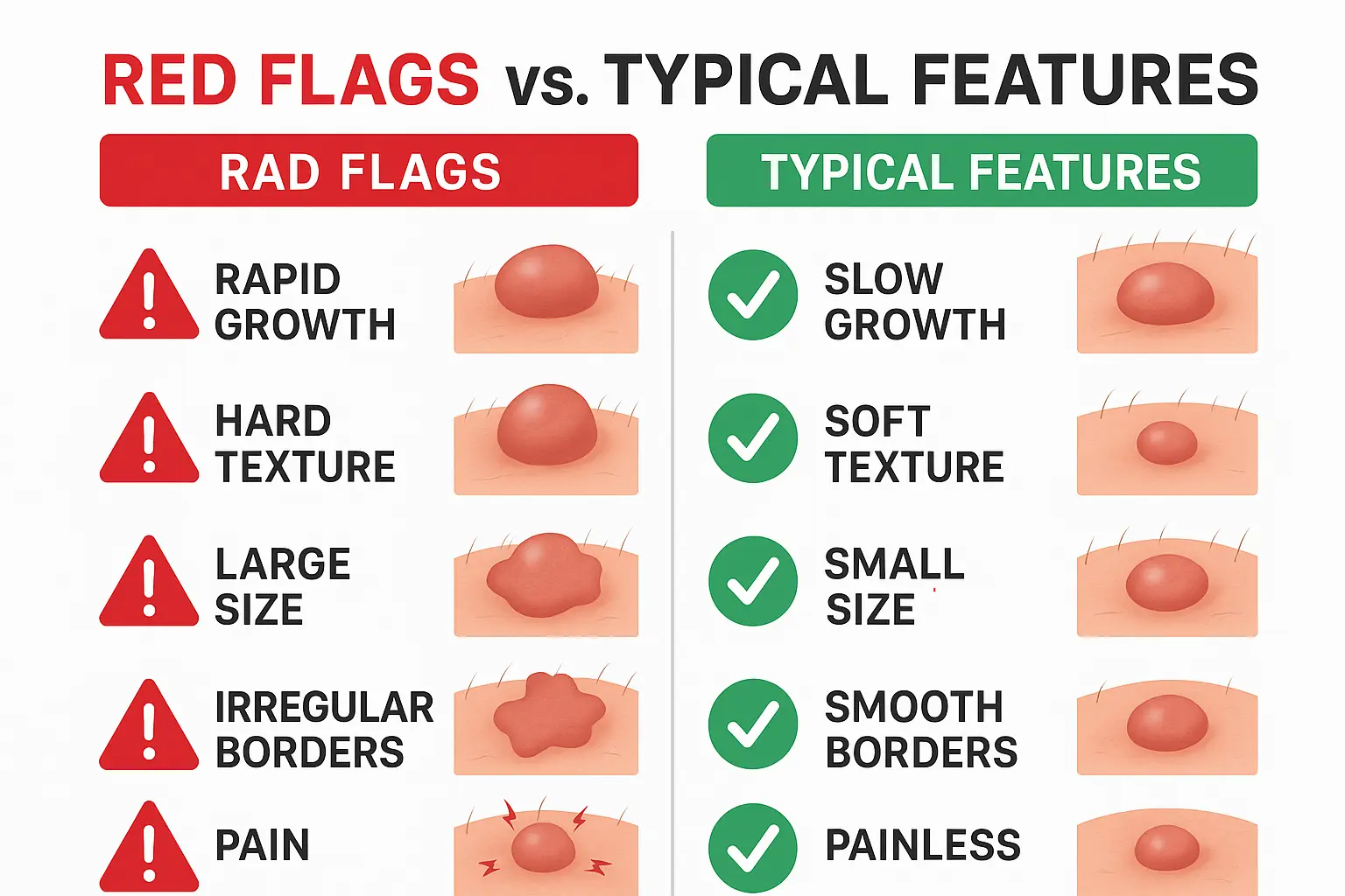
The most important takeaway is that most lumps under the skin are benign, and many don't require any imaging at all. When imaging is needed, starting with ultrasound makes sense both medically and financially for typical cases. Red flags like rapid growth, hard texture, large size, or associated symptoms warrant more urgent evaluation and potentially advanced imaging.
Your next steps should include:
Remember that good healthcare involves partnership between you and your medical team. By understanding the reasoning behind imaging recommendations, you can participate more effectively in decisions about your care while ensuring you receive appropriate evaluation for your specific situation.
For additional information about lumps, imaging, and treatment options, explore our comprehensive FAQ section or contact our team for personalized guidance.


