When Sarah noticed a small, scaly patch on her forearm that wouldn't heal after months of home remedies, she never imagined it could be skin cancer. Like many of us, she thought skin cancer would look obviously dangerous – perhaps a large, dark mole or an angry red growth. What she discovered changed her perspective entirely: squamous cell carcinoma can appear deceptively innocent in its early stages, making visual recognition absolutely crucial for early detection and successful treatment.
Understanding squamous cell carcinoma stages pictures isn't just about medical curiosity – it's about potentially saving lives. As the second most common form of skin cancer, affecting over 1 million Americans annually, squamous cell carcinoma (SCC) presents a unique challenge: it can look remarkably different depending on its stage of development. From barely noticeable rough patches to aggressive, ulcerated lesions, this cancer's appearance evolves dramatically as it progresses.
In this comprehensive guide, I'll walk you through the visual journey of squamous cell carcinoma stages, helping you understand what to look for, when to seek medical attention, and how early recognition can make all the difference in treatment outcomes. Whether you're concerned about a suspicious spot on your skin or simply want to be better informed about skin health, this visual exploration will equip you with the knowledge you need.
• Early-stage squamous cell carcinoma often appears as harmless rough patches, scaly spots, or persistent sores that don't heal within 2-3 weeks
• Advanced stages show dramatic changes including raised, warty growths, open ulcers, and potential bleeding or crusting
• Visual recognition is critical since SCC can develop anywhere on the body, not just sun-exposed areas
• Staging determines treatment – early detection typically requires simple surgical removal, while advanced cases may need extensive therapy
• Professional evaluation is essential for any suspicious skin changes, as self-diagnosis through pictures alone can be misleading
Before diving into the visual stages, let's establish a clear understanding of what we're dealing with. Squamous cell carcinoma develops in the squamous cells – thin, flat cells that make up the outermost layer of your skin. Think of these cells as the protective shingles on your body's roof, constantly renewing themselves to shield you from environmental damage.
When DNA damage accumulates in these cells (often from UV radiation, but also from other factors), they can begin growing uncontrollably. Unlike basal cell carcinoma, which rarely spreads, squamous cell carcinoma has the potential to metastasize if left untreated, making early detection absolutely critical.
Understanding who's at higher risk helps explain why visual monitoring becomes so important:

Medical professionals use specific staging systems to classify squamous cell carcinoma, and understanding this framework helps make sense of the visual changes we'll explore. The most commonly used system is the TNM staging system:
T (Tumor size and characteristics):
N (Lymph Node involvement):
M (Metastasis):
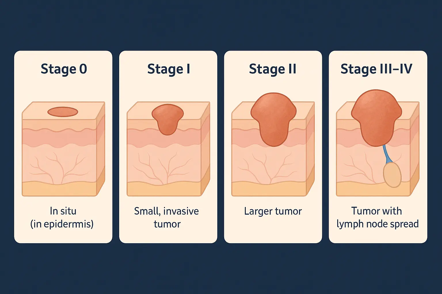
For practical understanding, these translate into stages that correlate with visual appearance:
StageDescriptionVisual CharacteristicsStage 0In situFlat, scaly patchesStage IEarly invasiveSmall raised lesionsStage IILocally advancedLarger, more obvious growthsStage IIIRegional spreadUlcerated lesions, possible lymph node involvementStage IVDistant metastasisExtensive local disease or distant spread
Stage 0 squamous cell carcinoma, also known as carcinoma in situ or Bowen's disease, represents the earliest detectable form of this cancer. At this stage, abnormal cells are confined entirely to the outermost layer of skin (epidermis) and haven't invaded deeper tissues.
The appearance of stage 0 SCC often leads to delayed diagnosis because it looks so benign:
Typical Appearance:
Common Locations:
I remember consulting with Maria, a 58-year-old gardener who had what she called her "stubborn patch" on her temple. For eight months, she treated it with over-the-counter creams, assuming it was dry skin from sun exposure. The patch would seem to improve with moisturizer, then return looking slightly different.
What made her finally seek medical attention wasn't dramatic change – it was the persistence. Stage 0 squamous cell carcinoma often presents this way: a seemingly innocent skin issue that just won't resolve completely.
When examining squamous cell carcinoma stages pictures of Stage 0, look for:
✅ Asymmetry – irregular shape or borders ✅ Persistence – doesn't heal or keeps returning ✅ Texture changes – rough, scaly, or thickened areas ✅ Color variation – patches of different colors within the lesion ✅ Slow growth – gradual increase in size over months
Stage I squamous cell carcinoma marks the transition from surface-only disease to actual invasion into deeper skin layers. This stage represents a critical window where treatment remains highly effective, but the visual changes become more apparent to both patients and healthcare providers.
At this stage, the cancer has broken through the basement membrane (the boundary between the epidermis and dermis) but remains relatively small and localized.
Medical Criteria:
The appearance becomes more obviously abnormal, which often prompts medical consultation:
Physical Characteristics:
Color Changes:
One of the most important things I've learned from reviewing squamous cell carcinoma stages pictures with patients is addressing the "mole myth." Unlike melanoma, which often develops from existing moles, SCC typically appears as entirely new growths or develops from precancerous lesions called actinic keratoses.
James, a 45-year-old construction worker, delayed seeking treatment because he was looking for changes in his existing moles. The raised, scaly bump on his nose seemed unrelated to skin cancer in his mind. This highlights why understanding the diverse appearances of SCC stages is so crucial.
Even in Stage I, certain characteristics indicate higher risk for progression:
🚨 Depth of invasion >2mm 🚨 Location on lips, ears, or genitals 🚨 Poorly defined borders 🚨 Rapid growth over weeks or months 🚨 Arising from chronic wounds or scars
Stage II squamous cell carcinoma represents a significant escalation in both appearance and medical urgency. At this stage, the cancer has either grown larger than 2 cm or displays multiple high-risk features that increase the likelihood of spread.
Stage II SCC meets one of these criteria:
The appearance of Stage II lesions often alarms patients, finally prompting urgent medical attention:
Prominent Features:
Size and Shape:
Dr. Patricia Williams, a dermatologist I frequently collaborate with, describes Stage II as the "suddenly worse" stage. Patients often report that a lesion they'd been watching for months suddenly changed dramatically over just a few weeks.
This was exactly what happened to Robert, a 62-year-old retiree. His small, scaly spot on his ear had been stable for over a year. Then, within six weeks, it doubled in size, began bleeding, and developed the characteristic "rolled border" appearance typical of Stage II SCC.
Squamous cell carcinoma stages pictures reveal that Stage II lesions can look quite different depending on their location:
Facial SCC:
Hand and Arm SCC:
Lip SCC:
Stage III squamous cell carcinoma represents advanced local disease with potential regional lymph node involvement. This stage requires immediate, aggressive treatment and carries significantly higher risks for complications and spread.
Stage III is defined by:
The appearance of Stage III lesions is typically unmistakable as serious disease:
Alarming Characteristics:
Associated Changes:
When reviewing squamous cell carcinoma stages pictures of Stage III disease, it's important to understand the human impact. These aren't just medical images – they represent people facing serious health challenges.
I worked with Margaret, a 71-year-old grandmother whose Stage III SCC on her scalp had grown to nearly 4 cm in diameter. The tumor had invaded through her skull bone, requiring extensive surgery involving both dermatologic and neurosurgical teams. Her case illustrates why early detection and treatment are so crucial.
Stage III SCC requires multidisciplinary care:
Surgical Options:
Additional Treatments:
Stage IV squamous cell carcinoma represents the most advanced form of this cancer, characterized by distant metastasis or extensive local invasion that makes cure difficult or impossible. While this stage is relatively uncommon for SCC (occurring in less than 5% of cases), understanding its presentation is crucial for comprehensive cancer awareness.
Stage IV SCC involves:
At this stage, the focus shifts from local appearance to systemic disease:
Primary Site Changes:
Systemic Signs:
While squamous cell carcinoma stages pictures of Stage IV disease can be disturbing, they serve an important educational purpose. These images remind us why prevention and early detection matter so much.
The transformation from a small, scaly patch (Stage 0) to life-threatening metastatic disease (Stage IV) typically occurs over months to years, providing multiple opportunities for intervention. This progression underscores why any persistent skin changes deserve professional evaluation.
Understanding how to interpret squamous cell carcinoma stages pictures requires knowing what visual clues to prioritize. Having reviewed thousands of clinical photographs over my career, I've learned that certain features consistently indicate the need for immediate medical attention.
While originally developed for melanoma, a modified ABCDE approach works well for SCC recognition:
A - Asymmetry and Area
B - Borders
C - Color
D - Diameter and Development
E - Evolution and Elevation
Dermatologists often use the "ugly duckling" principle when evaluating suspicious lesions. This means looking for spots that appear different from a person's other moles or skin markings.
When examining squamous cell carcinoma stages pictures, this principle becomes especially relevant because SCC often looks distinctly different from benign skin conditions:
SCC "Ugly Duckling" Features:
In 2025, several technological advances help with visual recognition:
Smartphone Apps:
Professional Tools:
Important Limitation: While technology assists in recognition, no app or photograph can replace professional medical evaluation. Squamous cell carcinoma stages pictures serve as educational tools, not diagnostic instruments.
Understanding who develops squamous cell carcinoma and why helps explain the importance of visual monitoring and early detection. The relationship between risk factors and cancer appearance provides crucial context for interpreting squamous cell carcinoma stages pictures.
Sun Exposure History:
Individual Characteristics:
Medical Factors:
Chemical Exposures:
Radiation History:
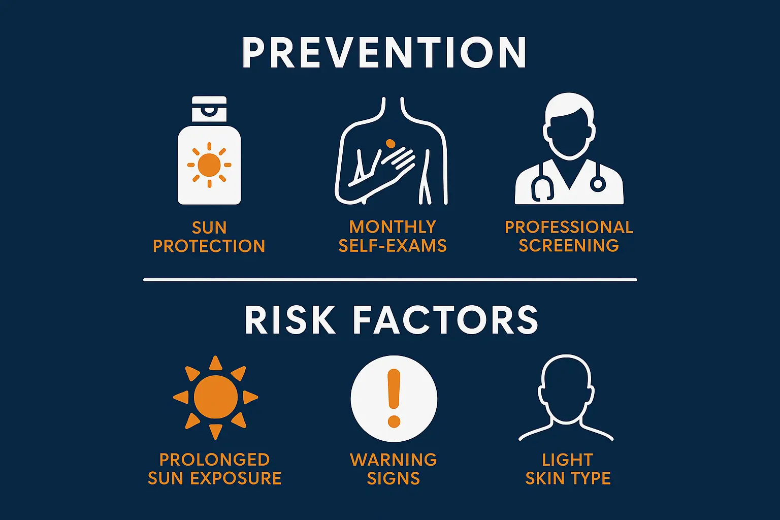
The most effective approach to dealing with SCC is preventing it entirely:
Sun Protection Strategies:
Regular Monitoring:
When I review squamous cell carcinoma stages pictures with patients, I always emphasize this crucial point: the difference between Stage 0 and Stage III isn't just medical staging – it's often the difference between a simple 15-minute office procedure and major surgery requiring reconstruction.
Consider these statistics:
Knowing when to move from self-monitoring to professional evaluation can literally save your life. Based on my experience reviewing squamous cell carcinoma stages pictures and working with patients, certain signs demand immediate medical attention.
Seek immediate evaluation for:
I recommend the two-week rule to my patients: if any skin change doesn't show improvement within two weeks of basic care (gentle cleansing, moisturizing, avoiding irritation), it deserves professional evaluation.
This rule helped save David, a 39-year-old teacher who noticed a small, scaly spot on his cheek. Initially dismissing it as dry skin from winter weather, he became concerned when two weeks of careful moisturizing showed no improvement. His dermatologist diagnosed Stage I SCC, which was successfully treated with a simple excision.
What to Bring:
What to Expect:
If a biopsy is performed, understanding the results helps you make informed treatment decisions:
Benign Results:
Malignant Results:
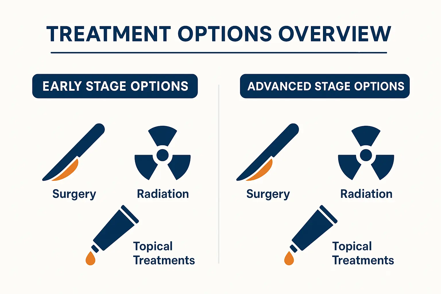
Treatment for squamous cell carcinoma varies dramatically based on the stage at diagnosis. Understanding these options helps explain why early detection – guided by recognition of squamous cell carcinoma stages pictures – makes such a significant difference in outcomes.
Topical Treatments:
Procedural Options:
Surgical Options:
Primary Treatment:
Success Rates:
95% cure rate with appropriate treatment
Surgical Management:
Adjuvant Therapy:
Stage III-IV Options:
Advanced SCC often requires reconstructive surgery:
Reconstruction Options:
A diagnosis of squamous cell carcinoma, regardless of stage, means lifelong vigilance and regular monitoring. Understanding what this means helps patients prepare for the journey ahead.
Post-Treatment Monitoring:
What Happens During Follow-up:
Monthly Self-Examinations:
Warning Signs to Report:
A cancer diagnosis affects more than just physical health:
Common Concerns:
Support Resources:
Having one SCC significantly increases the risk of developing others:
Enhanced Prevention:
In 2025, photography plays an increasingly important role in skin cancer detection and management. Understanding how to use squamous cell carcinoma stages pictures effectively can significantly improve outcomes.
Smartphone Photography Tips:
Organization Systems:
Clinical Documentation:
Telemedicine Applications:
Important Limitations:
The field of skin cancer detection and treatment continues to evolve rapidly. Understanding emerging trends helps patients and healthcare providers prepare for future advances.
Current Applications:
Future Developments:
Emerging Techniques:
Genetic Testing:
Targeted Therapies:
Understanding squamous cell carcinoma stages pictures represents far more than academic knowledge – it's about empowerment, early detection, and potentially life-saving awareness. Throughout this comprehensive exploration, we've journeyed from the deceptively innocent appearance of Stage 0 disease to the serious implications of advanced-stage SCC.
The visual progression of squamous cell carcinoma tells a story of opportunity. Each stage represents a window for intervention, with earlier detection consistently leading to better outcomes, simpler treatments, and higher cure rates. The difference between recognizing a Stage 0 lesion versus waiting until Stage III isn't just medical staging – it's often the difference between a simple office procedure and major surgery requiring reconstruction.
Key messages to remember:
🎯 Trust your instincts – if something looks different or concerning, seek professional evaluation 🎯 Document changes through photography and regular self-examinations 🎯 Understand your risk factors and adjust monitoring accordingly 🎯 Maintain sun protection as your primary defense against SCC 🎯 Follow through with recommended follow-up care after any skin cancer diagnosis
The stories I've shared – from Sarah's persistent patch to Margaret's advanced disease – illustrate real people facing real challenges. Their experiences remind us that behind every medical photograph is a human being whose life can be significantly impacted by early recognition and appropriate treatment.
As we move forward in 2025 and beyond, technological advances will continue improving our ability to detect and treat squamous cell carcinoma. However, the fundamental principle remains unchanged: your awareness and vigilance represent the first and most important line of defense.
If you're reading this article because of a concerning spot on your skin, don't wait. Schedule an appointment with a dermatologist or your primary care physician. If you're reading for general education, commit to regular self-examinations and annual professional skin checks if you're at higher risk.
Remember, squamous cell carcinoma stages pictures serve as educational tools to guide recognition, but they cannot replace professional medical evaluation. When in doubt, seek professional assessment. Early detection saves lives, preserves function, and maintains quality of life.
Your skin health journey starts with awareness, continues with vigilance, and succeeds through partnership with qualified healthcare providers. The knowledge you've gained here empowers you to be an active participant in protecting your health and potentially helping others recognize concerning changes in their skin.


