Squamous cell carcinoma stands as the second most common form of skin cancer, affecting millions of people worldwide each year. Understanding what this dangerous condition looks like through squamous cell carcinoma photos can literally save lives by enabling early detection and prompt treatment. For healthcare professionals, patients, and their families, visual recognition serves as the first line of defense against this potentially aggressive cancer.
The power of squamous cell carcinoma photos lies in their ability to educate and inform. These visual resources help individuals identify suspicious skin changes that might otherwise go unnoticed until the cancer progresses to more advanced stages. Early detection significantly improves treatment outcomes and reduces the risk of metastasis, making visual education through photographs an invaluable tool in the fight against skin cancer.
This comprehensive guide explores the critical role of squamous cell carcinoma photos in medical education, patient awareness, and clinical practice. From understanding the various presentations of this cancer to learning how photographs aid in diagnosis and treatment planning, readers will gain essential knowledge about this serious condition and the importance of visual documentation in modern dermatology.
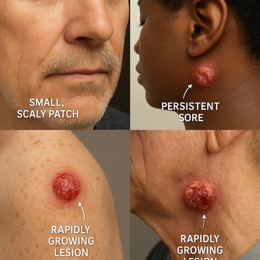
• Early Recognition Saves Lives: Squamous cell carcinoma photos help identify warning signs like persistent sores, scaly patches, and rapidly growing lesions before they become life-threatening
• Visual Diversity Matters: This cancer appears differently across skin types, body locations, and stages, making diverse photographic documentation essential for accurate recognition
• Professional Diagnosis Required: While photos aid in initial identification, only qualified medical professionals can provide definitive diagnosis and treatment recommendations
• Documentation Supports Treatment: Clinical photography plays a crucial role in tracking cancer progression, treatment response, and long-term monitoring
• Prevention Through Education: Understanding what squamous cell carcinoma looks like through photos empowers individuals to seek timely medical attention and practice better sun protection
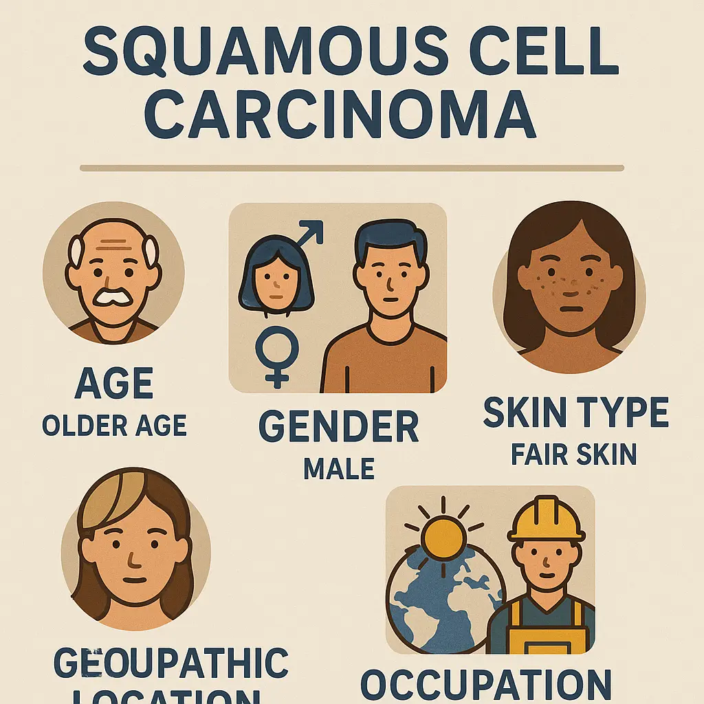
Squamous cell carcinoma develops in the squamous cells that make up the middle and outer layers of the skin. This type of cancer typically arises from prolonged exposure to ultraviolet radiation from the sun or tanning beds, though other factors can contribute to its development. Unlike some skin cancers that remain localized, squamous cell carcinoma has the potential to spread to other parts of the body if left untreated.
The importance of squamous cell carcinoma photos in understanding this condition cannot be overstated. These visual resources demonstrate how the cancer manifests in real patients, showing the wide range of appearances it can take. From small, scaly patches to large, ulcerated tumors, the photographic documentation helps bridge the gap between textbook descriptions and real-world presentations.
Medical professionals rely heavily on squamous cell carcinoma photos for educational purposes, using them to train new practitioners and update existing knowledge. The visual nature of dermatology makes photography an indispensable tool for learning and reference. Professional medical centers like The Minor Surgery Center utilize comprehensive photographic documentation to enhance patient care and education.
Understanding who develops squamous cell carcinoma helps explain why certain populations need more frequent monitoring and why squamous cell carcinoma photos from diverse patient groups are essential:
Squamous cell carcinoma photos serve multiple essential functions in modern healthcare. They provide a visual baseline for comparison, help track changes over time, and facilitate communication between healthcare providers. The systematic documentation of skin lesions through photography has revolutionized how medical professionals approach skin cancer diagnosis and management.
The standardization of squamous cell carcinoma photos has improved diagnostic accuracy and treatment planning. When healthcare providers can compare current lesions to previous photographs, they can better assess growth patterns, treatment responses, and potential complications. This visual timeline becomes particularly valuable when multiple providers are involved in a patient's care.
"A picture is worth a thousand words, but in dermatology, a photograph can be worth a life." - Leading dermatopathologist
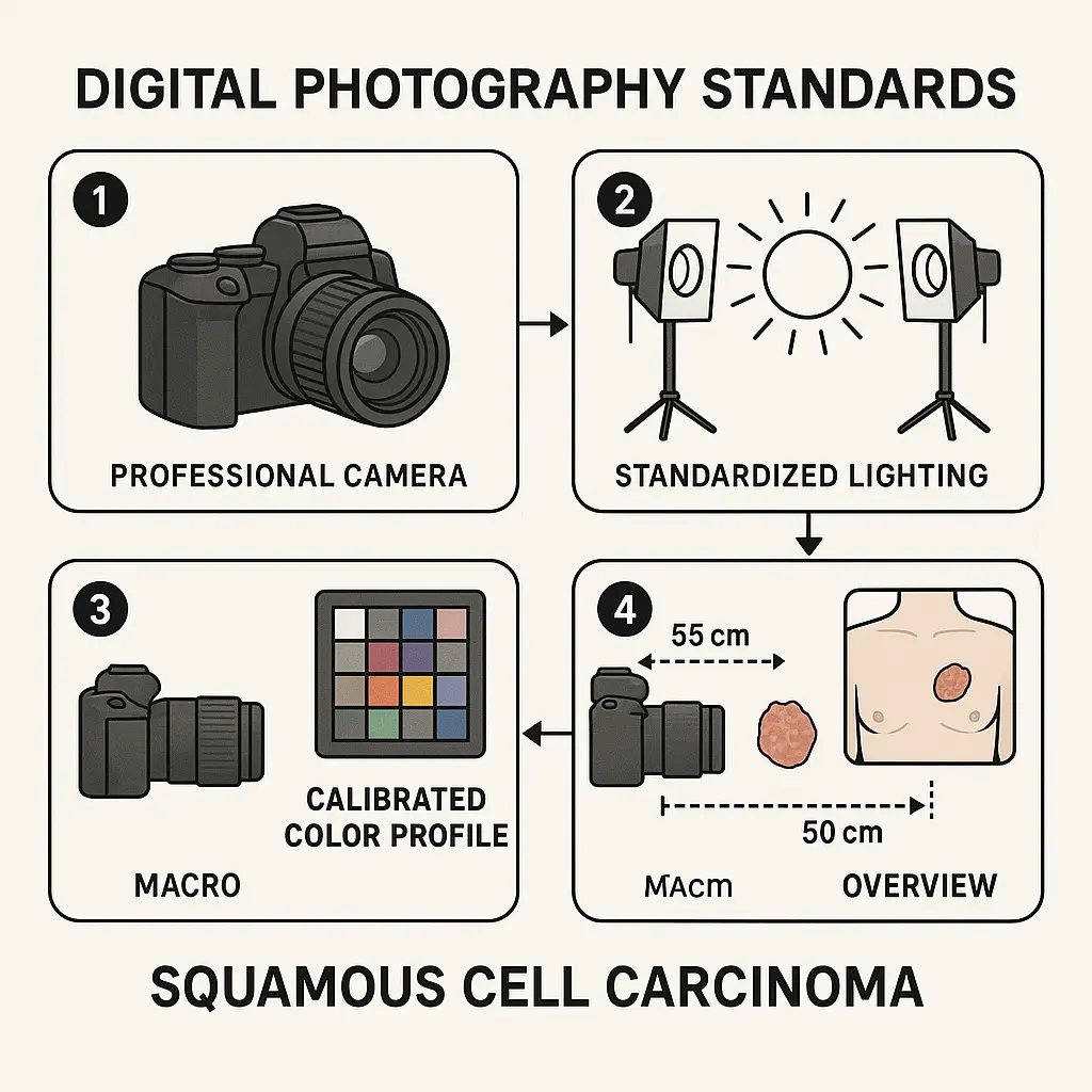
Modern medical facilities maintain strict protocols for capturing squamous cell carcinoma photos to ensure consistency and diagnostic value:
ParameterStandard RequirementResolutionMinimum 12 megapixelsLightingStandardized LED systemsDistanceConsistent macro and overview shotsColor AccuracyCalibrated color profilesDocumentationDetailed metadata and patient information
Recognizing the early signs of squamous cell carcinoma through photographic examples can significantly impact treatment outcomes. Squamous cell carcinoma photos typically show lesions that begin as small, red, scaly patches or persistent sores that refuse to heal. These early-stage photographs are particularly valuable because they demonstrate how subtle the initial signs can be.
Key visual characteristics commonly seen in squamous cell carcinoma photos include:
• Persistent scaling that doesn't respond to moisturizers
• Rough, crusty surfaces that may bleed when touched
• Raised borders around flat, reddish patches
• Rapid growth documented in sequential photos
• Ulceration or open sores that won't heal
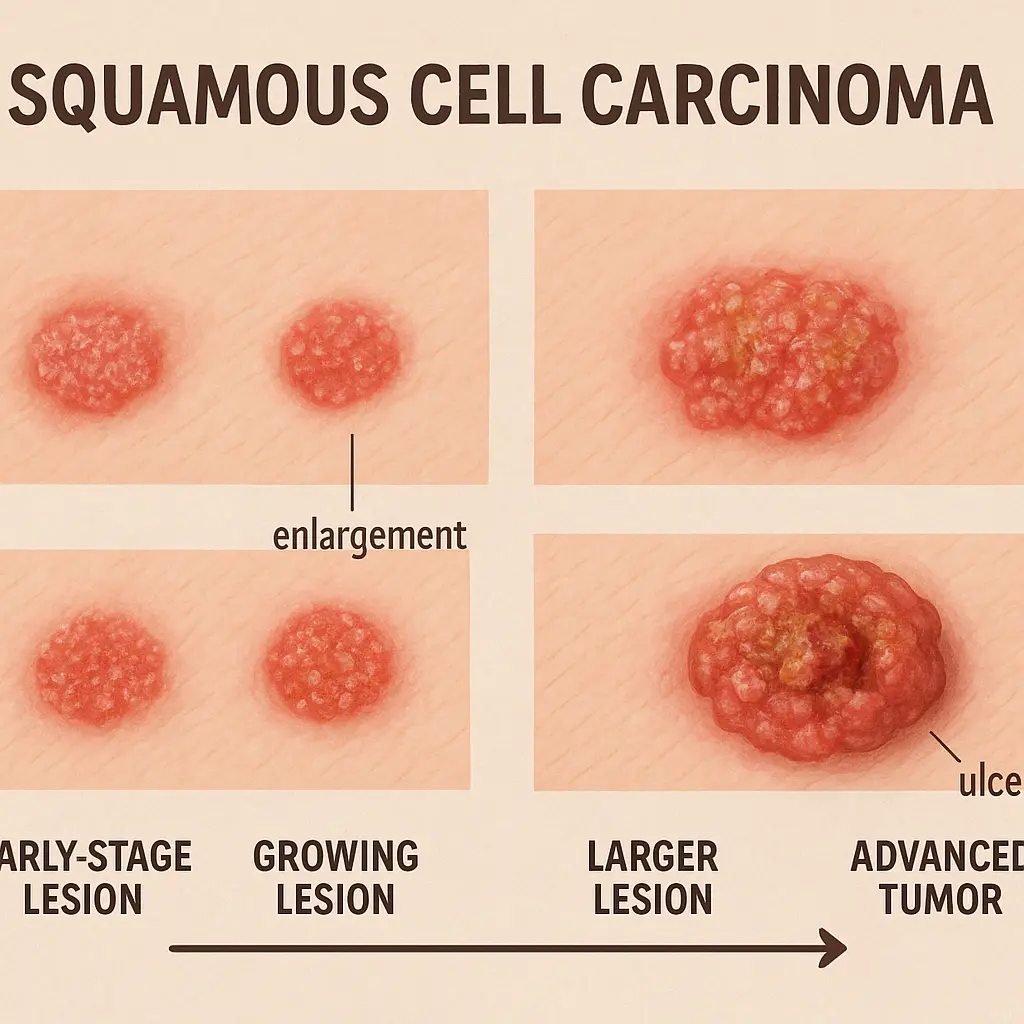
The progression of squamous cell carcinoma can be dramatic, and squamous cell carcinoma photos documenting this progression serve as powerful educational tools. Early-stage lesions may appear as minor skin irritations, but advanced cases can present as large, destructive tumors. This visual progression emphasizes the critical importance of early detection and intervention.
Healthcare providers often use squamous cell carcinoma photos to educate patients about self-examination techniques. By showing patients what to look for, these images empower individuals to become active participants in their healthcare. Regular self-examination, guided by knowledge gained from studying squamous cell carcinoma photos, can lead to earlier detection and better outcomes.
Squamous cell carcinoma photos reveal that this cancer can develop anywhere on the body, but certain locations are more frequently affected. Sun-exposed areas like the face, ears, neck, hands, and arms feature prominently in squamous cell carcinoma photos. However, the cancer can also develop in areas not typically exposed to sunlight, including the genital region and inside the mouth.
The appearance of squamous cell carcinoma varies significantly depending on its location. Squamous cell carcinoma photos of facial lesions often show well-defined, scaly patches that may be mistaken for age spots or seborrheic keratoses. In contrast, lesions on the hands or arms might appear more nodular or ulcerated in photographic documentation.
Different body areas present unique challenges for both photography and diagnosis:
Head and Neck Region 🔍
Extremities
Professional medical facilities, such as those detailed at The Minor Surgery Center conditions page, specialize in comprehensive evaluation and documentation of suspicious lesions across all body regions.
Medical photography for squamous cell carcinoma follows strict protocols to ensure diagnostic accuracy and legal compliance. Squamous cell carcinoma photos used in clinical practice must meet specific technical standards for lighting, resolution, and color accuracy. These requirements ensure that subtle details crucial for diagnosis are clearly visible and accurately represented.
Professional squamous cell carcinoma photos typically include both overview shots showing the lesion in context and close-up images revealing fine details. This dual approach provides comprehensive documentation that supports accurate diagnosis and treatment planning. The standardized approach to medical photography ensures consistency across different healthcare providers and facilities.
The capture of diagnostic-quality squamous cell carcinoma photos requires specialized equipment and training:
Quality assurance protocols ensure that squamous cell carcinoma photos meet the highest standards for clinical use. Regular equipment calibration, staff training updates, and image quality reviews maintain the integrity of the photographic documentation process.
Squamous cell carcinoma photos play a crucial role in cancer staging, which determines the extent of disease spread and guides treatment decisions. Sequential photography allows healthcare providers to track tumor growth, assess treatment response, and monitor for recurrence. This visual timeline becomes an essential component of the patient's medical record.
The staging process relies heavily on visual assessment, making squamous cell carcinoma photos invaluable for accurate classification. Early-stage tumors confined to the skin surface appear very different from advanced cancers that have invaded deeper tissues or spread to lymph nodes. Photographic documentation captures these differences with precision that written descriptions alone cannot achieve.
Staging Categories Visible in Photography:
• Stage 0 (In Situ): Abnormal cells confined to the epidermis
• Stage I: Small tumor confined to the skin
• Stage II: Larger tumor with high-risk features
• Stage III: Regional lymph node involvement
• Stage IV: Distant metastasis
Advanced squamous cell carcinoma photos often show dramatic changes in tissue architecture, with ulceration, bleeding, and destruction of normal skin structures. These images serve as powerful reminders of the importance of early detection and prompt treatment.
Squamous cell carcinoma photos serve as essential tools for documenting treatment progress and outcomes. Pre-treatment photographs establish baselines for comparison, while post-treatment images document healing progress and help identify potential complications. This visual record supports both immediate patient care and long-term follow-up monitoring.
Surgical treatment documentation through squamous cell carcinoma photos helps surgeons plan procedures and communicate with patients about expected outcomes. Before-and-after photographs demonstrate the effectiveness of various treatment approaches and help patients understand what to expect during recovery.
The documentation process extends beyond the immediate treatment period. Long-term follow-up squamous cell carcinoma photos help detect recurrence and monitor for new lesions in high-risk patients. This ongoing visual surveillance is particularly important for patients with multiple risk factors or previous skin cancers.
Different treatment approaches produce distinct visual results that are well-documented in medical photography:
Treatment TypeVisual CharacteristicsHealing TimelineSurgical ExcisionLinear scar formation2-4 weeks primary healingMohs SurgeryMinimal tissue removal1-3 weeks with optimal cosmesisRadiation TherapyGradual tissue changesSeveral monthsTopical TherapyInflammatory response4-8 weeks
Specialized medical centers, like those described at The Minor Surgery Center clinic, maintain comprehensive photographic records to track treatment outcomes and refine their approaches based on visual evidence.
Squamous cell carcinoma photos serve as powerful educational tools that help patients understand their condition and treatment options. Visual learning is particularly effective for medical conditions, as photographs can convey information that words alone cannot express. Patient education materials incorporating squamous cell carcinoma photos improve comprehension and compliance with treatment recommendations.
Educational squamous cell carcinoma photos must be carefully selected to inform without causing unnecessary alarm. Healthcare providers use these images to explain the importance of sun protection, regular skin examinations, and prompt medical attention for suspicious lesions. The visual impact of these photographs often motivates patients to take preventive measures more seriously.
Effective Patient Education Elements:
• Before-and-after treatment photos showing positive outcomes
• Comparison images distinguishing cancer from benign conditions
• Prevention examples demonstrating sun damage progression
• Self-examination guides with photographic references
• Risk factor illustrations showing high-risk lesion characteristics
The emotional impact of squamous cell carcinoma photos can be significant, requiring healthcare providers to present them with sensitivity and appropriate context. Professional counseling and support resources help patients process the information while maintaining hope for positive outcomes.
The use of squamous cell carcinoma photos in healthcare raises important ethical considerations regarding patient privacy, consent, and dignity. Medical facilities must maintain strict protocols for obtaining, storing, and using patient photographs to protect individual rights while advancing medical knowledge and education.
Informed consent for medical photography goes beyond simple permission to take pictures. Patients must understand how their squamous cell carcinoma photos will be used, who will have access to them, and what rights they retain regarding their images. This comprehensive consent process ensures ethical compliance and protects both patients and healthcare providers.
Modern healthcare facilities implement multiple layers of protection for squamous cell carcinoma photos:
Professional medical teams, such as those at The Minor Surgery Center team, receive ongoing training in ethical photography practices and patient privacy protection.
Advances in digital technology have revolutionized the capture and analysis of squamous cell carcinoma photos. High-resolution cameras, specialized lighting systems, and digital image processing tools enable healthcare providers to document skin lesions with unprecedented detail and accuracy. These technological improvements directly translate to better diagnostic capabilities and patient outcomes.
Artificial intelligence and machine learning algorithms are increasingly being applied to squamous cell carcinoma photos for automated screening and diagnosis support. While these tools cannot replace professional medical judgment, they can assist in identifying suspicious lesions and prioritizing cases for urgent evaluation.
Emerging Technologies in Medical Photography:
🔬 Dermoscopy Integration: Specialized cameras providing magnified, illuminated views 📱 Mobile Applications: Smartphone-based tools for preliminary screening 🤖 AI Analysis: Computer algorithms identifying suspicious features ☁️ Cloud Storage: Secure, accessible image repositories 📊 3D Imaging: Three-dimensional lesion documentation and analysis
The integration of these technologies with traditional squamous cell carcinoma photos creates comprehensive diagnostic tools that enhance clinical decision-making and improve patient care quality.
Squamous cell carcinoma photos contribute significantly to medical research and scientific advancement. Large databases of anonymized patient photographs enable researchers to study disease patterns, treatment outcomes, and risk factors across diverse populations. This research directly benefits future patients through improved diagnostic techniques and treatment protocols.
Clinical trials often rely on standardized squamous cell carcinoma photos to assess treatment effectiveness and compare different therapeutic approaches. The visual documentation provides objective measures of treatment response that complement other clinical assessments. This photographic evidence supports regulatory approvals for new treatments and influences clinical practice guidelines.
Medical photography databases containing squamous cell carcinoma photos support various research initiatives:
The cumulative knowledge gained from these research applications of squamous cell carcinoma photos continues to advance the field of dermatology and improve patient outcomes worldwide.
International variations in squamous cell carcinoma presentation and prevalence are well-documented through global photographic databases. Squamous cell carcinoma photos from different geographic regions reveal how factors like sun exposure, genetics, and healthcare access influence disease patterns. This global perspective informs prevention strategies and treatment approaches worldwide.
Cultural considerations also influence how squamous cell carcinoma photos are obtained and used in different regions. Healthcare providers must be sensitive to cultural norms regarding body exposure and medical photography while maintaining diagnostic accuracy and patient care standards.
Regional Variations in Presentation:
• Tropical regions: Higher incidence of aggressive subtypes
• High-altitude areas: Increased UV exposure effects
• Northern latitudes: Different seasonal exposure patterns
• Developing nations: Later-stage presentations due to limited access
• Urban vs. rural: Varying occupational and lifestyle risk factors
Understanding these global patterns through squamous cell carcinoma photos helps healthcare providers deliver culturally appropriate care while maintaining international standards for diagnosis and treatment.
Medical professionals require ongoing training in interpreting squamous cell carcinoma photos and understanding their clinical significance. Educational programs incorporate extensive photographic databases to teach recognition skills, diagnostic techniques, and treatment planning strategies. This visual education is essential for maintaining clinical competency in dermatology and related fields.
Continuing education programs often feature challenging cases documented through squamous cell carcinoma photos, allowing practitioners to test their diagnostic skills and learn from expert interpretations. These educational exercises improve diagnostic accuracy and help identify areas where additional training may be needed.
Training Program Components:
📚 Case-based learning using real patient photographs
🎯 Diagnostic challenges testing recognition skills
👥 Peer review sessions discussing difficult cases
🔄 Update seminars covering new diagnostic criteria
📈 Outcome studies reviewing treatment success rates
Professional development through photographic education ensures that healthcare providers maintain current knowledge and skills necessary for optimal patient care.
The future of squamous cell carcinoma photos lies in enhanced digital capabilities, artificial intelligence integration, and improved accessibility. Emerging technologies promise to make high-quality medical photography more widely available while maintaining the diagnostic accuracy essential for patient care.
Telemedicine applications increasingly rely on squamous cell carcinoma photos for remote consultations and screening programs. These applications require standardized photography protocols and secure transmission systems to ensure diagnostic quality and patient privacy protection.
Anticipated Developments:
🚀 Enhanced AI diagnostics providing real-time analysis
🌐 Global database integration enabling worldwide research collaboration
📱 Improved mobile technology democratizing access to quality photography
🔒 Advanced security measures protecting patient privacy
⚡ Faster processing capabilities enabling immediate clinical decisions
These technological advances will continue to improve the role of squamous cell carcinoma photos in healthcare delivery and patient outcomes.
Healthcare providers and patients commonly have questions about squamous cell carcinoma photos and their use in medical practice. Understanding these concerns helps ensure appropriate use of medical photography while maintaining patient comfort and trust.
For comprehensive answers to common questions about medical procedures and documentation, patients can refer to resources like The Minor Surgery Center FAQs which address many concerns about medical photography and patient privacy.
Common Patient Concerns:
❓ Privacy protection: How are photos stored and who can access them?
❓ Consent requirements: What rights do patients retain over their images?
❓ Usage limitations: How long are photos kept and for what purposes?
❓ Quality standards: What technical requirements ensure diagnostic accuracy?
❓ Cost implications: Are photography fees covered by insurance?
Addressing these concerns proactively helps maintain trust and ensures ethical compliance in medical photography practices.
Maintaining consistent quality in squamous cell carcinoma photos requires comprehensive quality assurance programs. These programs ensure that all photographic documentation meets clinical standards and provides reliable information for diagnosis and treatment planning.
Quality assurance protocols address technical aspects like lighting, resolution, and color accuracy, as well as procedural elements like patient positioning, consent documentation, and image storage. Regular audits and staff training maintain these standards over time.
Quality Metrics for Medical Photography:
These quality measures ensure that squamous cell carcinoma photos continue to serve their essential role in patient care while meeting professional and ethical standards.
Modern healthcare systems integrate squamous cell carcinoma photos directly into electronic health records, creating comprehensive patient documentation that supports continuity of care. This integration ensures that visual information is readily available to all authorized healthcare providers involved in patient care.
The technical challenges of integrating high-resolution medical photography with electronic health records include storage requirements, transmission speeds, and viewing capabilities. Healthcare systems must balance image quality with practical considerations of system performance and user accessibility.
The use of squamous cell carcinoma photos in healthcare is subject to various legal and regulatory requirements that protect patient rights and ensure appropriate medical practice. Healthcare providers must understand and comply with these requirements to avoid legal complications while providing optimal patient care.
Regulatory frameworks address issues like patient consent, image storage and transmission, access controls, and retention periods. Compliance with these regulations protects both patients and healthcare providers while enabling the beneficial uses of medical photography.
Key Regulatory Areas:
⚖️ HIPAA compliance protecting patient health information privacy ⚖️ State medical board regulations governing photography in clinical practice ⚖️ Institutional review board oversight for research applications ⚖️ International privacy laws affecting global data sharing ⚖️ Professional liability considerations related to diagnostic photography
Understanding and complying with these legal requirements ensures that squamous cell carcinoma photos can be used effectively while protecting all stakeholders' interests.
The implementation of comprehensive medical photography programs for documenting squamous cell carcinoma requires significant investment in equipment, training, and infrastructure. However, the long-term benefits often justify these costs through improved diagnostic accuracy, reduced liability, and enhanced patient outcomes.
Cost-effectiveness analyses of medical photography programs consider factors like equipment costs, staff training requirements, storage and maintenance expenses, and the value of improved clinical outcomes. These analyses help healthcare organizations make informed decisions about photography program implementation and expansion.
Economic Considerations:
💰 Initial equipment investment for cameras, lighting, and storage systems 💰 Ongoing maintenance costs including calibration and upgrades 💰 Staff training expenses ensuring competent operation 💰 Storage and security costs protecting patient information 💰 Liability reduction benefits through improved documentation
The economic benefits of squamous cell carcinoma photos often extend beyond direct medical care to include research contributions, educational value, and improved institutional reputation.
Squamous cell carcinoma photos represent far more than simple documentation; they serve as powerful tools for education, diagnosis, treatment planning, and research advancement. The comprehensive visual record provided by medical photography enhances every aspect of skin cancer care, from initial detection through long-term follow-up monitoring.
The evolution of medical photography technology continues to improve the quality and accessibility of squamous cell carcinoma photos, making these valuable resources available to more healthcare providers and patients worldwide. As artificial intelligence and digital innovations advance, the diagnostic and educational value of medical photography will only continue to grow.
Healthcare providers who understand the importance of high-quality squamous cell carcinoma photos and implement comprehensive photography programs position themselves to deliver superior patient care while contributing to the advancement of medical knowledge. The investment in proper equipment, training, and protocols pays dividends through improved outcomes and enhanced professional capabilities.
For patients and their families, understanding the role of squamous cell carcinoma photos in medical care can reduce anxiety and increase cooperation with documentation procedures. When patients understand how these images contribute to their care, they become active participants in the diagnostic and treatment process.
The future of skin cancer care will undoubtedly rely even more heavily on visual documentation and analysis. Healthcare organizations that establish strong foundations in medical photography today will be well-positioned to adopt emerging technologies and continue providing excellent patient care as the field evolves.
Take Action Today 🎯
If you have concerns about suspicious skin lesions or need professional evaluation of skin changes, don't wait for symptoms to worsen. Contact qualified healthcare professionals who can provide comprehensive assessment and documentation. Visit The Minor Surgery Center to learn more about professional skin cancer evaluation and treatment options, or reach out through their contact page to schedule a consultation.
Remember that early detection through professional evaluation and proper documentation with squamous cell carcinoma photos can make the difference between simple treatment and complex, life-threatening complications. Your health is worth the investment in professional care and comprehensive documentation that supports optimal outcomes.


