Understanding Skin Mole Types: A Complete Guide to Identifying and Managing Common and Concerning Moles

Picture this: you're getting ready for the day when you notice a small, dark spot on your shoulder that you've never seen before. Your mind immediately races with questions—is it dangerous? Should you be worried? Understanding skin mole types can help you distinguish between harmless beauty marks and spots that require medical attention. With millions of people developing new moles throughout their lives, knowing what to look for could literally save your life.

Key Takeaways

Common moles are typically harmless, symmetrical, and uniform in color, while atypical moles may show irregular features that warrant monitoring

A hyper-realistic image of a A hyper-realistic image of var...

• The ABCDE criteria (Asymmetry, Border, Color, Diameter, Evolving) helps identify potentially concerning mole changes

Congenital moles present at birth carry different risk factors than acquired moles that develop over time

Regular skin examinations by dermatologists are crucial for early detection of skin cancer, especially for high-risk individuals

Most moles are benign, but any changing or new moles after age 30 should be evaluated by a medical professional

What Are Moles and Why Do They Form?

Moles, medically known as nevi, are clusters of pigmented cells called melanocytes that appear as spots on the skin. These common skin growths develop when melanocytes grow in clusters instead of spreading evenly throughout the skin. Understanding the different skin mole types begins with recognizing that moles are incredibly common—most adults have between 10 to 40 moles on their body.

The formation of moles involves complex interactions between genetics, sun exposure, and hormonal changes. Melanocytes produce melanin, the pigment responsible for skin color and protection against UV radiation. When these cells cluster together, they create the various mole types we see on our skin.

Factors That Influence Mole Development

Several factors contribute to mole formation and the specific skin mole types that develop:

  • Genetics: Family history plays a significant role in mole development patterns
  • Sun exposure: UV radiation stimulates melanocyte activity and mole formation
  • Age: Most moles develop during childhood and adolescence
  • Hormones: Pregnancy and puberty can trigger new mole development
  • Skin type: Fair-skinned individuals often develop more moles

Common Benign Skin Mole Types

Most moles fall into the benign category, meaning they're harmless and pose no health risks. However, understanding the characteristics of different skin mole types helps distinguish normal variations from potentially concerning changes.

Junctional Moles

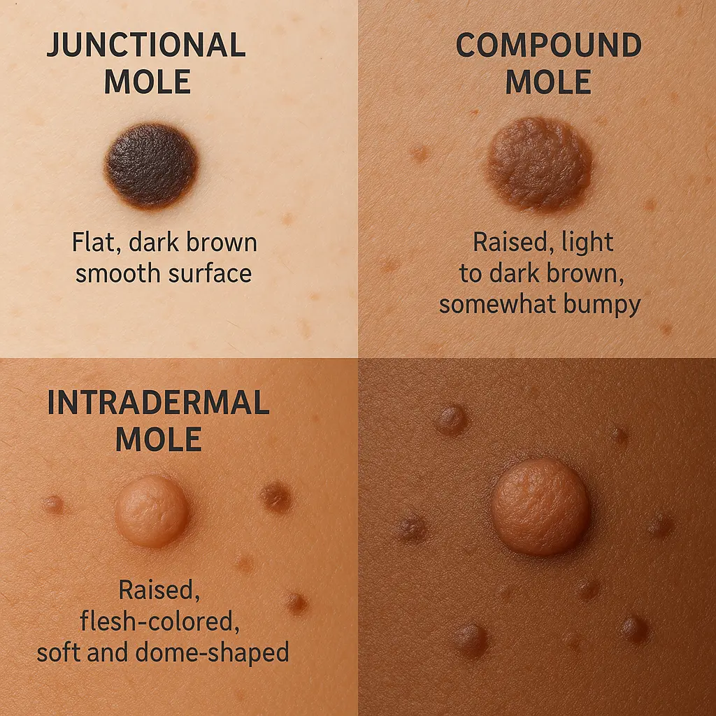
Junctional moles appear at the junction between the epidermis (outer skin layer) and dermis (deeper skin layer). These moles typically present as:

  • Flat or slightly raised surfaces
  • Dark brown or black coloration
  • Small size, usually less than 6mm in diameter
  • Smooth, well-defined borders
  • Common locations: face, trunk, and extremities

Junctional moles are among the most common skin mole types and rarely become problematic. They often develop during childhood and may fade or disappear with age.

Compound Moles

Compound moles contain melanocytes in both the epidermis and dermis, creating a distinctive appearance:

  • Raised or dome-shaped profile
  • Light to dark brown coloration
  • Smooth or slightly rough texture
  • Hair growth may occur from the mole
  • Stable appearance over time

These moles represent a natural progression from junctional moles as melanocytes migrate deeper into the skin. The presence of hair in a compound mole is actually a reassuring sign of benign behavior.

Intradermal Moles

Intradermal moles develop entirely within the dermis and represent the final stage of mole evolution:

  • Flesh-colored to light brown appearance
  • Raised, dome-shaped or pedunculated (on a stalk)
  • Soft, smooth texture
  • Hair growth commonly present
  • Typically develop in adulthood

These moles often lose their pigmentation over time and may become skin-colored. They're considered the most stable of all skin mole types and rarely undergo malignant transformation.

Atypical and Concerning Skin Mole Types

While most moles are harmless, certain skin mole types require closer monitoring due to their increased risk of developing into melanoma or other skin cancers.

Dysplastic Nevi (Atypical Moles)

Dysplastic nevi are unusual moles that share some characteristics with melanoma but aren't cancerous. Key features include:

  • Asymmetrical shape with irregular borders
  • Multiple colors within a single mole
  • Larger size, often exceeding 6mm in diameter
  • Flat areas mixed with raised portions
  • Rough or pebbly surface texture

🚨 Important: People with dysplastic nevi have a 4-12 times higher risk of developing melanoma compared to those without these atypical moles.

Congenital Melanocytic Nevi

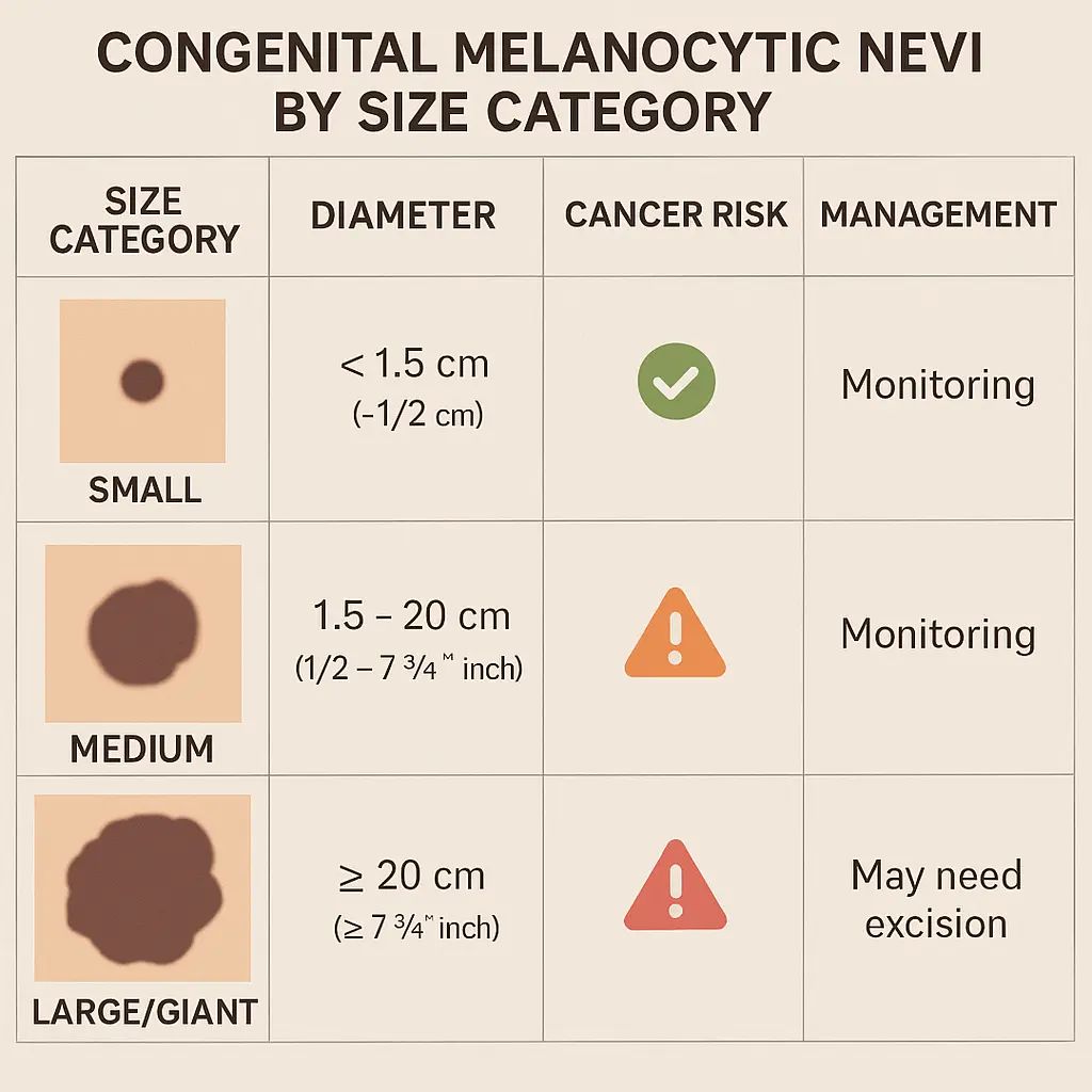
Congenital melanocytic nevi are moles present at birth, affecting approximately 1% of newborns. These skin mole types are classified by size:

Size CategoryDiameterCancer RiskManagementSmall<1.5 cmVery lowRoutine monitoringMedium1.5-19.9 cmLow to moderateRegular dermatology follow-upLarge/Giant≥20 cmModerate to highMultidisciplinary care

Large congenital nevi carry the highest risk of malignant transformation, with studies showing a 5-10% lifetime risk of developing melanoma.

Blue Nevi

Blue nevi are distinctive skin mole types characterized by their blue or blue-black coloration. They occur when melanocytes are trapped deep in the dermis, creating a blue appearance due to light scattering. Features include:

  • Blue, blue-black, or gray coloration
  • Small size, typically 2-10mm
  • Dome-shaped appearance
  • Stable over time
  • Common locations: scalp, face, hands, and feet

Most blue nevi are benign, but cellular blue nevi can occasionally undergo malignant transformation and require monitoring.

The ABCDE Method for Mole Evaluation

The ABCDE criteria provide a systematic approach to evaluating skin mole types and identifying potentially dangerous changes. This method helps both healthcare providers and patients recognize warning signs that warrant professional evaluation.

A hyper-realistic image of a An infographic illustrating th...

A - Asymmetry

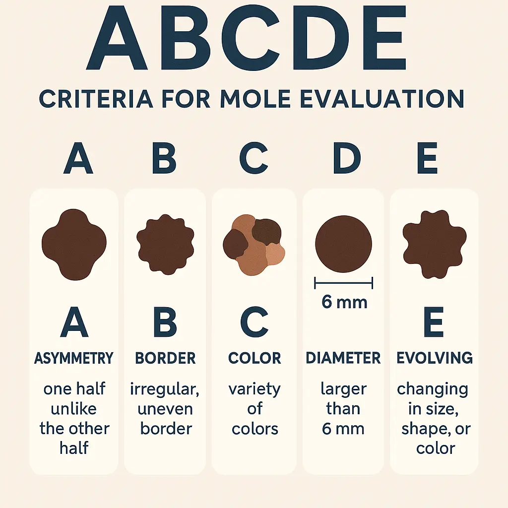
Asymmetrical moles have one half that doesn't match the other half. Benign moles are typically symmetrical, meaning if you draw a line through the middle, both sides should look similar.

  • Normal: Both halves mirror each other
  • Concerning: Irregular, lopsided appearance
  • Action: Compare mole halves regularly

B - Border Irregularity

Border characteristics provide important clues about mole behavior:

  • Normal borders: Smooth, well-defined edges
  • Irregular borders: Notched, scalloped, or poorly defined
  • Concerning features: Borders that seem to fade into surrounding skin

C - Color Variation

Color consistency is a key indicator of mole stability:

  • Normal coloration: Uniform brown, black, or flesh-colored
  • Concerning colors: Multiple colors within one mole
  • Warning signs: Red, white, blue, or very dark black areas

D - Diameter

Size considerations help identify problematic skin mole types:

  • Typical size: Most benign moles are smaller than 6mm (pencil eraser size)
  • Concerning size: Moles larger than 6mm diameter
  • Important note: Melanomas can be smaller than 6mm, so size alone isn't definitive

E - Evolving

Changes over time represent the most important warning sign:

  • Concerning changes: Size, shape, color, elevation, or texture modifications
  • New symptoms: Itching, bleeding, or tenderness
  • Timeline: Any change noticed over weeks to months

Special Considerations for Different Skin Mole Types

Understanding how various factors influence skin mole types helps create personalized monitoring strategies. Different populations and circumstances require tailored approaches to mole evaluation and management.

Moles in Children and Adolescents

Pediatric mole development follows predictable patterns that differ from adult skin mole types:

  • Birth to age 2: Congenital moles present; new moles rarely develop
  • Ages 2-10: Gradual mole development begins
  • Puberty: Rapid increase in mole number and size changes
  • Post-puberty: Mole development typically stabilizes

Key considerations for young patients:

  • Most childhood moles are benign
  • Melanoma is extremely rare before puberty
  • Family history of melanoma increases monitoring importance
  • Regular dermatological evaluations become essential for high-risk children

Pregnancy and Hormonal Changes

Hormonal fluctuations significantly impact skin mole types during pregnancy:

  • Existing moles may darken or enlarge
  • New moles can develop during pregnancy
  • Melasma (pregnancy mask) may be confused with moles
  • Postpartum changes usually return moles to pre-pregnancy appearance

Pregnant women should maintain regular skin monitoring while understanding that most changes are benign and hormone-related.

Sun-Damaged Skin and Mole Development

Chronic sun exposure influences both mole development and the appearance of other skin mole types:

  • Solar lentigines (age spots) may be confused with moles
  • Seborrheic keratoses can mimic concerning mole changes
  • Actinic keratoses require differentiation from atypical moles
  • Sun-damaged areas show increased mole density and variation

When to Seek Professional Evaluation

Knowing when to consult healthcare professionals about skin mole types can be lifesaving. While most moles are harmless, certain situations warrant immediate medical attention.

Urgent Warning Signs

Immediate medical evaluation is necessary for moles showing:

  • Rapid growth over days to weeks
  • Bleeding without trauma or injury
  • Ulceration or open sores that don't heal
  • Significant color changes, especially darkening or multiple colors
  • New symptoms like itching, pain, or tenderness

Routine Monitoring Schedule

Regular professional examinations should follow these guidelines:

Risk CategoryExamination FrequencySpecial ConsiderationsLow RiskAnnualRoutine skin checksModerate RiskEvery 6 monthsPersonal/family historyHigh RiskEvery 3-4 monthsMultiple risk factorsVery High RiskAs directedPrevious melanoma

Preparing for Your Appointment

Maximize your dermatology visit by:

  • Documenting changes with photographs and dates
  • Listing new moles or concerning symptoms
  • Bringing family history information
  • Removing nail polish for complete examination
  • Asking questions about your specific skin mole types

Professional evaluation at specialized clinics ensures comprehensive assessment using advanced diagnostic tools and expertise.

Mole Removal and Treatment Options

When skin mole types require intervention, several treatment options are available depending on the mole's characteristics, location, and underlying concerns.

Indications for Mole Removal

Medical reasons for mole removal include:

  • Suspicious features suggesting malignancy
  • Atypical characteristics requiring histological examination
  • Repeated irritation from clothing or jewelry
  • Bleeding or ulceration without clear cause
  • Patient anxiety about specific moles

Cosmetic considerations may also warrant removal:

  • Prominent location affecting appearance
  • Large size causing self-consciousness
  • Hair growth from moles in visible areas
  • Texture changes affecting comfort

Surgical Removal Techniques

Excisional biopsy represents the gold standard for suspicious skin mole types:

  • Complete removal with surrounding tissue margins
  • Histopathological examination of entire specimen
  • Definitive diagnosis with clear margins assessment
  • Minimal recurrence risk when properly performed

Shave excision may be appropriate for clearly benign moles:

  • Surface removal without deep tissue involvement
  • Minimal scarring for cosmetic procedures
  • Quick procedure with local anesthesia
  • Limited histological information available

Recovery and Follow-up Care

Post-procedure care ensures optimal healing:

  • Wound care instructions for incision sites
  • Activity restrictions during initial healing
  • Signs of infection to monitor
  • Suture removal timing when applicable
  • Pathology results discussion and implications

Professional surgical centers provide comprehensive care from initial evaluation through complete recovery.

Prevention and Risk Reduction Strategies

While genetic factors influence skin mole types, several strategies can reduce the risk of developing problematic moles and skin cancer.

A hyper-realistic image of a A detailed chart comparing con...

Sun Protection Fundamentals

Comprehensive sun protection forms the foundation of prevention:

  • Broad-spectrum sunscreen (SPF 30 or higher) applied regularly
  • Protective clothing including long sleeves and wide-brimmed hats
  • Shade seeking during peak UV hours (10 AM - 4 PM)
  • Sunglasses with UV protection for facial area coverage
  • UV index awareness for daily protection planning

Lifestyle Modifications

Behavioral changes that reduce mole-related risks:

  • Avoiding tanning beds completely due to concentrated UV exposure
  • Regular self-examinations for early change detection
  • Professional skin checks according to risk category
  • Photography documentation for comparison over time
  • Healthy lifestyle supporting overall skin health

High-Risk Population Strategies

Individuals with increased risk require enhanced prevention:

  • Fair skin types need extra protection measures
  • Family history of melanoma warrants closer monitoring
  • Multiple atypical moles require professional surveillance
  • Immunosuppression increases skin cancer risk
  • Previous skin cancer necessitates lifelong vigilance

Technology and Advanced Diagnostics

Modern technology has revolutionized how healthcare providers evaluate and monitor skin mole types, improving early detection and diagnostic accuracy.

Dermoscopy and Digital Imaging

Dermoscopy (dermatoscopy) provides magnified visualization of mole structures:

  • 10x magnification reveals subtle features invisible to naked eye
  • Pattern recognition helps distinguish benign from malignant features
  • Improved diagnostic accuracy compared to visual examination alone
  • Documentation capabilities for long-term monitoring

Digital dermoscopy mapping offers advanced tracking:

  • Total body photography establishes baseline documentation
  • Sequential imaging detects subtle changes over time
  • Computer-assisted analysis flags concerning developments
  • Patient education through visual comparison tools

Artificial Intelligence in Mole Assessment

AI-powered diagnostic tools are enhancing skin mole types evaluation:

  • Pattern recognition algorithms trained on thousands of images
  • Diagnostic accuracy approaching dermatologist-level performance
  • Accessibility improvements for underserved populations
  • Screening efficiency for large-scale programs

Confocal Microscopy

Reflectance confocal microscopy provides cellular-level imaging:

  • Non-invasive examination of mole architecture
  • Real-time imaging of cellular structures
  • Reduced unnecessary biopsies through improved accuracy
  • Immediate results during patient consultation

These advanced technologies are increasingly available at specialized treatment centers, improving patient outcomes through enhanced diagnostic capabilities.

Living with Multiple Moles

Individuals with numerous skin mole types face unique challenges in monitoring and managing their skin health. Understanding how to effectively track multiple moles while maintaining quality of life is essential.

Self-Examination Strategies

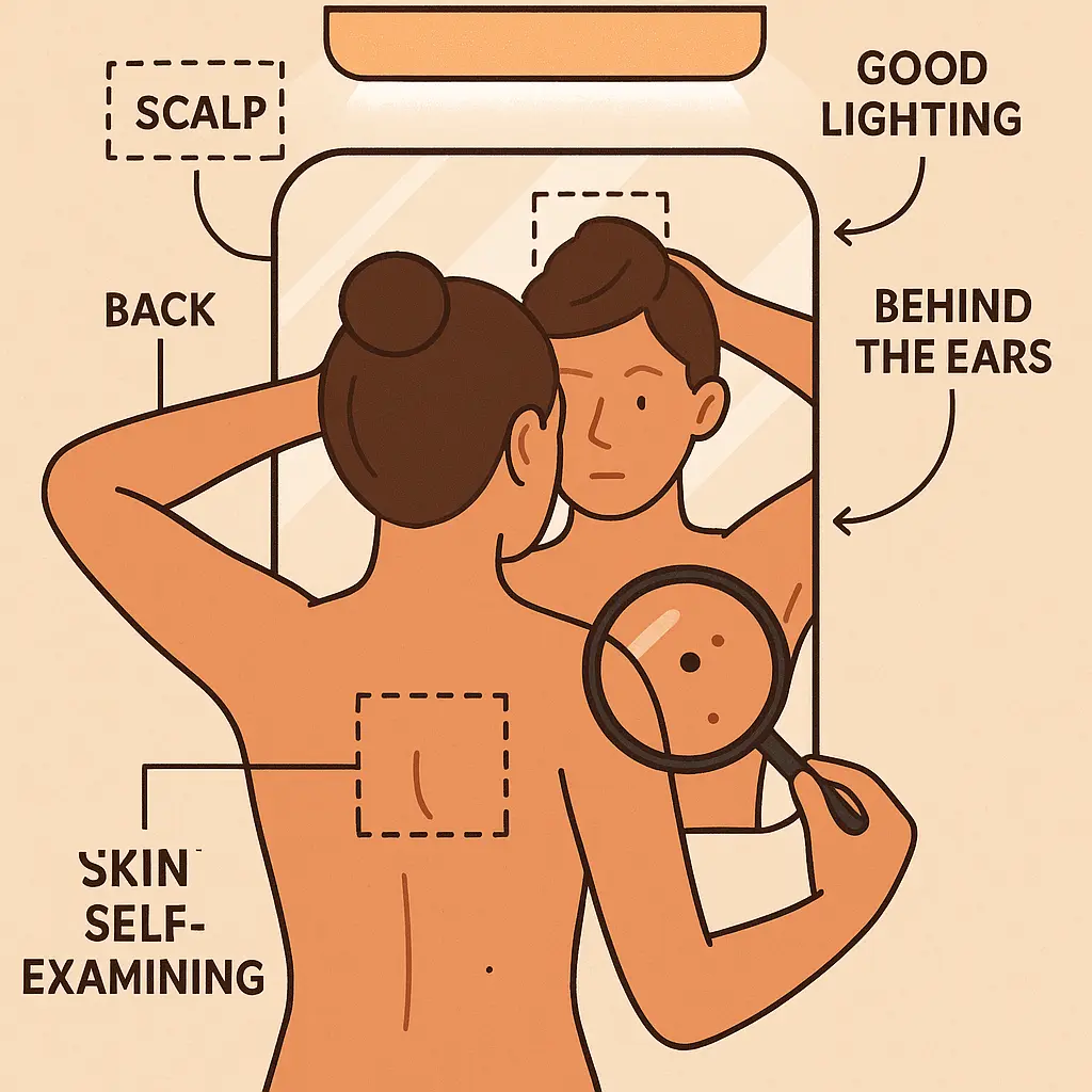
Systematic self-examination becomes crucial for people with many moles:

  • Monthly checks using consistent lighting and mirrors
  • Photography documentation of concerning areas
  • Partner assistance for hard-to-see locations
  • Mapping techniques to track individual moles
  • Change documentation with dates and descriptions

Psychological Considerations

Emotional aspects of living with multiple moles include:

  • Anxiety management about cancer risk
  • Body image concerns related to mole appearance
  • Hypervigilance leading to excessive worry
  • Social impacts affecting clothing choices and activities
  • Quality of life balance between monitoring and living normally

Support Resources

Professional support helps manage psychological aspects:

  • Dermatology counseling about realistic risks
  • Support groups for high-risk individuals
  • Mental health professionals familiar with medical anxiety
  • Educational resources providing accurate information
  • Family counseling for hereditary risk factors

Myths and Misconceptions About Skin Mole Types

Separating fact from fiction about skin mole types helps people make informed decisions about their skin health. Many persistent myths can lead to unnecessary anxiety or dangerous neglect.

Common Myths Debunked

Myth: "Hairy moles are always cancerous" Truth: Hair growth from moles actually indicates healthy, functioning follicles and is generally a reassuring sign of benign behavior.

Myth: "Cutting or irritating a mole causes cancer" Truth: Physical trauma doesn't cause mole transformation to cancer, though it may reveal pre-existing concerning features.

Myth: "All dark moles are dangerous" Truth: Color alone doesn't determine malignancy; many benign skin mole types are naturally dark.

Myth: "Moles that don't hurt are safe" Truth: Most melanomas are painless in early stages; absence of symptoms doesn't guarantee safety.

Myth: "Only sun-exposed areas develop dangerous moles" Truth: Melanoma can occur anywhere on the body, including areas never exposed to sun.

Scientific Evidence vs. Popular Beliefs

Evidence-based understanding of skin mole types reveals:

  • Genetic factors play larger roles than many people realize
  • UV protection is important but doesn't eliminate all risks
  • Regular monitoring is more valuable than obsessive daily checking
  • Professional evaluation remains the gold standard for assessment
  • Most moles remain stable and harmless throughout life

Addressing Patient Concerns

Healthcare providers should address common worries by:

  • Explaining normal variations in skin mole types
  • Providing realistic risk assessments based on individual factors
  • Teaching proper monitoring techniques
  • Addressing specific fears with factual information
  • Encouraging questions and open communication

Special Populations and Considerations

Different groups require tailored approaches to skin mole types monitoring and management based on unique risk factors and characteristics.

A hyper-realistic image of a A conceptual illustration depi...

Elderly Patients

Age-related changes affect mole evaluation in older adults:

  • Seborrheic keratoses may be confused with moles
  • Solar damage creates complex skin changes
  • Medication effects can alter skin appearance
  • Decreased immune function may increase cancer risk
  • Accessibility challenges for thorough self-examination

Immunocompromised Individuals

Reduced immune function significantly impacts skin mole types risk:

  • Increased cancer risk requiring more frequent monitoring
  • Atypical presentations of skin lesions
  • Faster progression of malignant changes
  • Multiple primary cancers possible
  • Enhanced prevention strategies necessary

Athletes and Outdoor Workers

High UV exposure groups need specialized strategies:

  • Occupational protection measures
  • Equipment modifications for sun safety
  • Frequent monitoring of exposed areas
  • Education programs for high-risk activities
  • Workplace policies supporting skin protection

Individuals with Fair Skin

Light-skinned populations face elevated risks:

  • Increased mole development from UV sensitivity
  • Higher melanoma incidence rates
  • Earlier onset of skin damage
  • Enhanced protection requirements
  • Genetic counseling considerations for families

Understanding these population-specific needs helps healthcare providers develop appropriate monitoring and treatment strategies.

Future Directions in Mole Research and Treatment

The field of skin mole types research continues evolving, with promising developments in prevention, diagnosis, and treatment approaching clinical application.

Genetic Research Advances

Molecular understanding of mole development reveals:

  • Genetic markers predicting mole behavior
  • Familial risk assessment tools
  • Personalized monitoring strategies
  • Targeted prevention approaches
  • Gene therapy possibilities for high-risk individuals

Immunotherapy Developments

Immune system modulation offers new treatment possibilities:

  • Checkpoint inhibitors for advanced melanoma
  • Vaccine development for prevention
  • Immune monitoring biomarkers
  • Combination therapies improving outcomes
  • Reduced toxicity treatment options

Telemedicine and Remote Monitoring

Digital health solutions expand access to specialized care:

  • Smartphone apps for mole photography
  • Remote consultation capabilities
  • AI-assisted screening programs
  • Population health monitoring systems
  • Underserved area access improvements

Biomarker Discovery

Molecular diagnostics enhance skin mole types evaluation:

  • Blood-based tests for melanoma detection
  • Tissue markers predicting behavior
  • Liquid biopsies for monitoring
  • Prognostic indicators guiding treatment
  • Minimal invasive testing options

These advancing technologies promise to improve outcomes while reducing the burden of monitoring multiple skin mole types.

Building Your Personal Mole Monitoring Plan

Creating an individualized approach to monitoring skin mole types helps ensure early detection while maintaining quality of life. Your personal plan should reflect your specific risk factors and circumstances.

Risk Assessment Components

Individual risk evaluation considers:

  • Family history of melanoma or atypical moles
  • Personal history of skin cancer or suspicious lesions
  • Skin type and sun sensitivity
  • UV exposure history and lifestyle
  • Number and types of existing moles

Customized Monitoring Schedule

Personalized surveillance based on risk level:

Low Risk Individuals:

  • Annual professional examinations
  • Quarterly self-checks
  • Immediate evaluation of changing moles
  • Standard sun protection measures

Moderate Risk Individuals:

  • Biannual professional examinations
  • Monthly self-checks with photography
  • Prompt evaluation of any changes
  • Enhanced sun protection strategies

High Risk Individuals:

  • Quarterly professional examinations
  • Monthly detailed self-examinations
  • Digital monitoring with dermoscopy
  • Aggressive prevention measures

Documentation Strategies

Effective tracking includes:

  • Baseline photography of all significant moles
  • Change logs with dates and descriptions
  • Professional examination records
  • Family history documentation
  • Treatment history if applicable

Emergency Action Plans

Clear protocols for concerning changes:

  • Contact information for dermatology providers
  • Urgent care options for immediate concerns
  • Documentation requirements for appointments
  • Insurance considerations for procedures
  • Support person involvement in decisions

Conclusion

Understanding skin mole types empowers individuals to take control of their skin health through informed monitoring and timely medical intervention. While the majority of moles remain harmless throughout life, recognizing the characteristics that distinguish benign from potentially dangerous lesions can literally save lives.

The key to effective mole management lies in balancing vigilant monitoring with realistic risk assessment. Most people can maintain excellent skin health through regular self-examinations, appropriate sun protection, and professional evaluations tailored to their individual risk factors. The ABCDE criteria provide a practical framework for identifying concerning changes, while understanding normal variations in skin mole types helps prevent unnecessary anxiety.

Modern advances in diagnostic technology, from dermoscopy to artificial intelligence, continue improving our ability to detect dangerous changes early when treatment is most effective. However, these tools complement rather than replace the fundamental importance of regular monitoring and professional evaluation.

Take action today by:

  • Scheduling your baseline dermatology examination if you haven't had one recently
  • Beginning monthly self-examinations using proper techniques
  • Documenting your current moles with photography for future comparison
  • Implementing comprehensive sun protection strategies
  • Discussing your family history and risk factors with healthcare providers

Remember that knowledge about skin mole types serves as your first line of defense against skin cancer. By staying informed, maintaining regular monitoring habits, and seeking professional guidance when needed, you can enjoy peace of mind while protecting your long-term health.

For comprehensive evaluation and treatment of concerning moles, consider consulting with specialists who can provide expert assessment and personalized monitoring strategies. Your proactive approach to understanding and monitoring skin mole types today contributes to a lifetime of healthy skin tomorrow.

Medical Review
Medical Review

Article Medically Reviewed By

Dr. Roberto Tutino

MD, CPSO #88547

Board-certified physician specializing in minor surgical procedures and dermatological treatments. Practicing across multiple locations in the Greater Toronto Area.

Verify CPSO Registration

Dr. Kunaal Jindal

MD, CPSO #88480

Licensed physician with expertise in minor surgery and skin procedures. Committed to providing comprehensive medical care and patient education across Ontario.

Verify CPSO Registration
Licensed by College of Physicians and Surgeons of Ontario
July 28, 2025