Have you ever noticed a small, flesh-toned bump on your skin that seemed to appear out of nowhere? You're not alone. Millions of people discover skin colored moles on their bodies each year, often causing concern and confusion about what these growths mean for their health.
Unlike the dark brown or black moles that most people recognize, a skin colored mole can be particularly puzzling because it blends seamlessly with the surrounding skin tone. These subtle growths often go unnoticed for months or even years, making them a unique category of skin lesions that deserves special attention.
Understanding the nature of skin colored moles is crucial for maintaining good skin health and knowing when to seek professional medical advice. These growths can range from completely harmless to potentially concerning, making proper identification and monitoring essential for everyone.
• Skin colored moles are common, benign growths that match your natural skin tone and can appear anywhere on the body
• These moles develop due to genetic factors, sun exposure, hormonal changes, and natural aging processes • Most skin colored moles are harmless, but changes in size, texture, or appearance warrant medical evaluation • Professional dermatological examination can distinguish between benign moles and potentially concerning lesions • Regular self-examinations and sun protection are key strategies for monitoring and preventing problematic mole development
A skin colored mole is a benign (non-cancerous) skin growth that appears in the same color as the surrounding skin tissue. Unlike traditional moles that contain high concentrations of melanin (the pigment that gives skin its color), these particular growths contain little to no melanin, resulting in their flesh-toned appearance.
Medical professionals often refer to skin colored moles by several technical terms, including:
These growths typically measure between 2-6 millimeters in diameter, though they can occasionally grow larger. The texture of a skin colored mole can vary significantly, ranging from completely smooth and flat to raised and slightly bumpy.
Skin colored moles possess several distinctive features that help differentiate them from other skin growths:
🔍 Color: Matches the exact tone of surrounding skin 🔍 Texture: Can be smooth, slightly rough, or have a "pebbly" surface 🔍 Shape: Usually round or oval with defined borders 🔍 Size: Typically small, ranging from 1-10 millimeters 🔍 Consistency: Firm to the touch but not hard like a cyst
The formation of a skin colored mole involves complex biological processes that occur within the skin's cellular structure. Understanding these mechanisms helps explain why some people develop more of these growths than others.
Skin colored moles develop when melanocytes (pigment-producing cells) cluster together in the skin but produce minimal or no melanin. This clustering can occur at different depths within the skin layers:
The depth of formation often influences the final appearance and texture of the skin colored mole.
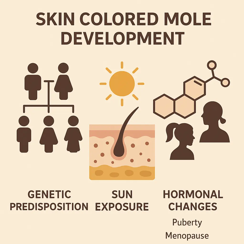
Several factors contribute to the development of skin colored moles:
Family history plays a significant role in mole development. Individuals with relatives who have numerous moles are more likely to develop skin colored moles themselves. Specific genetic mutations can affect how melanocytes function and cluster.
Ultraviolet (UV) radiation from sun exposure stimulates melanocyte activity and can trigger the formation of new moles. Interestingly, skin colored moles can develop even in areas with minimal sun exposure, suggesting other factors are involved.
Fluctuations in hormone levels, particularly during:
These hormonal shifts can stimulate new mole formation or cause existing moles to change appearance.
Most skin colored moles appear during childhood and young adulthood, though new ones can develop throughout life. The aging process affects skin cell behavior and can contribute to new growth formation.
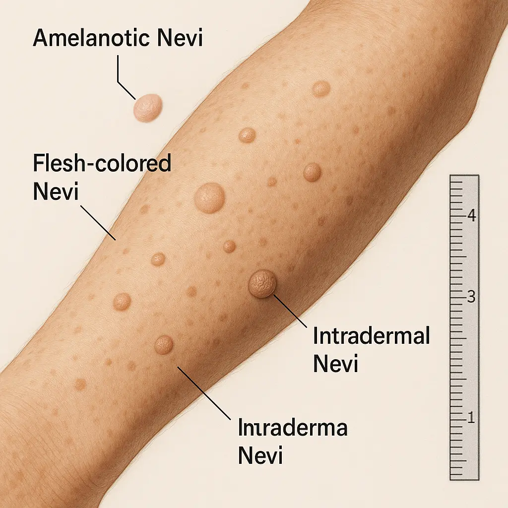
Not all skin colored moles are identical. Dermatologists classify these growths into several distinct categories based on their appearance, location, and cellular characteristics.
Intradermal nevi represent the most common type of skin colored mole. These growths develop entirely within the dermis layer and typically appear as:
Compound nevi span multiple skin layers and may display:
While technically not a true mole, dermatofibromas often get confused with skin colored moles. These benign growths feature:
Another growth sometimes mistaken for a skin colored mole, seborrheic keratoses exhibit:
While the vast majority of skin colored moles are completely harmless, certain warning signs warrant immediate medical attention. Understanding these red flags can help distinguish between normal, benign growths and potentially problematic lesions.
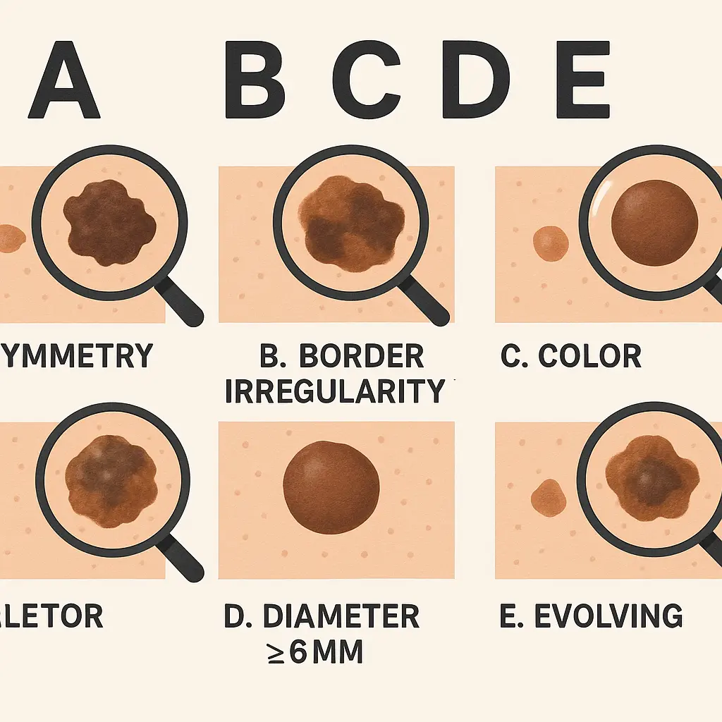
Medical professionals use the ABCDE rule to assess moles, including skin colored moles:
A - Asymmetry: One half doesn't match the other half B - Border: Irregular, scalloped, or poorly defined edges C - Color: Multiple colors or color changes within the mole D - Diameter: Larger than 6 millimeters (pencil eraser size) E - Evolving: Changes in size, shape, color, or texture
Pay particular attention to these concerning changes in a skin colored mole:
⚠️ Rapid growth over weeks or months ⚠️ Bleeding or oozing without trauma ⚠️ Itching or persistent irritation ⚠️ Color changes, especially darkening ⚠️ Surface changes like scaling or crusting ⚠️ Pain or tenderness ⚠️ Development of satellite lesions around the original mole
Although extremely uncommon, amelanotic melanoma can present as a skin colored mole. This type of skin cancer lacks the typical dark pigmentation associated with melanoma, making it particularly challenging to identify. Key characteristics include:
Important Note: Any skin colored mole that changes rapidly or exhibits concerning features should be evaluated by a dermatologist promptly.
When you notice a new skin colored mole or changes in an existing one, seeking professional medical evaluation provides peace of mind and ensures proper care.
A comprehensive skin colored mole evaluation typically includes:
The dermatologist will examine the mole using:
Your doctor will ask about:
Professional documentation may include:
When a skin colored mole appears suspicious, additional procedures might be necessary:
Dermoscopy (also called dermatoscopy) uses a specialized magnifying device to examine the mole's internal structure. This non-invasive technique can reveal:
If concerns persist, a biopsy may be recommended:
Shave Biopsy: Removes the surface portion for analysis Punch Biopsy: Extracts a small, round section of tissue Excisional Biopsy: Removes the entire mole and surrounding tissue
Tissue samples undergo microscopic examination to determine:
Most skin colored moles require no treatment and can be safely monitored over time. However, certain situations may warrant intervention.
The majority of skin colored moles benefit from a "watch and wait" approach:
Regular dermatological check-ups allow for:
Skin colored mole removal may be recommended for:
Surgical excision involves:
Shave excision works well for raised skin colored moles:
Laser therapy may be suitable for certain skin colored moles:
Post-treatment care ensures optimal healing:
Immediate Care:
Long-term Monitoring:
While you cannot prevent all skin colored moles from developing, certain strategies can reduce the risk of problematic changes and new growth formation.
Comprehensive sun protection significantly reduces mole-related risks:
Consistent monitoring helps catch changes early:
Perform thorough skin colored mole checks:
Regular dermatological examinations provide:
Certain lifestyle choices may influence skin colored mole development:
A diet rich in antioxidants may support skin health:
Proper skin care maintains healthy skin function:
Having skin colored moles is a normal part of life for many people. Understanding how to coexist peacefully with these growths reduces anxiety and promotes good skin health.
Creating a systematic approach to skin colored mole monitoring:
Skin colored mole anxiety is common and manageable:
Certain situations require prompt medical evaluation:
🚨 Rapid changes in mole appearance 🚨 Bleeding without obvious cause 🚨 Severe itching or pain 🚨 New symptoms developing suddenly 🚨 Multiple moles changing simultaneously
Advances in medical technology continue to improve skin colored mole diagnosis and treatment options.
AI-powered tools are revolutionizing mole assessment:
New imaging methods provide unprecedented detail:
Emerging treatments offer improved outcomes:
Future treatments may include:
Understanding skin colored moles empowers individuals to take an active role in maintaining their skin health. These common, typically benign growths require attention and monitoring but rarely cause serious health problems when properly managed.
The key to successful skin colored mole management lies in education, regular monitoring, and appropriate medical care when needed. By following the guidelines outlined in this comprehensive guide, individuals can confidently navigate the world of skin health while minimizing anxiety and maximizing protection.
Remember that most skin colored moles are harmless companions that will remain stable throughout life. However, staying vigilant for changes and maintaining open communication with healthcare providers ensures the best possible outcomes for long-term skin health.
Take charge of your skin health today by implementing a regular monitoring routine, practicing sun protection, and scheduling appropriate medical evaluations. Your skin will thank you for the attention and care you provide.


