Skin Cancer Symptoms Pictures: Your Complete Visual Guide to Early Detection 🔍
Every 36 seconds, someone in the United States is diagnosed with melanoma—the deadliest form of skin cancer. Yet, when caught early, skin cancer has a cure rate of nearly 99%. The difference between life and death often comes down to one crucial factor: knowing what to look for. Visual recognition of skin cancer symptoms can literally save your life, making skin cancer symptoms pictures one of the most valuable tools in early detection.
Key Takeaways
• Early detection saves lives: Skin cancer has a 99% cure rate when caught in its earliest stages
• Visual changes matter: New moles, changing existing spots, or unusual skin growths should always be evaluated by a professional
• The ABCDE rule helps: Asymmetry, Border irregularity, Color variation, Diameter changes, and Evolution are key warning signs
• Professional evaluation is essential: While pictures help with awareness, only a dermatologist can provide accurate diagnosis
• Regular self-exams are crucial: Monthly skin checks using visual guides can help identify concerning changes early
Understanding Skin Cancer: The Basics You Need to Know
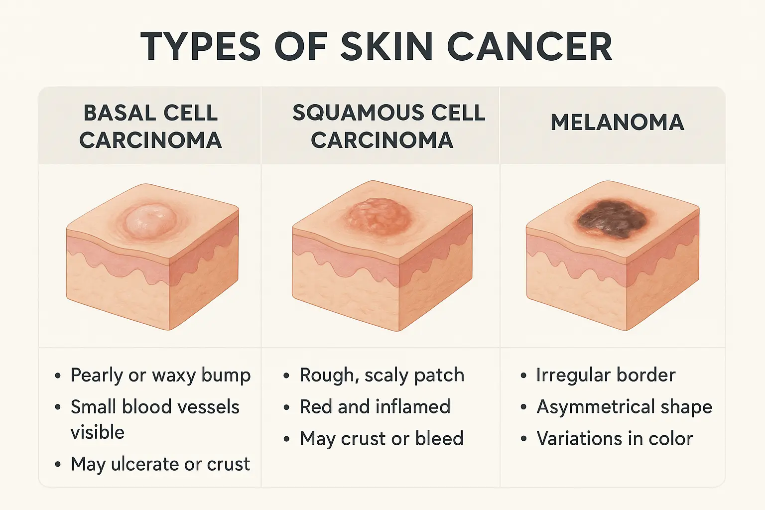
Skin cancer occurs when skin cells grow abnormally, typically due to DNA damage from ultraviolet (UV) radiation. Understanding the different types helps you know what warning signs to watch for in skin cancer symptoms pictures.
The Three Main Types of Skin Cancer
Basal Cell Carcinoma (BCC) 📊
Most common type (80% of all skin cancers)
Rarely spreads to other parts of the body
Often appears on sun-exposed areas like face, neck, and arms
Highly treatable when caught early
Squamous Cell Carcinoma (SCC)
Second most common type
Can spread if left untreated
Often develops on sun-damaged skin
More aggressive than basal cell carcinoma
Melanoma
Most dangerous form of skin cancer
Can spread rapidly to other organs
Often develops from existing moles
Requires immediate medical attention
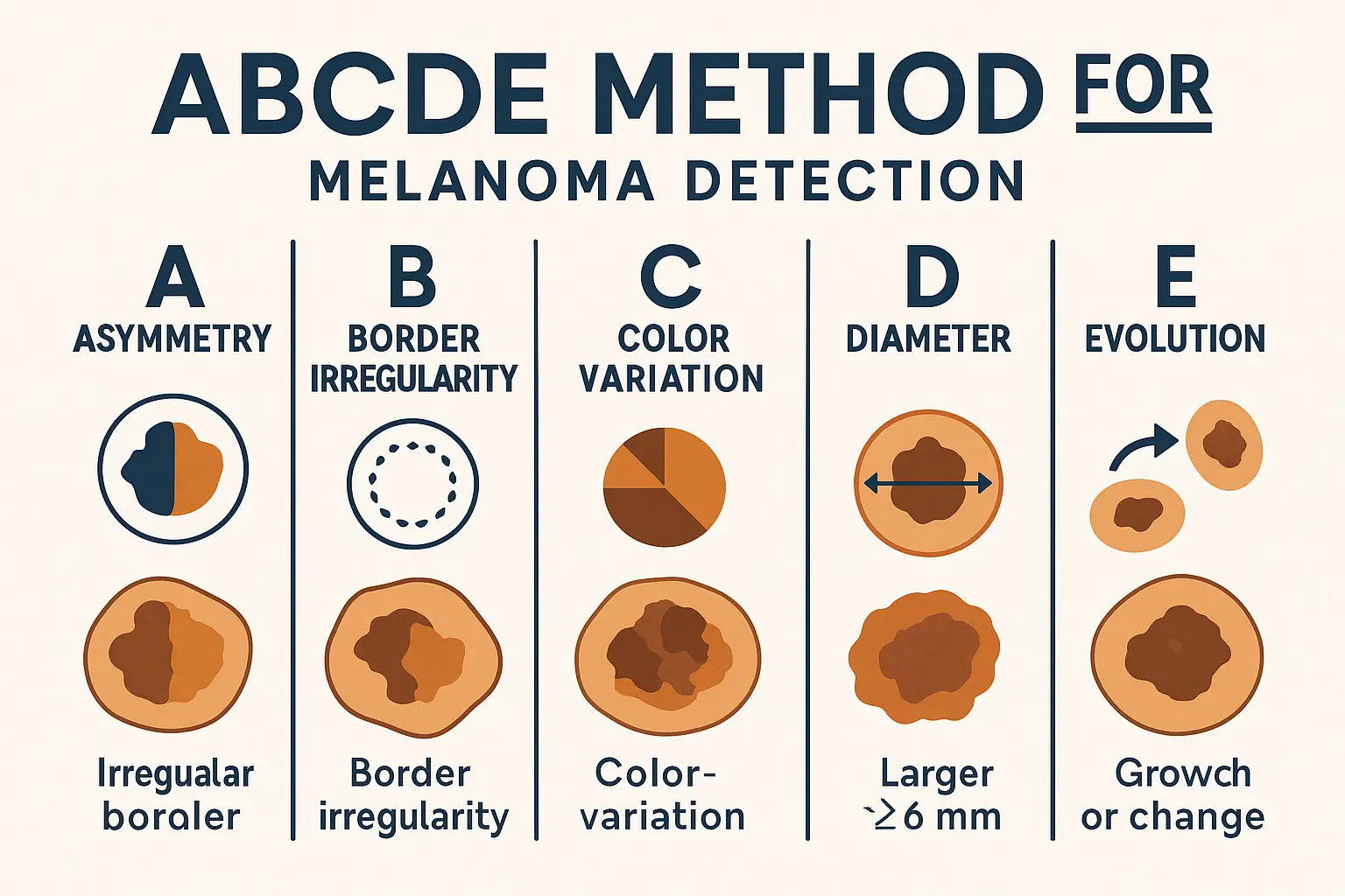
The ABCDE Method: Your Visual Detection System
The ABCDE method provides a systematic approach to evaluating suspicious spots on your skin. When examining skin cancer symptoms pictures or your own skin, remember these five critical signs:
A - Asymmetry
Normal moles are symmetrical—if you draw a line through the middle, both halves should match. Asymmetrical moles where one half doesn't match the other are a warning sign.
B - Border Irregularity
Healthy moles have smooth, even borders. Look for:
Scalloped edges
Notched borders
Blurred or poorly defined edges
Irregular or jagged outlines
C - Color Variation
Normal moles are typically one uniform color. Be concerned about:
Multiple colors within one mole
Uneven color distribution
Colors like red, white, blue, or black
Darkening or lightening of existing moles
D - Diameter
While not all dangerous moles are large, diameter changes are significant. Watch for:
Moles larger than 6mm (about the size of a pencil eraser)
Any mole that's growing in size
Small moles that suddenly become larger
E - Evolution
Perhaps the most important factor—any change in a mole's appearance warrants attention:
Changes in size, shape, or color
New symptoms like itching, bleeding, or crusting
Moles that look different from your other moles
Basal Cell Carcinoma: Visual Symptoms and Pictures
Basal cell carcinoma often appears deceptively harmless, which is why understanding its visual characteristics through skin cancer symptoms pictures is so important.
Common Appearances of Basal Cell Carcinoma
Pearly or Waxy Bumps ✨
Translucent appearance
May have visible blood vessels
Often flesh-colored, pink, or slightly darker
Can be mistaken for a pimple that won't heal
Flat, Scaly Patches
Brown or flesh-colored lesions
May appear slightly raised
Often found on the chest or back
Can resemble eczema or dry skin
Open Sores
Sores that bleed, ooze, or crust
May heal and return repeatedly
Often mistaken for minor injuries
Persist for weeks without proper healing
Red, Irritated Patches
May itch or be tender
Often appear on chest, shoulders, or limbs
Can be mistaken for rash or irritation
May have a slightly raised border
Squamous Cell Carcinoma: Recognizing the Warning Signs
Squamous cell carcinoma can be more aggressive than basal cell carcinoma, making early recognition through skin cancer symptoms pictures even more critical.
Visual Characteristics of Squamous Cell Carcinoma
Scaly, Red Patches 🔴
Rough, scaly surface texture
May bleed when scratched
Often appear on sun-exposed areas
Can be mistaken for dry skin or eczema
Open Sores with Raised Edges
Central depression or ulceration
Raised, irregular borders
May bleed or develop crusts
Often tender to touch
Wart-like Growths
Rough, elevated surface
May have a cauliflower-like appearance
Can develop from pre-existing warts
Often found on hands or feet
Horn-like Protrusions
Hard, pointed growths
Usually small but distinctive
May develop from actinic keratoses
Often found on ears, hands, or face
High-Risk Locations for Squamous Cell Carcinoma
Squamous cell carcinoma most commonly develops in areas with significant sun exposure:
Face and ears: Especially the rim of the ear and lower lip
Hands and forearms: Areas with chronic sun damage
Legs: Particularly in women, often on the lower legs
Genital area: Though less common, can be more aggressive
Melanoma: The Most Dangerous Form
Melanoma represents the most serious type of skin cancer, and recognizing its appearance in skin cancer symptoms pictures can be lifesaving.
Types of Melanoma and Their Appearances
Superficial Spreading Melanoma 🌊
Most common type (70% of melanomas)
Often develops from existing moles
Irregular shape with varied colors
May appear flat initially, then become raised
Nodular Melanoma
Fast-growing and aggressive
Often appears as a dark, raised bump
May be black, blue, or red
Can develop quickly over weeks or months
Lentigo Maligna Melanoma
Develops slowly over many years
Often appears as a large, flat, brown patch
Common in elderly individuals
Usually found on sun-damaged skin
Acral Lentiginous Melanoma
Occurs on palms, soles, or under nails
More common in people with darker skin
May appear as dark streaks under nails
Often mistaken for bruises or stains
The "Ugly Duckling" Sign
Beyond the ABCDE criteria, dermatologists often use the "ugly duckling" sign—any mole that looks different from your other moles deserves attention. This is particularly important because melanoma can sometimes break the traditional rules.
Pre-Cancerous Conditions: Early Warning Signs
Understanding pre-cancerous conditions helps identify potential problems before they become skin cancer. These conditions appear in many skin cancer symptoms pictures as important warning signs.
Actinic Keratoses (Solar Keratoses)
Visual Characteristics ☀️
Rough, scaly patches on sun-exposed skin
Pink, red, or brown coloration
Often feel rough or sandpaper-like
May be easier felt than seen
Significance
Considered pre-cancerous lesions
Can develop into squamous cell carcinoma
More common with age and sun exposure
Often multiple lesions present
Dysplastic Nevi (Atypical Moles)
Identifying Features
Larger than normal moles (usually over 5mm)
Irregular shape or color
May have some ABCDE characteristics
Often multiple in number
Risk Factors
Family history of melanoma
Multiple atypical moles
Fair skin and light eyes
History of severe sunburns
When to Seek Professional Help
While skin cancer symptoms pictures provide valuable guidance, they cannot replace professional medical evaluation. The Minor Surgery Center specializes in comprehensive skin cancer detection and treatment.
Red Flag Symptoms Requiring Immediate Attention ⚠️
For Any Skin Lesion:
Rapid growth or change in appearance
Bleeding, oozing, or crusting that doesn't heal
Itching, tenderness, or pain
Any spot that looks different from your other moles
Regular self-examinations using skin cancer symptoms pictures as reference guides can help you detect changes early. The key is consistency and knowing what to look for.
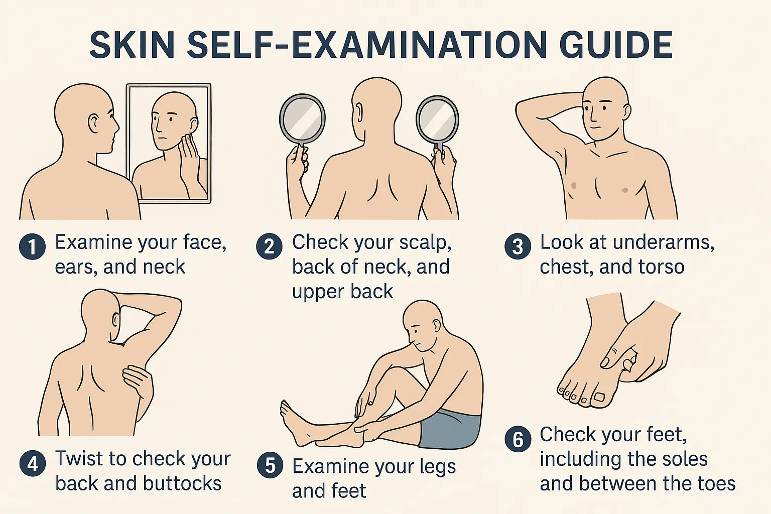
How to Perform a Thorough Skin Self-Exam
Preparation 📋
Choose a well-lit room with a full-length mirror
Have a hand mirror available for hard-to-see areas
Consider having a partner help with areas you can't see
Take photos of concerning spots for comparison
Systematic Approach
Face and scalp: Use a blow dryer to part hair and examine scalp thoroughly
Arms and hands: Check both sides, including palms and between fingers
Torso: Examine chest, abdomen, and sides with arms raised
Back: Use mirrors or ask for assistance
Legs and feet: Include soles, between toes, and toenails
Genital area: Often overlooked but important to check
Documentation Tips
Keep a body map of your moles
Take photos of any concerning spots
Note dates of any changes
Bring documentation to medical appointments
Monthly Self-Exam Schedule
Creating a routine helps ensure you don't skip this important health practice:
WeekFocus AreaSpecial AttentionWeek 1Head, neck, faceScalp and earsWeek 2Arms and torsoUnderarms and chestWeek 3Back and shouldersUse mirrors or partnerWeek 4Legs and feetSoles and between toes
Risk Factors and Prevention
Understanding risk factors helps you know if you're at higher risk and need more frequent monitoring using skin cancer symptoms pictures and professional evaluations.
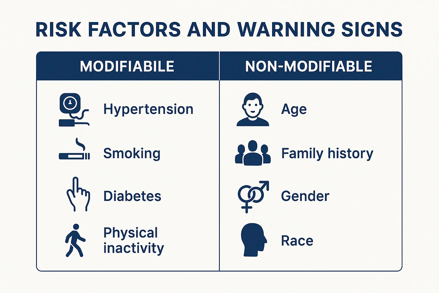
Major Risk Factors for Skin Cancer
Unmodifiable Risk Factors 🧬
Fair skin, light hair, and light eyes
Family history of skin cancer
Personal history of skin cancer
Age (risk increases with age)
Male gender (higher risk for melanoma)
Modifiable Risk Factors
UV exposure from sun or tanning beds
History of severe sunburns
Immunosuppression
Exposure to certain chemicals
Chronic wounds or scars
Prevention Strategies That Work
Sun Protection Basics ☂️
Use broad-spectrum SPF 30+ sunscreen daily
Reapply every 2 hours and after swimming/sweating
Wear protective clothing and wide-brimmed hats
Seek shade during peak hours (10 AM - 4 PM)
Advanced Protection Measures
UV-protective clothing with UPF ratings
Sunglasses with UV protection
Window films for cars and homes
Regular skin examinations by professionals
Lifestyle Modifications
Avoid tanning beds completely
Stay hydrated and maintain healthy skin
Eat antioxidant-rich foods
Consider vitamin D supplements instead of sun exposure
Special Populations and Considerations
Certain groups need special attention when it comes to skin cancer symptoms pictures and detection strategies.
Skin Cancer in People with Darker Skin Tones
Common Misconceptions 🌍
People with darker skin can and do get skin cancer
Melanoma is often more aggressive when it occurs
Acral lentiginous melanoma is more common
Often diagnosed at later stages due to delayed recognition
Key Areas to Monitor
Palms of hands and soles of feet
Under fingernails and toenails
Mucous membranes (mouth, nose, genital area)
Areas with less pigmentation
Pediatric Skin Cancer Considerations
Risk Factors in Children
Congenital moles (present at birth)
Family history of melanoma
Immunosuppression
Previous radiation therapy
Warning Signs
Any mole that changes rapidly
Bleeding or ulcerated lesions
Moles that look different from others
New moles appearing after puberty
Elderly Patients
Increased Risk Factors 👴
Cumulative sun damage over lifetime
Weakened immune system
Medications that increase photosensitivity
Difficulty performing self-examinations
Special Considerations
May need assistance with skin examinations
Higher likelihood of multiple skin cancers
Faster progression of some skin cancers
Importance of caregiver involvement
Technology and Skin Cancer Detection
Modern technology is revolutionizing how we use skin cancer symptoms pictures and detection methods.
Understanding treatment options helps reduce anxiety about skin cancer diagnosis and emphasizes the importance of early detection through skin cancer symptoms pictures.
Treatment for Basal Cell Carcinoma
Surgical Options ⚕️
Excision: Complete removal with clear margins
Mohs Surgery: Layer-by-layer removal with microscopic examination
Curettage and Electrodesiccation: Scraping and electrical destruction
Non-Surgical Options
Topical Medications: For superficial lesions
Radiation Therapy: For patients who cannot undergo surgery
Photodynamic Therapy: Light-activated treatment
Cryotherapy: Freezing with liquid nitrogen
Treatment for Squamous Cell Carcinoma
Standard Approaches
Surgical excision with appropriate margins
Mohs surgery for high-risk lesions
Radiation therapy for inoperable cases
Lymph node evaluation if spread is suspected
Success Rates
95-99% cure rate when caught early
Lower cure rates if lymph nodes involved
Importance of complete removal
Regular follow-up examinations essential
Melanoma Treatment
Staging Determines Treatment 📊
Stage 0-I: Surgical excision usually sufficient
Stage II-III: May require lymph node biopsy
Stage IV: Systemic therapy often needed
Advanced Cases: Immunotherapy and targeted therapy
Survival Rates by Stage
Stage I: 99% five-year survival
Stage II: 94% five-year survival
Stage III: 83% five-year survival
Stage IV: 27% five-year survival
These statistics underscore why recognizing early signs in skin cancer symptoms pictures is so crucial.
Living with Skin Cancer: Long-Term Management
After skin cancer treatment, ongoing vigilance and regular monitoring using skin cancer symptoms pictures as reference becomes even more important.
Follow-Up Care Requirements
Immediate Post-Treatment 🗓️
Wound care and healing monitoring
Watch for signs of infection
Activity restrictions as recommended
Pain management if needed
Long-Term Monitoring
Regular dermatology examinations
Self-examinations between visits
Photography documentation of new lesions
Prompt evaluation of concerning changes
Risk of Recurrence and New Cancers
Statistical Reality
40% chance of developing another skin cancer within 2 years
Higher risk with certain genetic factors
Importance of lifelong sun protection
Need for regular professional monitoring
Prevention Strategies
Enhanced sun protection measures
Regular dermatology visits
Careful self-monitoring
Family screening if genetic factors present
The Psychological Impact of Skin Cancer
Dealing with skin cancer affects more than just physical health, and understanding this helps patients cope better with their diagnosis and ongoing monitoring using skin cancer symptoms pictures.
Dispelling common myths helps people make better decisions about using skin cancer symptoms pictures and seeking appropriate care.
Common Myths Debunked
"Dark-skinned people don't get skin cancer" ❌
Reality: All skin types can develop skin cancer
Melanoma in darker skin is often more aggressive
Often diagnosed at later stages
Awareness and education are crucial
"Sunscreen prevents vitamin D production"
Reality: Most people get adequate vitamin D despite sunscreen use
Supplements are safer than sun exposure
Brief incidental exposure usually sufficient
Health benefits of sun protection outweigh risks
"A base tan protects against skin cancer"
Reality: Any tan indicates DNA damage
Provides minimal protection (SPF 2-4)
Increases long-term cancer risk
No such thing as a "safe" tan
"Skin cancer only occurs on sun-exposed areas"
Reality: Can occur anywhere on the body
Acral melanoma affects palms, soles, and nails
Mucosal melanoma affects mouth and genital areas
Complete body examinations are essential
Facts vs. Fiction in Prevention
MythFactExpensive sunscreens work betterSPF 30+ from any brand provides excellent protectionYou can't get burned on cloudy daysUp to 80% of UV rays penetrate clouds
| Tanning beds are safer than sun | Tanning beds increase melanoma risk by 75% | | Darker skin doesn't need sunscreen | All skin types benefit from sun protection |
Resources and Support Systems
Having access to reliable resources enhances your ability to use skin cancer symptoms pictures effectively and get appropriate care when needed.
The field of skin cancer detection continues to evolve, improving how we use skin cancer symptoms pictures and other diagnostic tools.
Emerging Technologies
Artificial Intelligence Advances 🤖
Machine learning algorithms for image analysis
Integration with smartphone cameras
Real-time risk assessment tools
Improved accuracy in detection
Genomic Testing
Genetic markers for cancer risk
Personalized screening recommendations
Targeted prevention strategies
Family risk assessment tools
Liquid Biopsies
Blood tests for cancer detection
Monitoring treatment response
Early recurrence detection
Less invasive than tissue biopsies
Research and Development
Current Research Focus
Improved imaging technologies
Better understanding of cancer biology
Novel treatment approaches
Prevention strategy development
Clinical Trials
New diagnostic methods
Innovative treatment options
Prevention studies
Quality of life research
Conclusion
Understanding skin cancer symptoms pictures and knowing what to look for can literally save your life. The key points to remember are:
Early Detection is Everything 🎯 With skin cancer having a 99% cure rate when caught early, your vigilance in recognizing warning signs makes all the difference. Use the ABCDE method, perform monthly self-examinations, and don't hesitate to seek professional evaluation for concerning changes.
Trust Your Instincts If something looks different or unusual, get it checked. The "ugly duckling" sign—any spot that looks different from your other moles—is often how people discover their skin cancer. Your instincts, combined with visual knowledge from skin cancer symptoms pictures, create a powerful detection system.
Professional Care is Essential While self-examination and symptom recognition are crucial, they cannot replace professional medical evaluation. Dermatologists and specialized clinics have the expertise and tools necessary for accurate diagnosis and treatment.
Prevention Remains Key Daily sun protection, avoiding tanning beds, and making smart lifestyle choices significantly reduce your risk of developing skin cancer. Remember that prevention is always easier than treatment.
Your Next Steps
Schedule a baseline skin examination with a dermatologist, especially if you have risk factors
Start monthly self-examinations using the guidance provided in this article
Implement comprehensive sun protection in your daily routine
Share this knowledge with family and friends—skin cancer awareness saves lives
Stay informed about new developments in detection and treatment
Remember, when it comes to skin cancer, you are your own best advocate. By staying informed, vigilant, and proactive about your skin health, you're taking the most important steps toward early detection and successful treatment. Your skin tells a story—make sure you're listening to what it's saying.