Pictures Of Wide Excision for Melanoma: What to Expect & See
When you or someone you care about receives a diagnosis of melanoma, a type of skin cancer, it can feel overwhelming. Many questions pop up, especially about treatment. One of the most common and effective treatments for melanoma is called a "wide excision." This surgery removes the melanoma along with a healthy border of skin around it. Understanding what this procedure involves, what it looks like, and what to expect during recovery can help ease your mind. This article aims to give you a clear picture of wide excision for melanoma, guiding you through the process step-by-step.
Key Takeaways
Wide excision is the main treatment for melanoma: It involves removing the cancerous mole and a safe amount of healthy skin around it to ensure all cancer cells are gone.
Surgical margins are key: The amount of healthy skin removed (the "margin") depends on how thick the melanoma is, helping prevent the cancer from coming back.
The procedure is usually quick and done as an outpatient: Most people go home the same day, often with local anesthesia to numb the area.
Recovery involves wound care and managing scarring: You'll need to keep the wound clean, and your doctor will discuss options for managing the appearance of the scar, which can vary in size.
Regular follow-ups are crucial: After surgery, you'll need regular check-ups to monitor your skin and overall health, ensuring any new concerns are caught early.
What Exactly Is Melanoma?
Melanoma is a serious type of skin cancer that starts in cells called melanocytes. These are the cells that make melanin, the pigment that gives your skin its color. While less common than other skin cancers like basal cell or squamous cell carcinoma, melanoma is more dangerous because it can spread to other parts of the body if not caught and treated early.
Why does melanoma happen? 🤔 Often, melanoma is linked to exposure to ultraviolet (UV) radiation from sunlight or tanning beds. But it can also happen in areas not exposed to the sun. Genetics can play a role too; if close family members have had melanoma, your risk might be higher.
Melanoma can appear in many ways:
A new, unusual-looking mole.
An existing mole that changes in size, shape, or color.
A sore that doesn't heal.
A dark spot under a fingernail or toenail.
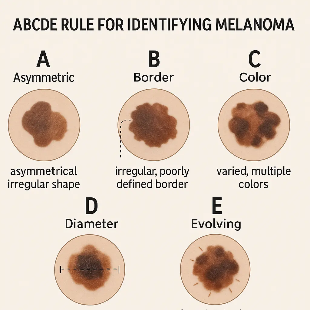
Doctors often use the "ABCDE" rule to help spot melanoma:
Asymmetry: One half of the mole doesn't match the other.
Border: The edges are irregular, ragged, notched, or blurred.
Color: The color is uneven, with shades of black, brown, and tan. It might also include white, red, or blue areas.
Diameter: Melanoma is often larger than 6 millimeters (about the size of a pencil eraser) when diagnosed, but it can be smaller.
Evolving: The mole is changing in size, shape, color, or elevation, or any new symptom appears, such as bleeding, itching, or crusting.
Catching melanoma early is incredibly important because it vastly improves the chances of successful treatment. This is why regular skin checks, both by yourself and a doctor, are so vital. If you have concerns about a mole or spot on your skin, don't hesitate to visit a healthcare professional. You can find more information about various skin conditions and their treatments at The Minor Surgery Center.
Why Is Wide Excision Necessary for Melanoma?
Once a skin biopsy confirms a diagnosis of melanoma, the next step is almost always a wide excision. Think of it as the primary way to "cut out" the problem. But why not just remove the visible melanoma?
Here's why wide excision is so important:
Complete Removal: Melanoma cells can sometimes spread slightly beyond the visible mole. These "invisible" cells are like tiny roots. A wide excision ensures that not just the main "plant" (the visible melanoma) but also its roots are removed.
Preventing Recurrence: By taking a margin of healthy tissue around the melanoma, doctors significantly reduce the chance of the cancer coming back in the same spot. This is called local recurrence.
Accurate Staging: The removed tissue, including the margin, is sent to a lab. Pathologists examine it closely to confirm that all cancer cells have been removed and to get more information about the melanoma's thickness and how deeply it has grown. This helps determine the "stage" of the cancer, which guides further treatment decisions.
A Doctor's Perspective:
"When we perform a wide excision, our goal is clear: to remove all melanoma cells while preserving as much healthy tissue as possible. It's a balance, but the priority is always the patient's long-term health." — Dr. Jane Doe, Dermatologic Surgeon
The size of the healthy skin margin taken depends on the thickness of the melanoma, also known as its Breslow depth. Thicker melanomas require wider margins to ensure all cancer cells are removed. Your doctor will discuss the exact margin needed for your specific case based on the pathology report from your initial biopsy.
The Wide Excision Procedure: A Closer Look
Understanding what happens during a wide excision can make the experience less daunting. While you won't literally see "pictures of wide excision for melanoma" during your procedure, we can walk you through the visual steps that doctors take.
Before the Surgery: Planning and Preparation
Before the day of your surgery, several important steps take place:
Diagnosis Confirmation: The initial biopsy confirms the melanoma and provides crucial details like its thickness.
Staging (if needed): For thicker melanomas, your doctor might order additional tests (like imaging scans or a sentinel lymph node biopsy) to see if the cancer has spread. This helps determine the best course of action.
Surgical Planning: Your surgeon will review your pathology report and decide on the appropriate "surgical margin." This means how much healthy skin around the melanoma needs to be removed.
For very thin melanomas (in situ or less than 1.0 mm thick), the margin might be 0.5 cm to 1.0 cm.
For thicker melanomas (1.0 mm to 2.0 mm thick), a 1.0 cm to 2.0 cm margin is common.
For melanomas over 2.0 mm thick, a 2.0 cm margin is often used.
Discussion with Your Surgeon: This is your chance to ask questions! Your surgeon will explain the procedure, what kind of scar to expect, and any specific instructions for before and after surgery. Don't be shy – it's important to feel comfortable and informed.
Pre-Op Instructions: You might be asked to stop certain medications (like blood thinners) a few days before surgery. You'll also get instructions on eating and drinking beforehand, especially if you're having sedation.
During the Surgery: Step-by-Step
Most wide excisions are outpatient procedures, meaning you go home the same day. They are often performed in a doctor's office or a minor surgery clinic. For more complex cases or those requiring general anesthesia, it might be done in a hospital. You can learn more about clinical settings and procedures at The Minor Surgery Center Clinic.
Here's a breakdown of what happens:
Anesthesia:
Local Anesthesia: This is the most common. The surgeon injects a numbing medicine (like lidocaine) directly into the skin around the melanoma. You'll feel a quick sting or pinch, but then the area will become numb. You'll be awake during the procedure but won't feel pain. You might feel some pressure or pulling.
Sedation: Sometimes, in addition to local anesthesia, you might receive a mild sedative to help you relax.
General Anesthesia: For very large or complex excisions, or if a skin graft or flap is needed, general anesthesia might be used, meaning you'll be completely asleep.
Marking the Area:
Once the area is numb, the surgeon will use a special pen to draw lines on your skin. These lines show the exact area that will be removed.
Visualizing the Area: Imagine the melanoma in the center. The surgeon draws an oval or ellipse shape around it, making sure the edges of the oval are the correct distance (the "margin") from the melanoma on all sides. This oval shape helps ensure the wound can be closed neatly. 📏
The Incision:
The surgeon uses a scalpel (a very sharp surgical knife) to make the incision along the marked lines. The cut goes through the skin and into the fat layer beneath.
What it looks like (conceptually): The incision creates an elongated oval or football shape. The exact depth and width depend on the melanoma's thickness and location. The goal is to remove the melanoma in one piece.
Tissue Removal:
The surgeon carefully removes the marked piece of skin and underlying tissue. They will ensure the entire "football" of tissue is removed cleanly.
Visualizing the Removed Tissue: Once removed, this piece of tissue looks like a small, elongated oval. The melanoma itself will be visible in the center of this removed tissue. This specimen is then sent to a pathology lab for detailed examination. The pathologist will check the "edges" (margins) of this removed tissue under a microscope to make sure no cancer cells are present at the very edge of the cut. This is called checking for "clear margins."
Bleeding Control:
Small blood vessels in the wound will bleed. The surgeon uses a tool called an electrocautery device (which uses heat) or sutures (stitches) to seal these vessels and stop the bleeding. You might hear a buzzing sound if electrocautery is used.
Wound Closure:
This is a crucial step for healing and appearance. The goal is to bring the edges of the wound together neatly.
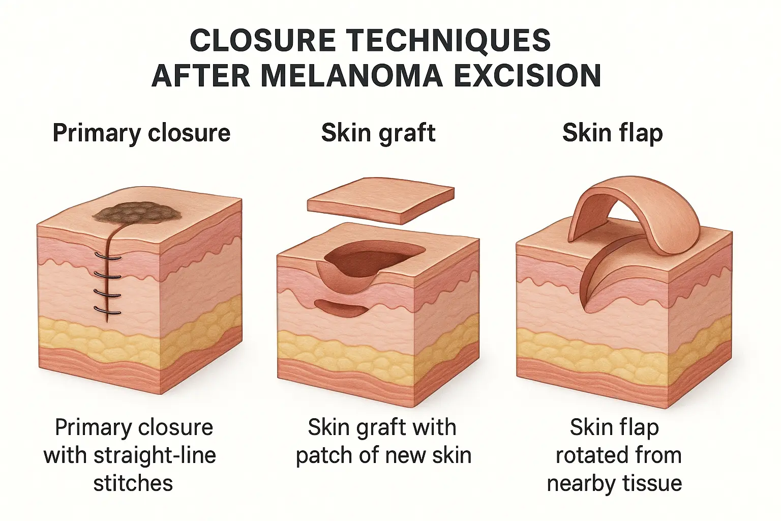
Primary Closure: For most wide excisions, especially on the torso or limbs, the surgeon can simply pull the edges of the oval-shaped wound together and close them with stitches. This turns the oval into a straight line.
Internal Stitches (dissolvable): These are placed deeper under the skin to bring the layers together and take tension off the surface stitches. They dissolve over time.
External Stitches (non-dissolvable): These are placed on the skin surface. They might be visible as small threads and usually need to be removed a week or two later. Sometimes, surgical glue or sterile strips (Steri-Strips) are used on top of or instead of external stitches.
Visualizing Primary Closure: Imagine pulling the two long sides of the "football" cut together. It becomes a straight line, like a neatly sewn seam. The length of this line will be longer than the original melanoma because of the oval shape of the excision.
Skin Graft: If the wound is too large or in an area where there isn't enough loose skin to pull together (like the shin or nose), a skin graft might be needed. This involves taking a thin layer of healthy skin from another part of your body (the "donor site," often the thigh or buttock) and carefully placing it over the wound. It's then stitched or stapled into place.
Visualizing a Skin Graft: Imagine a patch of new skin placed over the surgical hole, like a carefully fitted puzzle piece. The donor site will also need to heal, creating another wound.
Skin Flap: For very large or complex wounds, especially on the face, a skin flap might be used. This involves moving a piece of skin and underlying tissue that has its own blood supply from a nearby area to cover the wound. This is a more complex procedure than a skin graft.
Visualizing a Skin Flap: Think of it like rotating a piece of skin from next to the wound to cover the hole, rather than taking it from a distant site. This maintains blood supply, often leading to better cosmetic results for complex areas.
Dressing Application:
Once the wound is closed, a sterile dressing is applied to protect the area, absorb any drainage, and provide gentle pressure.
The entire procedure can take anywhere from 30 minutes to a few hours, depending on the size and location of the melanoma, and the type of closure needed.
Understanding Surgical Margins: Why They Matter So Much
You've heard us mention "surgical margins" several times. But what exactly are they, and why are they so crucial in the context of wide excision for melanoma?
What is a Surgical Margin? A surgical margin refers to the amount of healthy, normal-looking skin and tissue that is removed around the visible melanoma. It's the "buffer zone" taken to ensure that any microscopic melanoma cells that might have spread beyond the visible lesion are also removed.
Why are Margins Important?
Clearance: The primary goal is to achieve "clear margins." This means that when the pathologist examines the removed tissue under a microscope, they find no cancer cells at the very edges of the specimen. Clear margins are the best indicator that all the melanoma has been removed.
Reducing Recurrence: If the margins are "positive" (meaning cancer cells are found at the edge of the removed tissue), it suggests that some cancer cells might have been left behind. In such cases, a second surgery (re-excision) might be needed to remove more tissue and achieve clear margins. This greatly reduces the risk of the melanoma growing back in the same spot.
Tailored Treatment: The recommended margin width depends on the melanoma's thickness (Breslow depth).
Melanoma in situ (Stage 0): This is the earliest form, where cancer cells are only in the top layer of skin. A 0.5 cm margin is usually sufficient.
Melanoma < 1.0 mm thick (Stage I): A 1.0 cm margin is typically recommended.
Melanoma 1.01 mm to 2.0 mm thick (Stage I/II): A 1.0 cm to 2.0 cm margin is often used.
Melanoma > 2.0 mm thick (Stage II): A 2.0 cm margin is generally recommended.
The Role of the Pathologist: After your surgery, the removed tissue goes to a pathologist. They are like detectives with powerful microscopes! They meticulously examine slices of the tissue, especially focusing on the edges. They look for any stray melanoma cells. Their report will confirm whether your margins are clear or if further action is needed. This detailed examination is vital for your treatment plan.
Think of it like this: Imagine you have a spill of ink on a paper. You don't just cut out the visible ink stain; you cut out a bit of the clean paper around it, just to be sure you've gotten every last drop. The wider the spill, the more clean paper you'd cut out. Melanoma margins work similarly to ensure complete removal.
What to Expect Post-Surgery: Recovery and Care
Once the wide excision is complete, your journey shifts to recovery. Knowing what to expect can help you prepare for the days and weeks ahead.
Immediately After Surgery
Pain: You'll likely feel some soreness or discomfort as the local anesthetic wears off. Your doctor will advise on pain relief, often over-the-counter medications like ibuprofen or acetaminophen. For more significant pain, prescription medication might be provided.
Dressing: The surgical site will be covered with a sterile dressing. You'll get instructions on how to care for it.
Drainage: It's normal to have a small amount of clear or blood-tinged fluid drain from the wound for the first day or two. The dressing will absorb this.
Numbness: The area around the incision might feel numb for some time, due to nerve endings being cut during surgery. This usually improves over weeks or months, though some areas may have permanent numbness.
Wound Care: Your Daily Routine 🩹
Proper wound care is essential for healing and preventing infection. Your medical team will give you specific instructions, but generally, it involves:
Keeping it Clean and Dry: Keep the dressing clean and dry, especially for the first 24-48 hours. Avoid getting the wound wet during showers or baths for a period, as advised by your doctor.
Changing Dressings: You'll be told how often to change the dressing. This usually involves gently removing the old dressing, cleaning the wound (often with mild soap and water or saline solution), patting it dry, and applying a new sterile dressing.
Monitoring for Infection: Watch for signs of infection, such as:
Increased redness or warmth around the wound
Swelling that worsens
Pus or cloudy drainage
Fever
Increasing pain
If you notice any of these signs, contact your doctor right away!
Activity Restrictions
Rest: Get plenty of rest, especially in the first few days.
Avoid Strenuous Activity: Depending on the location of the excision, you'll need to limit activities that stretch or put tension on the wound. This means avoiding heavy lifting, vigorous exercise, or sudden movements for several weeks. Your doctor will give you specific guidance. For example, an excision on the back might limit bending or twisting, while one on the leg might limit long walks.
Elevation: If the excision is on an arm or leg, elevating it can help reduce swelling.
When Will Stitches Come Out?
If you have external stitches, they typically come out within 7 to 14 days, depending on the wound's location and tension. Your doctor or a nurse will remove them during a follow-up visit. This is usually a quick and relatively painless process.
Diet and Hydration
Maintain a healthy diet and stay well-hydrated. Good nutrition supports the body's healing process.
Scarring and Reconstruction: What to Expect Visually
One of the biggest concerns for many patients after a wide excision, especially on visible areas, is the resulting scar. While complete scar removal isn't possible, understanding what to expect and what options are available can help manage expectations.
How Scars Form
Any time the skin is cut, a scar will form as the body heals. The appearance of the scar depends on several factors:
Size and Depth of Excision: Larger and deeper excisions generally result in more noticeable scars.
Location: Scars tend to be more prominent in areas with high skin tension (e.g., shoulders, back, joints) or where there's less tissue to work with (e.g., shin, nose). Scars on the face often heal very well due to good blood supply.
Individual Healing: Everyone heals differently. Some people are prone to raised, thickened scars (hypertrophic scars or keloids).
Surgical Technique: A skilled surgeon uses techniques to minimize scarring, such as placing incisions along natural skin lines.
Visualizing the Scar:
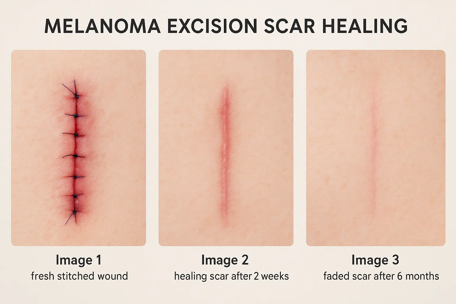
For primary closure, the scar will be a straight line, often longer than the original melanoma, running along the direction of the oval excision. Initially, it might be red and raised, but over months to a year, it usually flattens and fades to a lighter color.
For skin grafts, the scar will look like a patch of skin that might have a slightly different color or texture than the surrounding skin. The edges of the graft will also have a scar line. The donor site will have its own scar, which can be a straight line or a larger, lighter area if a very thin layer was taken.
For skin flaps, the scar can be more complex, reflecting the movement of tissue. These are often designed to blend in with natural features, especially on the face.
Options for Reconstruction
For larger excisions, especially on the face, hands, or lower legs, reconstructive options might be needed to close the wound or improve the cosmetic outcome.
Reconstruction MethodDescriptionCommon UsesScar AppearancePrimary ClosureDirectly stitching the wound edges together. The most common and simplest method.Smaller excisions, areas with loose skin (torso, upper limbs).A linear scar, often longer than the original lesion.Skin GraftTaking a thin layer of skin from one part of the body (donor site) and transplanting it to cover the wound.Large wounds, areas with little loose skin (shin, forehead), or where cosmetic outcome is less critical.A patched appearance, possibly different color/texture. Donor site also scars.Local FlapMoving a section of healthy skin and underlying tissue, with its own blood supply, from an area next to the wound to cover the defect.Larger defects, especially on the face, nose, or ears, where maintaining appearance and function is critical.Can be designed to blend well with facial features, but may still be noticeable. Often more complex scar patterns.Tissue ExpanderA balloon-like device placed under the skin near the wound. It's gradually filled with saline over weeks/months to stretch the skin.Used when a large amount of skin is needed for closure, often for very large defects or on the scalp.Can result in a very good color match, but requires multiple procedures and a temporary visible bulge.
Your surgeon will discuss the best reconstructive option for your specific case, considering the size and location of the melanoma, your overall health, and your cosmetic goals. Remember, the primary goal is always to ensure the complete removal of the melanoma. Cosmetic outcomes are important, but secondary to cancer clearance.
Potential Complications of Wide Excision
While wide excision is generally safe, like any surgery, it carries some potential risks. Being aware of these can help you recognize them early if they occur.
Bleeding: Some oozing from the wound is normal, but excessive bleeding (a dressing quickly soaking through) is a concern. This might require pressure, re-dressing, or in rare cases, a return to the clinic to have the bleeding stopped.
Infection: Despite sterile techniques, bacteria can sometimes get into the wound. Signs include increasing redness, warmth, swelling, pain, pus, or fever. Infections usually respond well to antibiotics.
Swelling and Bruising: These are very common around the surgical site and usually resolve within a few weeks. Elevating the affected area can help.
Seroma: This is a collection of clear fluid under the skin, forming a lump. It can happen if fluid builds up in the space where the tissue was removed. Small seromas often resolve on their own, but larger ones might need to be drained by your doctor.
Hematoma: This is a collection of blood under the skin, forming a painful, firm lump. Like a seroma, it might need to be drained by your doctor.
Nerve Damage: Small nerves in the skin can be cut during surgery, leading to temporary or permanent numbness, tingling, or altered sensation around the scar. Rarely, a motor nerve (controlling muscle movement) can be affected, leading to weakness.
Wound Dehiscence (Opening): Sometimes, especially if there's a lot of tension on the wound, or if an infection occurs, the stitches might break, and the wound edges can separate. This requires careful wound care and might need re-stitching.
Scarring Issues: As discussed, scars can sometimes become raised (hypertrophic) or grow beyond the original wound (keloid). These can be itchy or painful and may require specific treatments like steroid injections or laser therapy.
Allergic Reaction: Rarely, a patient might have an allergic reaction to the anesthetic, surgical materials (like sutures), or wound dressings.
Your surgical team will explain these risks to you and instruct you on what to watch for. Don't hesitate to contact them if you have any concerns during your recovery.
Follow-Up Care and Surveillance: Staying Vigilant
After your wide excision and once your wound has healed, your journey with melanoma doesn't end. Regular follow-up care, also known as surveillance, is a critical part of managing your health long-term.
Why is Follow-Up Important?
Early Detection of Recurrence: While wide excision aims to remove all cancer, there's always a small chance that some cells might have spread elsewhere or that a new melanoma could develop. Regular checks help catch any recurrence or new melanomas early, when they are most treatable.
Monitoring for New Melanomas: People who have had one melanoma are at a higher risk of developing another.
Overall Skin Health: Your doctor will also check for other types of skin cancer and provide advice on sun protection.
What Does Follow-Up Involve?
Regular Skin Exams: This is the most important part. Your dermatologist will perform a thorough head-to-toe skin examination, looking for any suspicious moles or lesions. This includes examining areas you can't easily see yourself.
Frequency: The frequency of these exams depends on the stage of your melanoma. For early-stage melanoma, you might have checks every 3-6 months for the first few years, then less frequently if all remains clear. For higher-risk melanoma, checks might be more frequent and continue for a longer period.
Lymph Node Checks: Your doctor will also feel the lymph nodes near where your melanoma was removed to check for any swelling, which could indicate spread.
Self-Skin Exams: You'll be taught how to perform regular self-skin exams at home. This means checking your entire skin surface monthly, using a mirror for hard-to-see areas. Look for any new moles, or changes in existing ones (remember the ABCDEs!).
Imaging or Blood Tests (for higher stages): For more advanced melanomas, or if there's concern about spread, your doctor might order imaging scans (like CT, PET, or MRI scans) or blood tests to check for cancer in other parts of the body.
Sun Protection Education: Your doctor will reinforce the importance of lifelong sun protection, as UV exposure is a major risk factor for new melanomas. This includes:
Using broad-spectrum sunscreen with an SPF of 30 or higher daily.
Avoiding tanning beds.
Who Will Provide Your Follow-Up Care? Often, a dermatologist will lead your follow-up care. However, depending on the stage of your melanoma, you might also see an oncologist (cancer specialist) or a surgical oncologist. It's a team effort to ensure your long-term health.
When to Seek Medical Attention After Wide Excision
While your surgical team will provide detailed post-operative instructions, it's vital to know when to contact them immediately. Don't wait if you experience any of the following:
Signs of Infection:
Fever (temperature over 100.4°F or 38°C)
Increased redness, warmth, or swelling around the incision that is spreading
Pus or thick, foul-smelling discharge from the wound
Increasing pain that is not relieved by pain medication
Excessive Bleeding:
If your dressing becomes quickly soaked with blood, or if bleeding doesn't stop after applying firm pressure for 15-20 minutes.
Wound Opening (Dehiscence):
If the edges of your incision separate or the wound opens up.
Severe or Persistent Pain:
Pain that is severe, getting worse, or not manageable with prescribed pain relief.
New or Worsening Numbness/Weakness:
Especially if it affects your ability to move a limb or part of your face.
Swelling with Red Streaks:
Red streaks spreading from the wound, which could indicate a serious infection (cellulitis).
Any Other Major Concern:
If something just "doesn't feel right" or you have a new symptom that worries you.
It's always better to be safe and call your healthcare provider if you have any doubts or concerns about your recovery. They are there to help you through this process. For general medical concerns or to find a clinic that can help, you can visit The Minor Surgery Center.
Living with a Melanoma Diagnosis
A melanoma diagnosis and the subsequent wide excision can be a life-changing event. Beyond the physical recovery, there's often an emotional and psychological journey.
Emotional Support
It's completely normal to feel a range of emotions: fear, anxiety, sadness, anger, or even relief that the cancer has been removed.
Talk About It: Share your feelings with trusted family members, friends, or a support group. Connecting with others who have gone through similar experiences can be incredibly helpful.
Seek Professional Help: If you find yourself struggling with persistent anxiety, depression, or difficulty coping, consider talking to a counselor, therapist, or psychologist. Many cancer centers offer psychological support services.
Patient Advocacy Groups: Organizations dedicated to melanoma patients can provide resources, connect you with support networks, and offer valuable information.
Lifestyle Adjustments
Sun Protection is Paramount: This becomes a lifelong commitment. You've already learned about the importance of sun-safe habits. Make them a part of your daily routine. This isn't just about preventing new melanomas but also protecting your overall skin health.
Regular Self-Exams: Make checking your skin a monthly habit. Get to know your moles and skin spots so you can quickly spot any changes.
Healthy Living: A balanced diet, regular exercise (once cleared by your doctor), and avoiding smoking can contribute to your overall well-being and recovery.
Stress Management: Find healthy ways to manage stress, whether through mindfulness, meditation, yoga, hobbies, or spending time in nature.
Living with a melanoma diagnosis means being proactive about your health and vigilant about your skin. It's a journey, but with proper care and support, you can lead a full and healthy life.
Conclusion
A diagnosis of melanoma can be frightening, but understanding the treatment process, especially wide excision, can empower you. We've explored what "pictures of wide excision for melanoma" truly mean: not just the visual aspects of the surgery itself, but the meticulous planning, the careful removal of tissue with precise margins, and the journey of healing and recovery.
Wide excision is a highly effective treatment that aims to remove the cancer completely, giving you the best chance for a healthy future. While the procedure leaves a scar, this scar is a testament to a successful battle against a serious disease. Remember that comprehensive follow-up care is just as important as the surgery itself, ensuring that you remain vigilant and proactive about your skin health. By staying informed, adhering to your follow-up schedule, and practicing sun safety, you can confidently move forward on your path to recovery and long-term well-being.
Melanoma Wide Excision Recovery Timeline
Important Note: This timeline provides general guidance. Every person's healing journey is unique. Always follow your surgeon's specific instructions for your recovery.