Understanding Pictures of Merkel Cell Carcinoma: A Visual Guide to Recognition and Diagnosis

Merkel cell carcinoma (MCC) represents one of the most aggressive forms of skin cancer, yet many people remain unfamiliar with its appearance. Understanding what this rare cancer looks like through pictures of Merkel cell carcinoma can be crucial for early detection and treatment. This comprehensive guide explores the visual characteristics, diagnostic features, and important warning signs that everyone should know about this serious skin condition.
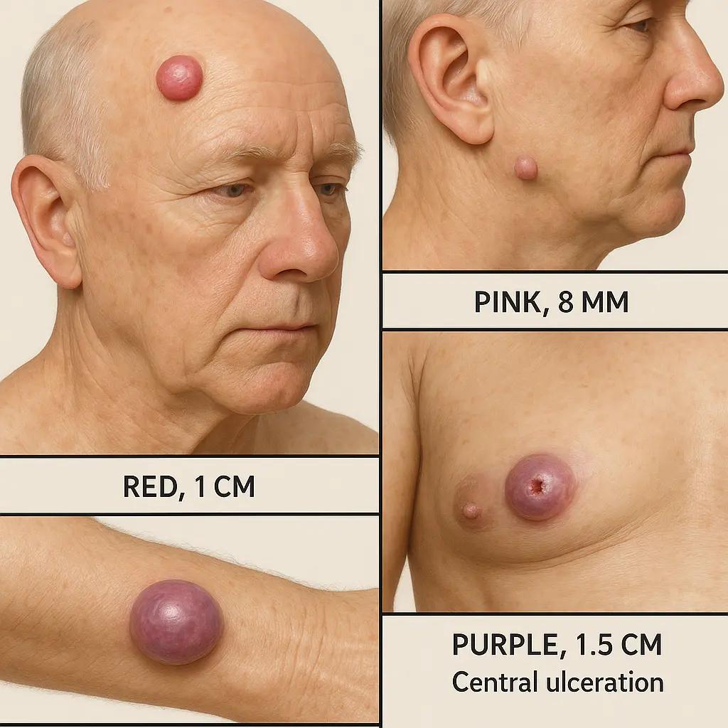
Key Takeaways 📌

Pictures of Merkel cell carcinoma typically show firm, painless nodules that appear red, pink, or purple in color

A hyper-realistic image of a A hyper-realistic image showin...

• Early detection through visual recognition can significantly improve treatment outcomes and survival rates • MCC often appears on sun-exposed areas but can develop anywhere on the body • The cancer grows rapidly and requires immediate medical attention when suspected • Professional dermatological evaluation is essential for accurate diagnosis

What Is Merkel Cell Carcinoma?

Merkel cell carcinoma stands as a rare but highly aggressive form of skin cancer that develops from Merkel cells located in the skin's outer layer. These specialized cells, discovered by German anatomist Friedrich Merkel in 1875, function as touch receptors that help the body sense light touch and texture.

When examining pictures of Merkel cell carcinoma, medical professionals and patients often notice distinct characteristics that set this cancer apart from other skin conditions. The disease affects approximately 3,000 people annually in the United States, making visual recognition skills particularly valuable for healthcare providers and individuals monitoring their skin health.

The Importance of Visual Recognition

Understanding the appearance of MCC through clinical photographs and medical imaging plays a vital role in:

  • Early detection and intervention
  • Patient education and awareness
  • Medical training for healthcare professionals
  • Differential diagnosis from other skin conditions
  • Treatment planning and monitoring

Visual Characteristics in Pictures of Merkel Cell Carcinoma

Primary Visual Features

When studying pictures of Merkel cell carcinoma, several distinctive features typically emerge:

1. Color Variations 🎨

  • Red or pink hues (most common)
  • Purple or violet tones
  • Flesh-colored appearance
  • Occasionally bluish tint

2. Size and Growth Pattern

  • Initial size: 0.5-2 cm diameter
  • Rapid growth over weeks to months
  • Can reach 5+ cm if untreated
  • Dome-shaped or nodular appearance

3. Texture and Surface

  • Smooth, shiny surface
  • Firm to touch
  • Non-tender or painless
  • May develop central ulceration

Location Patterns

Analysis of pictures of Merkel cell carcinoma reveals common locations:

Body AreaFrequencySun ExposureHead/Neck50%HighArms20%HighLegs15%ModerateTrunk10%VariableOther areas5%Low

Distinguishing Features in Clinical Photography

Comparison with Other Skin Conditions

Professional pictures of Merkel cell carcinoma help distinguish MCC from similar-appearing conditions:

Basal Cell Carcinoma Differences:

  • MCC grows more rapidly
  • Lacks pearly borders
  • More uniform coloration
  • Firmer consistency

Melanoma Differences:

  • MCC typically lacks dark pigmentation
  • More symmetrical appearance
  • Smoother surface texture
  • Different growth pattern

Cyst Differences:

  • MCC feels firmer
  • Doesn't move freely under skin
  • Rapid growth rate
  • Vascular appearance

Dermoscopic Features

Advanced imaging techniques in pictures of Merkel cell carcinoma reveal:

  • Milky-red areas indicating vascular patterns
  • Polymorphous vessels of varying sizes
  • Absence of pigment network
  • White structureless areas

Stages and Progression Shown in Pictures

Stage I: Early Disease

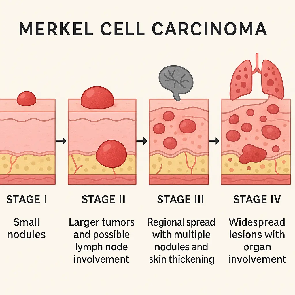
Early-stage pictures of Merkel cell carcinoma typically show:

  • Small nodules (< 2 cm)
  • Localized to primary site
  • No lymph node involvement
  • Clear surrounding skin

Stage II: Larger Primary Tumors

Progressive pictures of Merkel cell carcinoma demonstrate:

  • Tumors > 2 cm diameter
  • Possible satellite lesions
  • Increased vascular appearance
  • Beginning tissue distortion

Stage III: Regional Spread

Advanced pictures of Merkel cell carcinoma reveal:

  • Lymph node enlargement
  • Multiple nodules
  • Skin thickening
  • Possible ulceration

Stage IV: Distant Metastasis

Late-stage imaging shows:

  • Multiple organ involvement
  • Widespread skin lesions
  • Significant tissue changes
  • Complex treatment areas

Risk Factors and Visual Associations

Understanding risk factors helps interpret pictures of Merkel cell carcinoma in context:

A hyper-realistic image of a A detailed infographic compari...

Sun Damage Indicators 🌞

Visual signs often accompanying MCC:

  • Actinic keratoses (rough, scaly patches)
  • Solar lentigines (age spots)
  • Telangiectasias (visible blood vessels)
  • Skin texture changes

Immunosuppression Effects

Patients with weakened immune systems may show:

  • Multiple skin lesions
  • Faster tumor growth
  • Atypical presentations
  • Poor wound healing

Diagnostic Imaging Beyond Surface Pictures

Specialized Imaging Techniques

While surface pictures of Merkel cell carcinoma provide valuable information, additional imaging includes:

1. Dermoscopy

  • Magnified skin surface examination
  • Vascular pattern analysis
  • Structural detail assessment

2. Ultrasound Imaging

  • Tumor depth measurement
  • Lymph node evaluation
  • Blood flow assessment

3. PET/CT Scans

  • Whole-body tumor detection
  • Metastasis identification
  • Treatment response monitoring

When to Seek Medical Evaluation

Warning Signs Requiring Professional Assessment

Anyone noticing features similar to pictures of Merkel cell carcinoma should seek immediate evaluation at The Minor Surgery Center if experiencing:

"A rapidly growing, firm, painless nodule on sun-exposed skin warrants immediate dermatological evaluation, especially in individuals over 50 or those with compromised immune systems."

Red Flag Symptoms:

  • ⚠️ New nodule appearing within weeks
  • ⚠️ Rapid size increase
  • ⚠️ Firm, non-tender mass
  • ⚠️ Red, pink, or purple coloration
  • ⚠️ Location on sun-exposed areas

Documentation for Medical Visits

When consulting healthcare providers about suspicious lesions:

  1. Photograph the lesion with size reference
  2. Note growth timeline and changes
  3. Document any symptoms or sensations
  4. List current medications and health conditions
  5. Prepare family history information

Treatment Visualization and Outcomes

Surgical Excision Images

Post-treatment pictures of Merkel cell carcinoma sites show:

  • Wide excision margins
  • Reconstructive results
  • Healing progression
  • Scar formation patterns

Radiation Therapy Effects

Visual documentation during treatment reveals:

  • Skin reaction patterns
  • Treatment field markings
  • Response indicators
  • Side effect management

Prevention and Monitoring Strategies

Self-Examination Guidelines

Regular skin checks comparing findings to pictures of Merkel cell carcinoma should include:

Monthly Self-Exam Steps:

  1. Full-body inspection in good lighting
  2. Use mirrors for hard-to-see areas
  3. Document changes with photos
  4. Note new growths or changes
  5. Schedule regular check-ups at The Minor Surgery Center's clinic

Sun Protection Measures 🛡️

Preventive strategies include:

  • Daily SPF 30+ sunscreen application
  • Protective clothing and wide-brimmed hats
  • Shade seeking during peak hours
  • UV-blocking sunglasses
  • Regular skin examinations

Educational Resources and Support

Professional Resources

Healthcare providers studying pictures of Merkel cell carcinoma can access:

A hyper-realistic image of a A conceptual diagram illustrat...
  • Medical image databases
  • Dermatology atlases
  • Case study collections
  • Training modules
  • Peer-reviewed publications

Patient Education Materials

Individuals concerned about skin changes can find resources through:

Technology and Future Directions

Artificial Intelligence in Image Analysis

Emerging technologies analyzing pictures of Merkel cell carcinoma include:

AI Applications:

  • Automated lesion detection
  • Risk assessment algorithms
  • Growth tracking software
  • Diagnostic assistance tools
  • Treatment planning systems

Telemedicine and Remote Monitoring

Digital health platforms enable:

  • Remote consultation capabilities
  • Image sharing with specialists
  • Progress monitoring
  • Follow-up scheduling
  • Second opinion access

Special Populations and Considerations

Elderly Patients

Pictures of Merkel cell carcinoma in older adults often show:

  • Multiple comorbid skin conditions
  • Delayed presentation
  • Atypical locations
  • Treatment complications

Immunocompromised Individuals

Special considerations include:

  • More aggressive disease course
  • Unusual presentations
  • Multiple primary tumors
  • Complex treatment decisions

Research and Clinical Trials

Visual Documentation in Research

Clinical trials utilizing pictures of Merkel cell carcinoma focus on:

  • Treatment response assessment
  • New therapy development
  • Biomarker identification
  • Outcome prediction
  • Quality of life measures

Emerging Therapies

Visual monitoring of new treatments includes:

  • Immunotherapy responses
  • Targeted therapy effects
  • Combination treatment outcomes
  • Side effect profiles

Living with Merkel Cell Carcinoma

Survivorship and Follow-Up

Long-term monitoring through pictures of Merkel cell carcinoma sites helps:

  • Detect recurrence early
  • Monitor treatment effects
  • Document healing progress
  • Guide rehabilitation
  • Support psychological adjustment

Quality of Life Considerations

Visual changes affecting patients include:

  • Surgical scarring
  • Radiation effects
  • Lymphedema development
  • Cosmetic concerns
  • Functional limitations

Conclusion

Understanding pictures of Merkel cell carcinoma empowers both healthcare providers and patients in the fight against this aggressive cancer. Visual recognition remains a cornerstone of early detection, which significantly improves treatment outcomes and survival rates. The distinctive appearance of MCC—typically presenting as a rapidly growing, firm, painless nodule with red, pink, or purple coloration—makes photographic documentation invaluable for diagnosis and monitoring.

As medical technology advances, the role of visual documentation continues to expand, from AI-assisted diagnosis to telemedicine consultations. However, nothing replaces the importance of regular skin examinations and prompt medical evaluation of suspicious lesions.

Anyone noticing skin changes resembling the characteristics described in this guide should seek immediate professional evaluation. Early detection and treatment remain the most powerful tools in combating Merkel cell carcinoma. For expert evaluation and treatment of suspicious skin lesions, consider scheduling an appointment with The Minor Surgery Center's experienced team.

Remember: when it comes to skin cancer, vigilance saves lives. Regular self-examinations, sun protection, and professional skin checks form the foundation of effective prevention and early detection strategies.

June 6, 2025