Nail Apparatus Melanoma: Current Management and Future Perspectives

You might think melanoma only appears on sun-exposed skin, but nail apparatus melanoma (NAM) is a hidden threat that often goes unnoticed until it reaches advanced stages. This rare subtype of melanoma develops beneath or around your nails, masquerading as harmless discoloration or injury. The stakes are high: early detection can mean the difference between a simple surgical procedure and extensive treatment involving amputation.

In 2025, advances in diagnostic technology and surgical techniques are transforming how we approach this condition. Whether you're a patient who's noticed suspicious nail changes or simply want to understand this complex condition, this comprehensive guide will walk you through everything you need to know about nail apparatus melanoma—from recognizing early warning signs to understanding cutting-edge treatment options.

Key Takeaways

  • Nail apparatus melanoma is rare but serious: It accounts for only 0.7-3.5% of all melanomas but carries significant diagnostic challenges due to its subtle presentation
  • Early recognition saves lives: The ABCDEF rule helps identify suspicious nail changes, with Hutchinson's sign being a critical red flag requiring immediate evaluation
  • Dermoscopy revolutionizes diagnosis: Modern imaging techniques combined with timely biopsy significantly improve early detection rates
  • Surgical precision matters: Wide local excision with appropriate margins remains the gold standard, though functional preservation is increasingly prioritized
  • New treatments offer hope: Immunotherapy and targeted therapies are showing promise for advanced cases, changing the landscape of NAM management
Nail Melanoma ABCDEF Assessment Tool

Nail Melanoma ABCDEF Assessment Tool

Use this interactive tool to evaluate suspicious nail changes based on the ABCDEF criteria. Check all boxes that apply to your situation.

A
Age and Ancestry

Higher risk in individuals aged 50-70 and those of African, Asian, Native American, or Hispanic descent.

B
Band (Brown-Black)

Pigmented band 3mm or wider with irregular borders and color variation.

C
Change

Any change in size, shape, or color of the pigmentation.

D
Digit

Affects the thumb, index finger, or big toe (most common sites).

E
Extension (Hutchinson's Sign)

Pigmentation extending onto the nail fold, cuticle, or fingertip—a critical warning sign.

F
Family or Personal History

Previous melanoma diagnosis or family history of melanoma.

Assessment Result

Select criteria above
Check the boxes above that apply to your nail changes to receive a risk assessment and recommendations.
⚠️ Important Disclaimer: This tool is for educational purposes only and does not replace professional medical evaluation. If you have any concerning nail changes, consult a dermatologist or qualified healthcare provider immediately. Early detection saves lives.

Understanding Nail Apparatus Melanoma: The Hidden Melanoma

What Makes Nail Apparatus Melanoma Different?

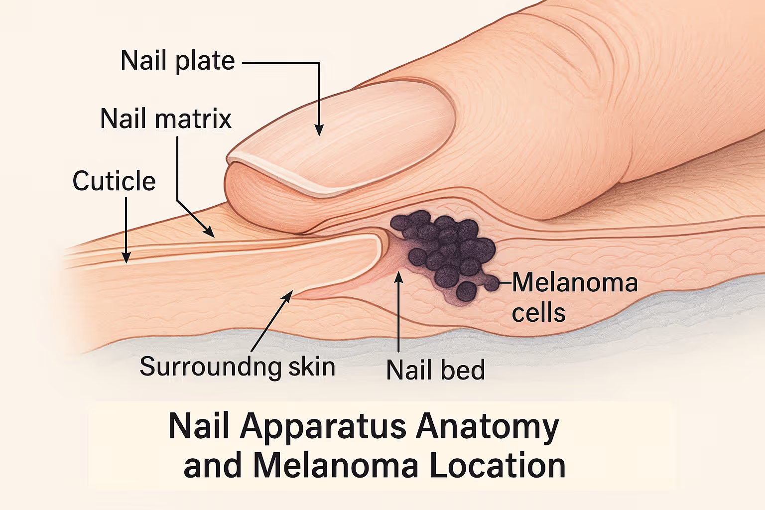
Nail apparatus melanoma develops in the melanocytes—pigment-producing cells—located in the nail matrix, nail bed, or surrounding skin. Unlike melanomas that appear on your arms or back, NAM hides beneath the protective covering of your nail plate, making it incredibly difficult to spot in its early stages.

Here's what makes NAM particularly challenging:

  • 🔍 Hidden location: The nail plate obscures direct visualization of the underlying tissue
  • Delayed diagnosis: Average time from symptom onset to diagnosis is often 2-3 years
  • 🎭 Master of disguise: NAM mimics benign conditions like fungal infections, warts, or bruises
  • 📊 Higher risk in certain populations: More common in individuals with darker skin tones, where it can represent up to 75% of all melanomas

The nail apparatus consists of several components: the nail matrix (where nail growth begins), the nail bed (tissue beneath the nail plate), the nail plate itself, and the surrounding folds of skin. Melanoma can arise in any of these structures, though it most commonly originates in the nail matrix.

Who's at Risk?

While anyone can develop nail apparatus melanoma, certain factors increase your risk:

Demographics and Risk Factors:

Risk FactorDetailsAgeMost common in people aged 50-70 yearsEthnicityHigher proportional incidence in Asian, African, and Hispanic populationsLocationThumb and big toe account for 75-90% of casesPrevious traumaHistory of nail injury may increase risk, though connection remains controversialGenetic factorsFamily history of melanoma increases overall riskRadiation exposurePrevious radiation treatment to hands or feet

It's crucial to understand that nail apparatus melanoma doesn't follow the same sun-exposure pattern as cutaneous melanoma. You won't prevent it by applying sunscreen to your nails. Instead, vigilance and early recognition of suspicious changes are your best defense.

Recognizing the Warning Signs: The ABCDEF Rule for Nails

Infographic in 1536x1024 landscape format showing the ABCDEF criteria for nail melanoma. Six distinct sections arranged in a grid (2 rows, 3

Early detection dramatically improves outcomes for nail apparatus melanoma. That's why understanding the warning signs is so important—for you, your loved ones, and even your healthcare providers.

The ABCDEF Rule: Your Diagnostic Roadmap

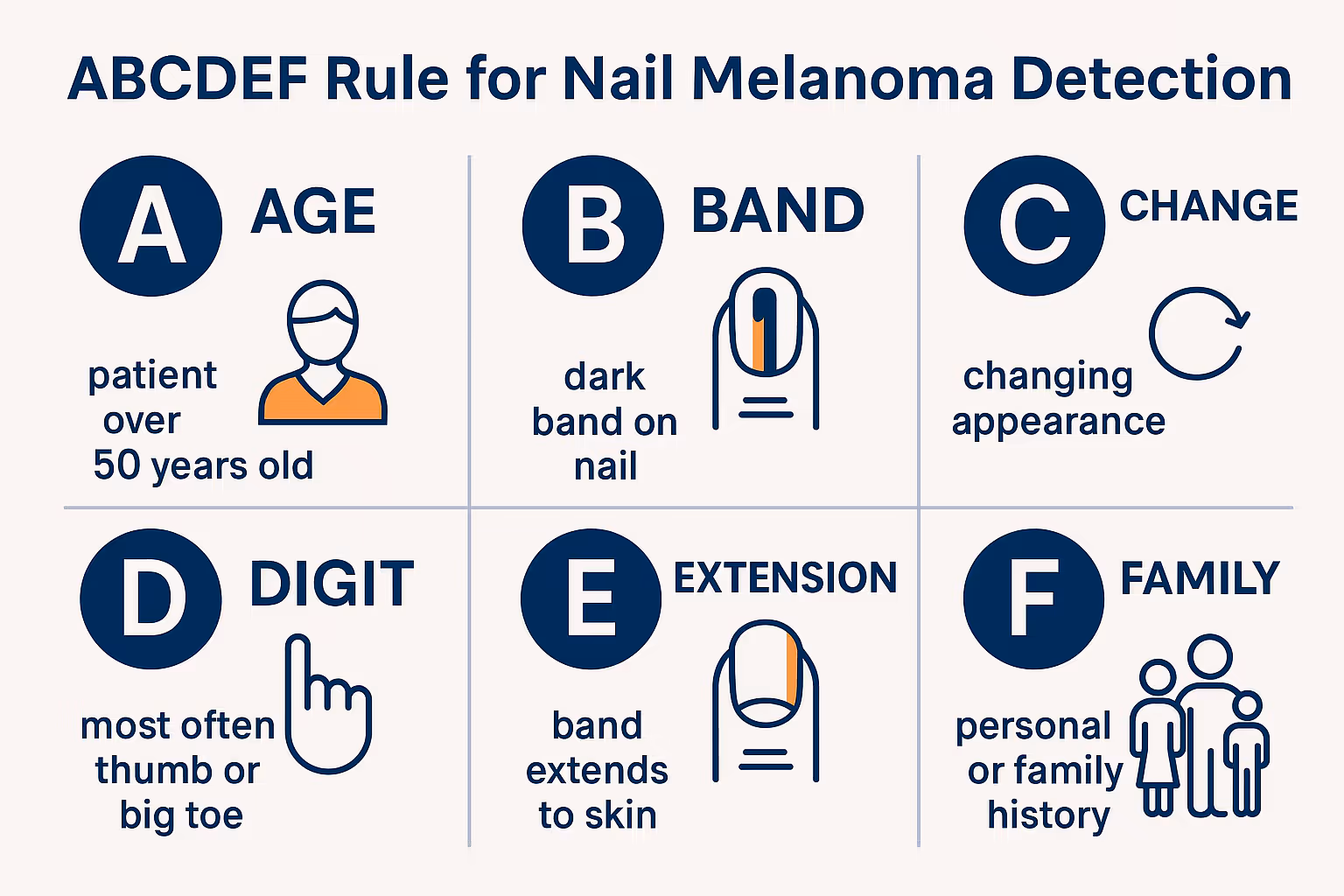
Medical professionals use the ABCDEF criteria to evaluate suspicious nail pigmentation:

A - Age and Ancestry

  • Peak incidence in individuals aged 50-70
  • Higher proportional rates in African, Asian, Native American, and Hispanic populations

B - Band (Brown-Black)

  • Pigmented band measuring 3mm or wider
  • Irregular borders with color variation
  • Darkening or widening over time

C - Change

  • Any change in size, shape, or color
  • Rapid growth or evolution of the pigmented area
  • New symptoms like bleeding or pain

D - Digit

  • Thumb, index finger, or big toe most commonly affected
  • Single digit involvement (multiple digits suggest benign causes)

E - Extension

  • Hutchinson's sign: Pigmentation extending onto the nail fold, cuticle, or fingertip
  • This is one of the most specific indicators of melanoma

F - Family or Personal History

  • Previous melanoma diagnosis
  • Family history of melanoma or dysplastic nevus syndrome

"Hutchinson's sign—pigmentation spreading beyond the nail onto the surrounding skin—should always prompt immediate medical evaluation. It's not something to watch and wait on." — Expert consensus from dermatologic oncology

Beyond the ABCDEF: Other Red Flags

Pay attention to these additional warning signs:

  • 🩸 Nail dystrophy: Splitting, cracking, or destruction of the nail plate
  • 📏 Width progression: A streak that widens from cuticle to nail tip
  • 🎨 Color variation: Multiple shades within the same band (brown, black, blue, or gray)
  • 💧 Spontaneous bleeding: Without obvious trauma
  • 🔴 Amelanotic presentation: Rare cases appear pink or flesh-colored rather than pigmented

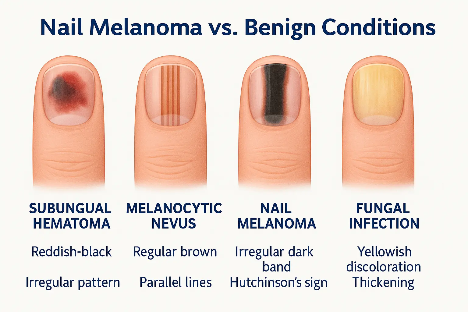
Not every dark streak on your nail is melanoma. In fact, most aren't. Benign conditions like subungual hematoma (blood under the nail), fungal infections, and melanocytic nevi are far more common. However, when in doubt, get it checked out. The Minor Surgery Center specializes in evaluating and treating concerning skin lesions, including those affecting the nail apparatus.

Differential Diagnosis: What Else Could It Be?

One of the greatest challenges in managing nail apparatus melanoma is distinguishing it from benign conditions that look remarkably similar. Understanding these lookalikes helps explain why diagnosis can be so challenging.

Common Benign Conditions That Mimic NAM

1. Subungual Hematoma (Blood Under the Nail)

  • Caused by trauma or repetitive pressure
  • Typically red-purple initially, darkening to brown-black
  • Moves forward as the nail grows
  • Dermoscopy shows reddish-black globules

2. Melanocytic Nevus (Mole)

  • Benign collection of melanocytes
  • Usually presents as regular, evenly spaced brown streaks
  • Stable over time
  • No Hutchinson's sign or nail dystrophy

3. Onychomycosis (Fungal Infection)

  • Yellow-brown discoloration
  • Nail thickening and crumbling
  • Often affects multiple nails
  • May have associated scaling

4. Subungual Bowen's Disease

  • A form of squamous cell carcinoma in situ
  • Can present with melanonychia
  • Often accompanied by nail deformity
  • Requires biopsy for definitive diagnosis

5. Onychopapilloma

  • Benign tumor of the nail bed
  • Irregular melanonychia in the lateral nail fold
  • Localized nail splitting
  • Dermoscopy shows specific patterns

6. Drug-Induced Melanonychia

  • Medications like antimalarials, chemotherapy agents, or antiretrovirals
  • Usually affects multiple nails
  • Develops after starting medication
  • Resolves after discontinuation

Why Dermoscopy Changes Everything

Dermoscopy—examination using a specialized magnifying device—has revolutionized the evaluation of nail pigmentation. This non-invasive technique allows your doctor to see patterns invisible to the naked eye.

Key dermoscopic features suggesting melanoma:

  • 🔬 Irregular pigmentation: Chaotic arrangement of brown-black lines
  • 📊 Color variation: Multiple colors within the band
  • 🎯 Background pigmentation: Diffuse pigmentation between lines
  • Hutchinson's sign: Extension onto periungual tissue
  • 🔄 Micro-Hutchinson's sign: Visible only with dermoscopy

Benign conditions typically show more regular patterns with evenly spaced, parallel lines and consistent coloration. However, dermoscopy alone cannot definitively rule out melanoma—when suspicion is high, biopsy remains essential.

For concerning skin changes anywhere on your body, including nails, our experienced team can provide expert evaluation and guidance on next steps.

Diagnostic Approach: From Suspicion to Confirmation

Medical illustration in 1024x1024 square format showing a detailed anatomical cross-section of a fingernail and nail apparatus. Clearly labe

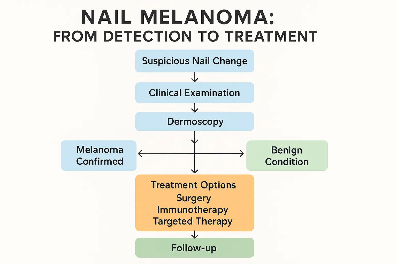
Diagnosing nail apparatus melanoma requires a systematic approach combining clinical examination, imaging, and histopathological confirmation. Here's how the diagnostic journey typically unfolds.

Step 1: Clinical Evaluation

Your doctor will begin with a thorough history and physical examination:

History Questions:

  • When did you first notice the discoloration?
  • Has it changed in size, shape, or color?
  • Any history of trauma to the nail?
  • Are you taking any medications?
  • Do you have a personal or family history of melanoma?

Physical Examination:

  • Inspection of all nails on hands and feet
  • Dermoscopic examination of the affected nail
  • Assessment for Hutchinson's sign
  • Evaluation of regional lymph nodes
  • Full skin examination for other melanomas

Step 2: Dermoscopy and Imaging

Modern diagnostic approaches incorporate several imaging techniques:

Dermoscopy

  • First-line imaging tool
  • Identifies specific patterns associated with melanoma vs. benign conditions
  • Allows documentation for monitoring changes over time

Reflectance Confocal Microscopy (RCM)

  • Emerging technology providing cellular-level imaging
  • Non-invasive "optical biopsy"
  • Particularly useful for monitoring borderline cases

Optical Coherence Tomography (OCT)

  • Provides cross-sectional imaging
  • Helps visualize depth of pigmentation
  • Useful for planning biopsy approach

Step 3: Biopsy—The Gold Standard

When clinical and dermoscopic findings raise suspicion for melanoma, biopsy is essential. However, nail biopsies are technically challenging and should be performed by experienced practitioners.

Biopsy Techniques:

TechniqueWhen UsedAdvantagesDisadvantagesNail matrix biopsySuspected matrix melanomaDirect sampling of origin siteRisk of permanent nail dystrophyNail bed biopsyPigmentation visible after plate removalGood tissue sampleRequires nail plate removalExcisional biopsyHigh suspicion, small lesionComplete removal, definitive diagnosisMore invasive, potential functional impactPunch biopsyLower suspicion, diagnostic uncertaintyLess invasive, preserves functionMay miss diagnosis if sampling error

The biopsy should include adequate depth to assess invasion and should be performed in a way that preserves nail function when possible. At The Minor Surgery Center, procedures are performed with precision and attention to both diagnostic accuracy and cosmetic outcomes.

Step 4: Histopathological Examination

Once tissue is obtained, a dermatopathologist examines it under the microscope to determine:

  • Diagnosis: Melanoma vs. benign condition
  • Breslow thickness: Depth of invasion (critical prognostic factor)
  • Ulceration: Presence or absence affects staging
  • Mitotic rate: Number of dividing cells per area
  • Margins: Whether the lesion was completely removed

Immunohistochemistry (special staining techniques) may be used to confirm the diagnosis, particularly in challenging cases. Markers like S100, SOX10, Melan-A, and HMB-45 help identify melanocytic cells.

Step 5: Staging Workup

If melanoma is confirmed, staging determines the extent of disease and guides treatment:

Local Assessment:

  • Clinical examination of the primary site
  • Assessment of regional lymph nodes

Imaging for Advanced Disease:

  • Sentinel lymph node biopsy: For lesions >1mm thick or with high-risk features
  • CT scans: Chest, abdomen, pelvis for distant metastases
  • PET/CT: May be used for stage III-IV disease
  • MRI: Brain imaging if neurological symptoms or high-risk disease
  • Blood tests: LDH (lactate dehydrogenase) as prognostic marker

Staging follows the American Joint Committee on Cancer (AJCC) TNM system, which considers tumor thickness, nodal involvement, and metastases.

Current Management Strategies: The Treatment Landscape in 2025

Treatment for nail apparatus melanoma has evolved significantly over the past decade. The approach depends on the stage at diagnosis, tumor characteristics, and patient factors. Let's break down the current treatment options.

Surgical Management: The Cornerstone of Treatment

Surgery remains the primary treatment for localized nail apparatus melanoma. The goal is complete tumor removal with adequate margins while preserving as much function as possible.

Wide Local Excision

Margin Recommendations:

Breslow ThicknessRecommended MarginIn situ (confined to epidermis)5-10 mm≤1.0 mm1 cm1.01-2.0 mm1-2 cm>2.0 mm2 cm

For nail apparatus melanoma, achieving these margins often requires:

Digit-Preserving Surgery:

  • Removal of the entire nail apparatus
  • Excision of underlying bone if involved
  • Reconstruction with skin grafts or local flaps
  • Preservation of digit length and function when oncologically safe

Amputation:

  • Required when adequate margins cannot be achieved otherwise
  • Level depends on tumor extent:
    • Distal amputation: Through distal phalanx
    • Middle or proximal amputation: For more extensive disease
  • Modern techniques prioritize functional preservation

"The decision between digit preservation and amputation requires careful consideration of tumor extent, functional outcomes, and patient preferences. When possible, we aim to preserve function without compromising oncologic outcomes." — Surgical oncology perspective

Mohs Micrographic Surgery:

An emerging approach for select cases of nail apparatus melanoma:

  • Layer-by-layer removal with immediate microscopic examination
  • Allows maximum tissue preservation with complete margin assessment
  • Requires specialized expertise in melanoma Mohs surgery
  • Best suited for in situ or thin melanomas

Sentinel Lymph Node Biopsy (SLNB)

For melanomas >1mm thick or with high-risk features (ulceration, high mitotic rate), sentinel lymph node biopsy is recommended:

The Procedure:

  • Radioactive tracer and/or blue dye injected near the primary site
  • Identifies the first lymph node(s) to receive drainage
  • Removed and examined for microscopic metastases
  • Positive findings upstage disease and may affect treatment

Controversy and Evolution:

Recent studies (MSLT-II trial) showed that completion lymph node dissection (removing all regional nodes) after positive SLNB doesn't improve survival compared to observation with ultrasound surveillance. This has shifted practice toward more conservative lymph node management.

Adjuvant Therapy: Reducing Recurrence Risk

For patients with high-risk melanoma (stage IIB-IV), adjuvant therapy after surgery reduces the risk of recurrence and improves survival.

Immunotherapy Options

Pembrolizumab (Keytruda)

  • PD-1 immune checkpoint inhibitor
  • FDA-approved for adjuvant treatment of stage IIB-IV melanoma
  • Given for one year
  • Significantly reduces recurrence risk

Nivolumab (Opdivo)

  • Another PD-1 inhibitor
  • FDA-approved for adjuvant treatment of stage III-IV melanoma
  • Alternative to pembrolizumab

How They Work: These medications release the "brakes" on your immune system, allowing it to recognize and attack melanoma cells more effectively.

Common Side Effects:

  • Fatigue
  • Skin rash
  • Diarrhea
  • Immune-related inflammation (thyroid, liver, lungs, colon)

Most side effects are manageable, but immune-related adverse events require prompt recognition and treatment.

Targeted Therapy

For melanomas with BRAF V600 mutations (found in about 50% of cutaneous melanomas):

Combination Therapy:

  • Dabrafenib + Trametinib (Tafinlar + Mekinist)
  • Vemurafenib + Cobimetinib (Zelboraf + Cotellic)
  • Encorafenib + Binimetinib (Braftovi + Mektovi)

These combinations target specific genetic mutations driving melanoma growth.

Important Notes:

  • BRAF mutation testing required before starting therapy
  • Given for one year in the adjuvant setting
  • Different side effect profile than immunotherapy
  • Effective but typically reserved for BRAF-mutated tumors

Treatment of Advanced Disease

For stage IV melanoma (distant metastases), treatment has been revolutionized by immunotherapy and targeted therapy:

First-Line Options:

  • Immunotherapy (pembrolizumab, nivolumab, or nivolumab + ipilimumab combination)
  • Targeted therapy for BRAF-mutated tumors
  • Clinical trials of novel agents

Response Rates in 2025: Modern therapies achieve objective response rates of 40-60%, with some patients experiencing durable complete responses lasting years.

Radiation Therapy:

  • Used for symptomatic metastases (brain, bone, soft tissue)
  • Palliative treatment to improve quality of life
  • Stereotactic radiosurgery for brain metastases

For comprehensive evaluation and treatment of skin concerns, including suspicious lesions that require expert care, explore our services designed to provide accessible, expert surgical care.

Prognosis and Survival: Understanding the Numbers

Comparison infographic in 1536x1024 landscape format showing four nail conditions side by side. Each shows a realistic illustration of a fin

Prognosis for nail apparatus melanoma depends heavily on the stage at diagnosis. Let's look at the survival data and factors that influence outcomes.

Survival by Stage

Five-Year Survival Rates (Approximate):

StageDescription5-Year SurvivalStage 0In situ (confined to epidermis)~99%Stage IThin melanoma, no spread90-95%Stage IIThicker melanoma, no spread65-90%Stage IIIRegional lymph node involvement40-70%Stage IVDistant metastases15-30%

Important Context: These numbers represent historical data and don't fully reflect the impact of modern immunotherapy and targeted therapy, which have significantly improved outcomes for advanced disease since 2015.

Prognostic Factors

Several factors influence prognosis:

Tumor Characteristics:

  • Breslow thickness: Single most important factor
  • Ulceration: Presence worsens prognosis
  • Mitotic rate: Higher rates indicate more aggressive disease
  • Lymphovascular invasion: Indicates higher metastatic potential

Patient Factors:

  • Age (younger patients generally have better outcomes)
  • Overall health and ability to tolerate treatment
  • Immune system function

Treatment-Related Factors:

  • Adequacy of surgical margins
  • Response to adjuvant therapy
  • Access to specialized care

The Challenge of Late Diagnosis

Unfortunately, nail apparatus melanoma is often diagnosed at more advanced stages compared to cutaneous melanoma:

  • Average diagnostic delay: 2-3 years from symptom onset
  • Proportion presenting with stage III-IV disease: 20-35%
  • Reason: Misdiagnosis as benign conditions, low awareness, hidden location

This underscores the critical importance of awareness, early recognition, and prompt evaluation of suspicious nail changes.

Recurrence and Follow-Up

After treatment, regular follow-up is essential:

Surveillance Schedule:

  • Stage 0-IIA: Every 6-12 months for 5 years, then annually
  • Stage IIB-IV: Every 3-6 months for 2 years, then every 6 months for 3 years, then annually

What Follow-Up Includes:

  • Complete skin examination
  • Examination of surgical site and regional lymph nodes
  • Imaging (CT, PET/CT) for high-risk disease
  • Patient education on self-examination

Recurrence Patterns:

  • Local recurrence at the surgical site
  • In-transit metastases (between primary site and regional nodes)
  • Regional lymph node metastases
  • Distant metastases (lungs, liver, brain, bone)

If you have questions about follow-up care or need guidance on concerning changes, our team is here to help provide clarity and expert evaluation.

Future Perspectives: What's on the Horizon?

The field of melanoma treatment is rapidly evolving. Here are the most promising developments that may change how we manage nail apparatus melanoma in the coming years.

Advanced Diagnostic Technologies

Artificial Intelligence and Machine Learning

  • AI algorithms analyzing dermoscopic images
  • Potential to improve early detection accuracy
  • May help non-specialists identify high-risk lesions
  • Several systems in development and early clinical testing

Molecular Diagnostics

  • Gene expression profiling to predict prognosis
  • Circulating tumor DNA (ctDNA) for monitoring treatment response
  • Liquid biopsies to detect recurrence before clinical manifestation

Enhanced Imaging

  • Line-field confocal optical coherence tomography (LC-OCT)
  • In vivo cellular imaging without biopsy
  • Real-time assessment of treatment response

Novel Therapeutic Approaches

Oncolytic Virus Therapy

  • T-VEC (talimogene laherparepvec): Already FDA-approved for injectable melanoma metastases
  • Viruses engineered to selectively infect and kill cancer cells
  • Stimulates immune response against melanoma
  • Future applications may include adjuvant settings

Cellular Therapies

  • Tumor-infiltrating lymphocyte (TIL) therapy: Harvesting, expanding, and reinfusing patient's own anti-tumor immune cells
  • CAR-T cell therapy: Engineering T cells to target melanoma antigens
  • Showing promise in clinical trials for advanced melanoma

Next-Generation Immunotherapy

  • Novel checkpoint inhibitors (LAG-3, TIM-3, TIGIT)
  • Combination strategies to overcome resistance
  • Personalized neoantigen vaccines
  • Oncolytic viruses combined with checkpoint inhibitors

Targeted Therapy Advances

  • Inhibitors for non-BRAF mutations (NRAS, KIT, NF1)
  • Overcoming resistance mechanisms
  • Combination strategies targeting multiple pathways

Precision Medicine and Biomarkers

The future of melanoma treatment is increasingly personalized:

Genomic Profiling:

  • Comprehensive tumor sequencing to identify actionable mutations
  • Matching patients to targeted therapies based on their tumor's genetic profile
  • Identifying resistance mechanisms before they cause treatment failure

Predictive Biomarkers:

  • Identifying who will respond to immunotherapy
  • Tumor mutational burden (TMB)
  • PD-L1 expression
  • Microbiome analysis
  • Circulating immune cells

Response Monitoring:

  • ctDNA to detect minimal residual disease
  • Early identification of treatment resistance
  • Real-time treatment adjustment

Surgical Innovation

Minimally Invasive Techniques:

  • Refined digit-sparing approaches
  • Improved reconstruction methods
  • Better functional outcomes without compromising oncologic control

Intraoperative Imaging:

  • Real-time margin assessment
  • Fluorescence-guided surgery
  • Confocal microscopy during surgery

Prevention and Early Detection

Screening Programs:

  • Targeted screening for high-risk populations
  • Mobile dermoscopy clinics
  • Telemedicine for remote evaluation

Public Awareness:

  • Education campaigns about nail melanoma
  • Training primary care providers and nail technicians
  • Self-examination tools and apps

Risk Stratification:

  • Genetic testing for high-risk individuals
  • Polygenic risk scores
  • Personalized surveillance strategies

Clinical Trial Landscape

Numerous clinical trials are currently investigating:

  • Neoadjuvant immunotherapy (before surgery)
  • Novel drug combinations
  • Biomarker-driven treatment selection
  • Minimal residual disease monitoring
  • Quality of life interventions

Participating in clinical trials may provide access to cutting-edge treatments while advancing scientific knowledge. Ask your oncologist about appropriate trials for your situation.

Living with Nail Apparatus Melanoma: Practical Guidance

Treatment pathway flowchart in 1536x1024 landscape format. Start with "Suspicious Nail Change" at top, flowing down through decision points

A diagnosis of nail apparatus melanoma brings not only medical challenges but also emotional, functional, and practical considerations. Here's guidance for navigating life during and after treatment.

Coping with Diagnosis

Receiving a melanoma diagnosis is overwhelming. Here are strategies that help:

Emotional Support:

  • 💬 Connect with support groups (in-person or online)
  • 🧠 Consider counseling or therapy
  • 👨‍👩‍👧‍👦 Lean on family and friends
  • 📚 Educate yourself, but avoid information overload

Practical Steps:

  • Create a medical binder with all records, test results, and treatment plans
  • Bring a trusted person to appointments for support and note-taking
  • Write down questions before appointments
  • Keep a symptom diary

Trusted Resources:

  • American Cancer Society
  • Melanoma Research Foundation
  • Skin Cancer Foundation
  • Cancer Support Community

Managing Treatment Side Effects

Different treatments come with different challenges:

Surgical Recovery:

  • Follow wound care instructions carefully
  • Elevate the affected extremity to reduce swelling
  • Attend all follow-up appointments
  • Engage in recommended physical or occupational therapy

Immunotherapy Side Effects:

  • Report new symptoms promptly
  • Carry a card identifying you as an immunotherapy patient
  • Stay hydrated and maintain good nutrition
  • Don't ignore seemingly minor symptoms—they may indicate immune-related inflammation

Targeted Therapy Side Effects:

  • Manage skin changes with gentle skincare
  • Protect from sun exposure
  • Report fever or signs of infection
  • Maintain regular monitoring appointments

Functional Adaptation

Amputation or extensive surgery may require adjustments:

Occupational Therapy:

  • Learn adaptive techniques for daily tasks
  • Explore assistive devices
  • Maintain independence and quality of life

Prosthetics:

  • Cosmetic prosthetics for fingers or toes
  • Functional prosthetics for improved grip or balance
  • Custom-designed options available

Psychological Adjustment:

  • Body image concerns are normal
  • Professional counseling can help
  • Connect with others who've had similar experiences

Surveillance and Self-Examination

After treatment, you play a crucial role in monitoring for recurrence:

Self-Examination:

  • Examine all nails monthly
  • Check surgical site for changes
  • Perform full-body skin checks
  • Use the ABCDE criteria for new or changing lesions

What to Report Immediately:

  • New lumps or bumps near the surgical site
  • New or changing skin lesions anywhere
  • Persistent cough, headaches, or bone pain
  • Any concerning symptoms

Sun Protection: While nail melanoma isn't caused by sun exposure, protecting your skin from UV damage reduces risk of other skin cancers:

  • Use broad-spectrum SPF 30+ sunscreen
  • Wear protective clothing
  • Avoid tanning beds
  • Seek shade during peak sun hours

Nutrition and Lifestyle

While no specific diet prevents melanoma recurrence, healthy lifestyle choices support overall health:

Nutrition:

  • Eat a balanced diet rich in fruits, vegetables, and whole grains
  • Stay well-hydrated
  • Maintain a healthy weight
  • Limit alcohol consumption

Physical Activity:

  • Regular exercise improves outcomes and quality of life
  • Start slowly and build gradually
  • Choose activities you enjoy
  • Consult your doctor before starting new exercise programs

Stress Management:

  • Practice relaxation techniques (meditation, yoga, deep breathing)
  • Maintain social connections
  • Engage in hobbies and activities you enjoy
  • Get adequate sleep

For any concerning skin changes or questions about minor surgical procedures, reach out to our team for compassionate, expert guidance.

Frequently Asked Questions About Nail Apparatus Melanoma

Can nail melanoma be cured?

Yes, when caught early. In situ melanoma (confined to the top layer) has a cure rate near 99%. Even invasive melanomas have excellent cure rates when diagnosed at stage I or II. The key is early detection and appropriate treatment.

Does removing the nail help diagnose melanoma?

Sometimes nail plate removal (avulsion) is necessary to visualize the underlying nail bed and matrix. This allows better examination and, if needed, biopsy. The nail will regrow, though it may take 4-6 months.

Will I lose my finger or toe?

Not necessarily. Many nail melanomas can be treated with digit-preserving surgery, especially when caught early. Amputation is reserved for cases where adequate margins cannot be achieved otherwise or when the tumor extensively involves bone or other structures.

How is nail melanoma different from regular melanoma?

The biology is similar, but the location creates unique challenges. Nail melanoma is hidden beneath the nail plate, making early detection difficult. It's also more common in people with darker skin tones, where it represents a higher proportion of all melanomas.

Can nail polish or gel manicures cause melanoma?

There's no strong evidence that nail polish or gel manicures cause melanoma. However, nail polish can hide suspicious changes, and UV lamps used for gel nails expose skin to UV radiation (though the cancer risk appears very low). If you have a suspicious nail change, remove polish for medical evaluation.

Should I see a dermatologist or a surgeon first?

Start with a dermatologist for evaluation. If melanoma is suspected or confirmed, you'll likely be referred to a surgical oncologist or Mohs surgeon for definitive treatment. A multidisciplinary approach often provides the best outcomes.

What if my biopsy comes back as "atypical" or "uncertain"?

Nail lesions can be challenging to diagnose pathologically. If the diagnosis is uncertain, options include:

  • Second opinion from a specialized dermatopathologist
  • Complete excision for definitive diagnosis
  • Close monitoring with serial dermoscopy
  • Molecular testing to help clarify diagnosis

Can melanoma come back after treatment?

Yes, melanoma can recur. That's why regular follow-up is essential. Recurrence risk depends on the original stage—higher stage disease has greater recurrence risk. Modern adjuvant therapies significantly reduce recurrence rates.

Is nail melanoma genetic?

While most cases aren't inherited, having a family history of melanoma increases your risk. Genetic syndromes like familial atypical multiple mole melanoma (FAMMM) syndrome increase melanoma risk, though they more commonly affect skin than nails.

Can children get nail melanoma?

Extremely rarely. Nail melanoma is predominantly a disease of adults, with peak incidence in the 50-70 age range. Pigmented bands in children are almost always benign, but any concerning changes should still be evaluated.

If you have additional questions or concerns about nail changes or other skin lesions, our FAQ section provides helpful information, and our team is always available for consultation.

Taking Action: Your Next Steps

Modern medical graphic in 1024x1024 square format showing future perspectives. Center image of a stylized DNA helix or molecular structure,

Knowledge is power, but only when combined with action. If you've read this far, you're already taking an important step in protecting your health. Here's what to do next.

If You Notice Suspicious Nail Changes

Don't wait and watch indefinitely. While not every dark streak is melanoma, concerning features warrant prompt evaluation:

  1. Document the change: Take clear photos with good lighting
  2. Note the timeline: When did you first notice it? Has it changed?
  3. Schedule an evaluation: See a dermatologist or qualified healthcare provider
  4. Be prepared: Bring your photos, list of medications, and relevant medical history

Red flags requiring urgent evaluation:

  • Hutchinson's sign (pigmentation extending beyond the nail)
  • Rapid growth or change
  • Bleeding or ulceration
  • Width ≥3mm
  • Irregular borders or multiple colors

If You've Been Diagnosed

Take a deep breath. A melanoma diagnosis is serious, but you're not alone, and treatment options are better than ever.

Immediate steps:

  1. Assemble your team: Dermatologist, surgical oncologist, medical oncologist
  2. Get all the information: Pathology report, staging workup results
  3. Explore treatment options: Understand risks and benefits of each approach
  4. Seek support: Connect with loved ones, support groups, counseling
  5. Consider a second opinion: Especially for complex cases

Questions to ask your doctors:

  • What stage is my melanoma?
  • What are my treatment options?
  • What are the risks and benefits of each option?
  • What is my prognosis?
  • Should I consider a clinical trial?
  • What follow-up will I need?

For Everyone: Prevention and Awareness

Even if you don't currently have concerning nail changes, awareness helps:

Monthly self-examination:

  • Examine all nails on hands and feet
  • Look for new pigmentation or changes to existing marks
  • Check the skin around nails for unusual changes
  • Photograph anything concerning

Annual professional skin examinations:

  • Full-body skin check by a dermatologist
  • Especially important if you have risk factors
  • Provides baseline for comparison

Spread awareness:

  • Share information about nail melanoma with friends and family
  • Nail technicians can be important partners in early detection
  • Educate others about the ABCDEF rule

Finding Expert Care

The Minor Surgery Center specializes in the evaluation and treatment of skin lesions, providing expert care in a comfortable, accessible setting. Whether you need evaluation of a concerning nail change, removal of a suspicious lesion, or guidance on next steps, our experienced team is here to help.

What sets us apart:

  • ✅ Expert surgical care without long wait times
  • ✅ Clear, compassionate communication
  • ✅ Comprehensive evaluation and treatment planning
  • ✅ Focus on both medical outcomes and cosmetic results
  • ✅ Coordination with specialists when needed

Ready to take the next step? Contact us to schedule a consultation. Your skin health deserves expert attention, and we're here to provide it.

Conclusion: Hope Through Knowledge and Action

Nail apparatus melanoma may be rare, but its impact on those affected is profound. The hidden nature of this disease, the diagnostic challenges it presents, and the potential for serious outcomes make it a formidable adversary. Yet, the landscape of melanoma care in 2025 offers genuine hope.

Early detection remains your most powerful weapon. The difference between a curable in situ melanoma and advanced disease often comes down to awareness and timely action. By understanding the warning signs—particularly the ABCDEF criteria and Hutchinson's sign—you can catch this disease when it's most treatable.

Modern treatment options are transforming outcomes. Surgical techniques continue to improve, balancing oncologic control with functional preservation. Immunotherapy and targeted therapy have revolutionized the treatment of advanced disease, turning what was once a rapidly fatal diagnosis into a manageable chronic condition for many patients. Ongoing research promises even better treatments in the years ahead.

You're not alone in this journey. Whether you're monitoring a suspicious nail change, recovering from surgery, or managing advanced disease, a community of healthcare providers, researchers, survivors, and supporters stands ready to help. The Minor Surgery Center is proud to be part of that community, offering expert care delivered with compassion and clarity.

The story of Sarah, which opened this article, has a hopeful ending. Caught at stage IB, her melanoma was successfully treated with digit-preserving surgery. Three years later, she remains cancer-free, vigilant about self-examination, and passionate about sharing her story to help others recognize the warning signs she initially missed.

Your story can have a hopeful ending too. Stay vigilant, seek prompt evaluation of concerning changes, and remember that knowledge combined with action saves lives.

Your next step is clear: If you have any concerning nail changes, don't wait. Schedule an evaluation today. If you're already in treatment, stay committed to your care plan and follow-up schedule. If you're cancer-free, maintain awareness and regular self-examinations.

Your skin tells a story. Make sure you're listening.

For more information about skin conditions, minor surgical procedures, and expert care, visit our blog for helpful resources and updates.

October 9, 2025
🇨🇦 Our clinic currently provides care to patients within Canada only. We apologize for any inconvenience this may cause.