Moles on the Neck and Chest: Cosmetic Concerns vs. Warning Signs
The appearance of moles on the skin is a common human experience. For many, these small, pigmented spots are simply a part of their unique physical landscape, often prompting little more than a passing glance in the mirror. However, when these moles appear on highly visible areas such as the neck and chest, they can become a source of cosmetic concern. Beyond aesthetics, there's a more critical question that often arises: are these moles merely harmless marks, or could they be warning signs of something more serious, like skin cancer? This article, "Moles on the Neck and Chest: Cosmetic Concerns vs Warning Signs," delves into this crucial distinction, providing comprehensive information to help you understand, monitor, and address any concerns you may have about moles in these prominent areas.
Understanding the nature of moles, especially those on the neck and chest, is paramount for both peace of mind and proactive health management. While most moles are benign, it's vital to be aware of the signs that could indicate a need for professional evaluation. This guide will equip you with the knowledge to differentiate between typical moles and those that might require medical attention, ensuring that cosmetic worries don't overshadow potential health risks, and conversely, that every mole isn't a source of undue anxiety.
Key Takeaways
Most moles are harmless: The vast majority of moles on the neck and chest are benign (non-cancerous) and pose no health risk, though they may be a cosmetic concern.
Sun exposure is a factor: Areas like the neck and chest are often exposed to the sun, increasing the risk of both benign and atypical mole development, and emphasizing the need for regular self-checks.
The ABCDE rule is crucial: Familiarize yourself with the ABCDE criteria (Asymmetry, Border, Color, Diameter, Evolving) for identifying potentially dangerous moles, regardless of location.
Cosmetic removal options exist: For benign moles that are bothersome due to their appearance or location, various safe and effective cosmetic removal procedures are available.
Professional evaluation is key: Any mole that exhibits changes, new growth, or fits the ABCDE criteria should be promptly evaluated by a dermatologist or healthcare professional.
Understanding Normal Moles on the Neck and Chest: The Cosmetic Perspective
The neck and chest are areas frequently exposed to sunlight and are often part of our daily visual assessment, making moles here particularly noticeable. Understanding what constitutes a "normal mole" is the first step in differentiating between a cosmetic concern and a potential health issue. Most moles, medically known as nevi (singular: nevus), are common growths on the skin that develop when pigment-producing cells, called melanocytes, grow in clusters. They can appear anywhere on the body, but their presence on the neck and chest carries unique considerations.
What are Moles? A Basic Overview
Moles can vary widely in appearance, even when they are perfectly benign. They can be flat or raised, smooth or rough, and range in color from light tan to dark brown, or even black. Some moles may have hair growing from them. The average adult has between 10 and 40 moles, many of which develop during childhood and adolescence. While some moles are present at birth (congenital nevi), most appear later in life (acquired nevi).
On the neck and chest, moles are particularly common due to sun exposure. The skin here is often thinner and more sensitive than other areas, and clothing choices can also influence friction, leading to irritation or changes in appearance.
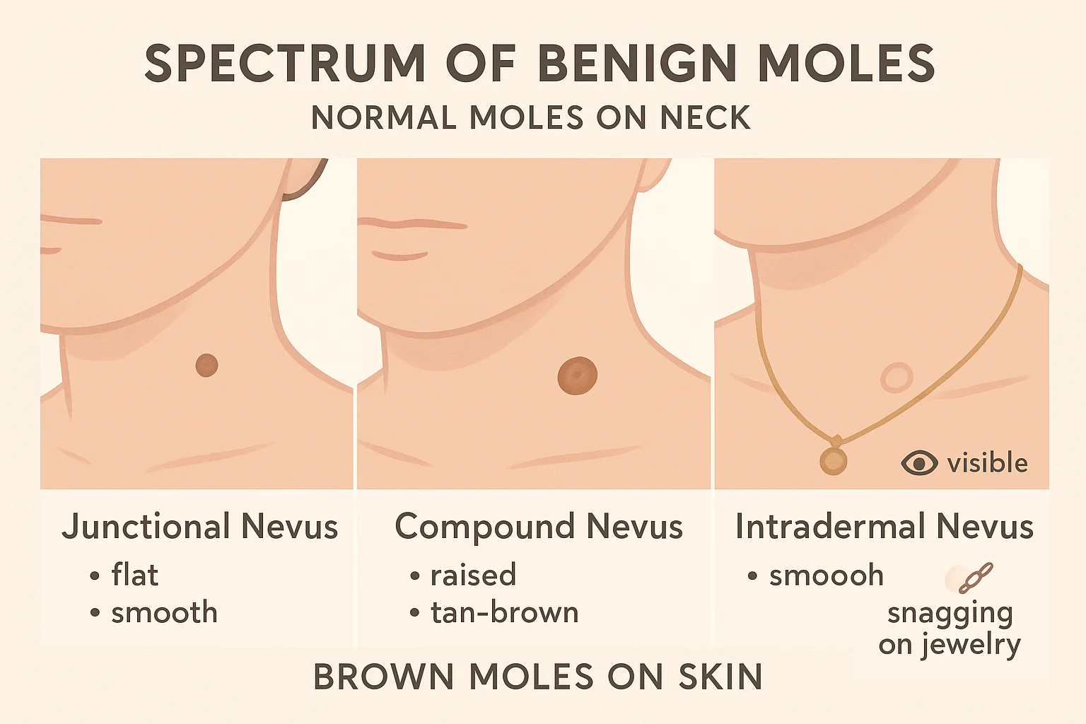
Common Types of Benign Moles on the Neck and Chest
There are several types of benign moles that commonly appear on the neck and chest:
Junctional Nevi: These moles are typically flat or slightly raised, usually round or oval, and uniformly brown or black. They occur at the junction of the epidermis (outer layer of skin) and the dermis (inner layer).
Compound Nevi: These moles have cells in both the epidermis and the dermis, making them slightly raised, often dome-shaped, and usually light to dark brown. They may become lighter or more flesh-colored over time.
Intradermal Nevi: Located entirely within the dermis, these moles are often raised, dome-shaped, and can be flesh-colored, tan, or brown. They may sometimes have hairs growing out of them and tend to soften and lose pigment with age.
Seborrheic Keratoses: While not technically moles, these common, benign skin growths are often mistaken for moles. They are usually waxy, scaly, and slightly raised, appearing in various colors from light tan to black. On the neck and chest, they can be quite common, especially as people age.
Skin Tags (Acrochordons): These are small, soft, benign growths that often hang off the skin by a stalk. They are very common on the neck, armpits, and chest, especially in areas of friction. They are not moles but are often confused with them. If you're concerned about skin tags, you can learn more about banishing flat black skin tags with your complete removal guide.
Why Moles on the Neck and Chest Become Cosmetic Concerns
For many individuals, the primary concern with moles on the neck and chest is their visibility. Unlike moles on the back or legs, those in these areas are often exposed, making them a focal point for cosmetic worries.
Aesthetic Impact: A mole that is large, unusually dark, or raised can be perceived as visually unappealing, especially if it's prominently located on the décolletage or neck.
Interference with Jewelry and Clothing: Moles on the neck can often get snagged by necklaces, causing irritation, bleeding, or discomfort. Similarly, moles on the chest might rub against clothing, leading to similar issues. This constant friction can sometimes cause changes in the mole's appearance, which, while usually benign, can lead to anxiety.
Social and Psychological Effects: Individuals may feel self-conscious about highly visible moles, affecting their confidence in social settings, particularly when wearing certain types of clothing.
When Cosmetic Removal is an Option
If a mole on your neck or chest is benign but causing cosmetic distress or physical irritation (like snagging on jewelry), removal is often an option. It's crucial to first have the mole examined by a healthcare professional to confirm its benign nature. Once confirmed, various removal methods are available.
Surgical Excision: This involves cutting out the mole and a small margin of surrounding skin. The wound is then closed with stitches. This method is often preferred for moles that are raised or larger, and it allows for pathological examination of the mole. Learn more about mole removal procedures.
Shave Excision: For raised moles, a shave excision involves using a surgical blade to "shave off" the mole so it's level with the surrounding skin. This method typically doesn't require stitches and results in a flat scar.
Laser Removal: Some flat, brown moles on the skin that are purely cosmetic concerns may be suitable for laser treatment, which uses focused light energy to break down the pigment. This is less common for suspicious moles.
It's important to discuss the pros and cons of each method with a qualified practitioner, including potential scarring, recovery time, and cost. Always ensure that any mole removal is performed by a professional, especially for visible areas like the neck and chest, to minimize scarring and ensure proper diagnosis. For those seeking expert care, centers like The Minor Surgery Center offer specialized services for mole and cyst removal.
Identifying Warning Signs: Moles on the Neck and Chest as Indicators of Concern
While many moles on the neck and chest are harmless, it's critically important to be vigilant for changes or characteristics that could signal a more serious underlying issue, such as melanoma—the most dangerous form of skin cancer. Due to the high sun exposure these areas often receive, they are particularly susceptible to sun damage, which is a significant risk factor for skin cancer development. Knowing how to identify the warning signs is essential for early detection and successful treatment.
The Importance of Self-Checks and Professional Screenings
Regular self-skin exams are vital. Ideally, you should check your skin head-to-toe once a month, paying close attention to moles on the neck and chest. Due to the difficulty of examining certain areas of your back or scalp, consider enlisting a partner or using a full-length mirror and a hand mirror.
In addition to self-checks, annual professional skin screenings with a dermatologist are highly recommended, especially for individuals with multiple moles, a history of sunburns, or a family history of skin cancer. A dermatologist can identify suspicious lesions that might be missed during a self-exam. For those in the Vaughan area, you might consider visiting the best skin cancer clinic for expert evaluation.
The ABCDE Rule: Your Guide to Detecting Suspicious Moles
The universally recognized ABCDE rule is a simple yet powerful tool for identifying potential signs of melanoma. This mnemonic helps you remember what to look for when examining moles on your neck, chest, or any other part of your body.
A – Asymmetry:
What it means: Benign moles are typically symmetrical, meaning if you draw a line through the middle, both halves would match. A mole that is asymmetrical—where one half does not match the other—is a warning sign.
Visual Cue: Imagine folding the mole in half; if the two sides look different, it's asymmetrical.
B – Border Irregularity:
What it means: Normal moles usually have smooth, even borders. Melanoma often presents with irregular, notched, scalloped, or poorly defined borders.
Visual Cue: Look for borders that are fuzzy, jagged, or fading into the surrounding skin rather than having a clear, sharp edge.
C – Color Variation:
What it means: A benign mole is usually a single shade of brown or tan. A mole with color variation—different shades of brown, tan, black, red, white, or blue within the same lesion—is a significant warning sign.
Visual Cue: Be suspicious of moles that are multi-colored or have uneven distribution of pigment.
D – Diameter:
What it means: Melanomas are often, but not always, larger than 6 millimeters (about the size of a pencil eraser) when diagnosed. While benign moles can also be large, a mole that is growing in size or is larger than 6mm warrants attention.
Visual Cue: Use a pencil eraser or a ruler to measure moles that appear suspicious.
E – Evolving (Changing):
What it means: This is arguably the most crucial criterion. Any change in an existing mole—in size, shape, color, elevation, or any new symptoms like bleeding, itching, or crusting—is a serious warning sign. This also applies to new moles that appear and change rapidly.
Visual Cue: If a mole on your neck or chest suddenly feels different (itchy, tender), looks different (changing color, growing), or bleeds without trauma, it needs immediate professional evaluation. This 'E' also encompasses 'Evolution' over time.
It's important to remember that not every mole exhibiting one of these signs is cancerous, but it does warrant a professional check. Conversely, a mole can be melanoma even if it doesn't fit all the ABCDE criteria. Therefore, any suspicious mole or new growth should be seen by a doctor.
Other Red-Flag Features and Symptoms
Beyond the ABCDEs, there are other characteristics and symptoms that should prompt a visit to a healthcare provider:
New Moles Appearing in Adulthood: While new moles can appear, especially in young adults, a significant number of new moles appearing later in life, particularly if they are atypical, should be monitored.
"Ugly Duckling" Sign: This concept suggests that a mole that looks different from all the other moles on your body – an "ugly duckling" among a flock of "swans" – is a cause for concern.
Soreness, Itching, or Tenderness: Benign moles typically do not cause discomfort. If a mole on your neck or chest starts to itch, become tender to the touch, or feels sore, it needs to be checked.
Bleeding, Crusting, or Oozing: Moles that bleed spontaneously (without being picked or scratched), crust over, or ooze fluid are highly suspicious and require immediate medical attention.
Moles that Feel Lumpy or Firm: A mole that changes in texture, becoming firm, hard, or lumpy, especially if it was previously soft or flat.
Types of Skin Cancer to Be Aware Of
While melanoma is the most concerning, other types of skin cancer can also appear as moles or lesions on the neck and chest:
Basal Cell Carcinoma (BCC): This is the most common type of skin cancer. It often appears as a pearly or waxy bump, a flat, flesh-colored or brown scar-like lesion, or a bleeding sore that heals and then returns. On the neck and chest, it can sometimes be mistaken for a normal mole or a persistent pimple. You can learn more about Basal Cell Carcinoma treatment.
Squamous Cell Carcinoma (SCC): The second most common type, SCC often appears as a firm, red nodule, or a flat, scaly, crusty lesion. It can be painful or tender and may bleed. These are also common in sun-exposed areas like the neck and chest. For more information, refer to actinic keratosis to SCC: understanding pre-cancerous lesions.
Melanoma: This is the most dangerous form of skin cancer, as it can spread aggressively if not detected and treated early. It often resembles an atypical mole, exhibiting the ABCDE signs. Early detection is critical for survival. Understanding advanced melanoma stages highlights the importance of early diagnosis.
Risk Factors for Skin Cancer on the Neck and Chest
Several factors increase the risk of developing skin cancer, especially in sun-exposed areas:
Excessive Sun Exposure: Frequent sunburns, particularly during childhood, and cumulative sun exposure over time significantly increase risk. The neck and chest are often unprotected.
Fair Skin and Light Hair/Eye Color: Individuals with lighter skin tones, red or blonde hair, and blue or green eyes have less protective melanin and are more susceptible to UV damage.
Numerous Moles or Atypical Moles: Having many moles (over 50) or several atypical (dysplastic) moles increases your risk of melanoma.
Family History of Melanoma: If a close family member (parent, sibling, child) has had melanoma, your risk is elevated.
Weakened Immune System: Conditions or medications that suppress the immune system can increase skin cancer risk.
Age: The risk of skin cancer generally increases with age, though melanoma can occur at any age.
Previous Skin Cancer: If you've had one skin cancer, you're at higher risk for developing another.
Given the prevalence of moles on the neck and chest and the significant impact of sun exposure on these areas, diligent monitoring and prompt professional evaluation of any suspicious changes are paramount. Early detection is truly the best defense against skin cancer.
Diagnosis and Management of Moles on the Neck and Chest
When you notice a mole on your neck or chest that raises concerns, whether cosmetic or medical, the next step is typically a professional evaluation. A dermatologist is the most appropriate specialist to assess skin lesions and provide an accurate diagnosis. Understanding the diagnostic process and available management options can help alleviate anxiety and guide your decisions.
What to Expect During a Dermatological Examination
During a visit to a dermatologist for a mole assessment, you can expect a thorough examination:
Medical History: The dermatologist will ask about your personal and family history of skin cancer, your sun exposure habits, and any changes you've noticed in the mole(s) in question.
Visual Inspection: The doctor will perform a full-body skin examination, not just focusing on the mole you're concerned about. They will visually inspect your entire skin surface, often using a bright light.
Dermoscopy: For suspicious moles, the dermatologist will use a handheld device called a dermatoscope. This tool magnifies the mole and illuminates its internal structures, allowing for a more detailed assessment of pigment patterns, blood vessels, and other features that are not visible to the naked eye. This non-invasive procedure significantly aids in differentiating benign moles from malignant ones.
Photography (Optional): Some clinics use mole mapping technology or take clinical photographs of suspicious moles to monitor changes over time, particularly for individuals with many moles. You can explore whether 3-D mole mapping apps are reliable.
When a Biopsy is Necessary
If, after the visual and dermoscopic examination, a mole on your neck or chest is deemed suspicious, the dermatologist will likely recommend a skin biopsy. A biopsy is the only definitive way to diagnose skin cancer.
Procedure: A biopsy involves removing a small sample of the mole (or the entire mole) for microscopic examination by a pathologist. This is usually a quick, in-office procedure performed under local anesthesia.
Types of Biopsies:
Shave Biopsy: For raised lesions, a thin slice of the mole is shaved off the skin's surface.
Punch Biopsy: A circular tool is used to remove a small cylinder of tissue, including deeper layers of skin.
Excisional Biopsy: The entire mole, along with a small margin of surrounding healthy skin, is surgically removed. This is often preferred for highly suspicious lesions or suspected melanomas, as it provides the most comprehensive tissue sample for diagnosis. This is often the procedure performed at centers like The Minor Surgery Center.
Results: The pathologist's report will determine if the mole is benign, atypical (dysplastic nevus), or cancerous (basal cell carcinoma, squamous cell carcinoma, or melanoma).
Interpreting Biopsy Results and Next Steps
The biopsy results will dictate the subsequent course of action:
Benign Mole: If the mole is confirmed benign, no further treatment may be necessary, especially if it's not causing cosmetic distress or irritation. If it was removed due to cosmetic concerns, the procedure is complete.
Atypical Nevus (Dysplastic Mole): These moles are not cancerous but have features that are abnormal and could potentially evolve into melanoma over time. Depending on the degree of atypia and the clarity of the margins, your doctor might recommend a wider excision to ensure all atypical cells are removed, or simply recommend regular monitoring.
Skin Cancer Diagnosis: If the biopsy confirms skin cancer (BCC, SCC, or melanoma), further treatment will be necessary.
Basal Cell Carcinoma and Squamous Cell Carcinoma: These are typically treated by surgical excision, Mohs surgery (a specialized technique that removes cancerous tissue layer by layer while sparing healthy tissue), or other methods like cryotherapy, curettage and electrodesiccation, or topical medications, depending on the type, size, and location of the cancer.
Melanoma: Treatment for melanoma usually involves surgical removal of the melanoma with a wider margin of healthy tissue (wide local excision). In some cases, sentinel lymph node biopsy, radiation therapy, chemotherapy, targeted therapy, or immunotherapy may be required, particularly for more advanced stages. Early detection is critical for managing melanoma effectively. Finding the best melanoma specialists in Toronto can be crucial for optimal outcomes.
Post-Removal Care and Scarring Considerations
Regardless of whether a mole is removed for cosmetic reasons or due to a cancer diagnosis, proper post-removal care is essential for healing and minimizing scarring.
Wound Care: Follow your doctor's instructions meticulously regarding wound cleaning, dressing changes, and activity restrictions. This is especially important for moles on the neck and chest, where movement can affect healing.
Sun Protection: Protect the healing area from direct sun exposure, as UV radiation can worsen scarring and potentially cause discoloration. Use sunscreen (SPF 30 or higher) and protective clothing.
Scar Management: Scarring is an inevitable part of any surgical procedure. The extent of the scar depends on the size and depth of the mole, the removal method, the location on the body, and individual healing characteristics. On the neck and chest, scars can sometimes be more noticeable due to skin tension.
Massage: Gently massaging the scar once the wound has healed can help improve its appearance and flexibility.
Silicone Products: Silicone sheets or gels can be effective in reducing scar prominence.
Topical Treatments: Certain creams and oint recommended by your doctor may aid in scar maturation.
Advanced Scar Revision: For problematic or highly visible scars, procedures like laser therapy, steroid injections, or surgical scar revision may be considered after the initial healing period. More information on improving older scars can be found in our blog.
The Role of Prevention: Protecting Your Skin
Prevention is always better than cure, especially when it comes to skin cancer. Adopting sun-safe practices is the most effective way to reduce your risk of developing new atypical moles or skin cancers on your neck and chest.
Seek Shade: Especially between 10 AM and 4 PM when UV rays are strongest.
Wear Protective Clothing: Broad-brimmed hats, sunglasses, and clothing with UPF (Ultraviolet Protection Factor) can shield your neck and chest.
Use Sunscreen Daily: Apply broad-spectrum sunscreen with an SPF of 30 or higher liberally to all exposed skin, including your neck and chest, every day, even on cloudy days. Reapply every two hours, or more often if swimming or sweating.
Avoid Tanning Beds: Tanning beds emit harmful UV radiation and significantly increase the risk of skin cancer.
Regular Self-Exams: Continue to perform monthly self-checks to monitor existing moles and detect new ones.
Annual Professional Skin Exams: Schedule annual check-ups with a dermatologist, particularly if you have risk factors.
By understanding the diagnostic process, being aware of management options, and prioritizing preventive measures, you can effectively address concerns about moles on your neck and chest, ensuring both your cosmetic preferences and your health are well-protected.
Conclusion
Moles on the neck and chest are a common feature of human skin, often presenting as harmless spots that might occasionally cause cosmetic concern or minor irritation. However, it is crucial to recognize that these highly visible and sun-exposed areas can also harbor moles that signal a more serious underlying health risk, specifically skin cancer. Navigating the distinction between benign cosmetic concerns and potentially dangerous warning signs is a vital aspect of proactive skin health management in 2025.
The journey begins with education and self-awareness. Familiarizing yourself with the characteristics of normal moles on the neck and brown moles on the skin, as discussed in this article, provides a baseline for understanding your own skin. The "ugly duckling" sign and the comprehensive ABCDE rule—Asymmetry, Border irregularity, Color variation, Diameter greater than 6mm, and most critically, Evolving changes—are indispensable tools for identifying moles that warrant closer inspection. Any mole that deviates from its previous appearance or from the typical presentation of your other moles should immediately raise a red flag.
While cosmetic considerations for benign moles on the neck and chest are perfectly valid and can be addressed through various removal methods, they should never overshadow the importance of health vigilance. If a mole is bothering you aesthetically, always consult a medical professional first to confirm its benign nature before proceeding with any removal. Options ranging from surgical excision to shave excision or laser removal are available at specialized centers like The Minor Surgery Center, but a proper diagnosis is the foundational step.
Conversely, if a mole on your neck or chest exhibits any of the warning signs discussed, prompt professional evaluation is not just recommended, it is essential. Early detection of skin cancer, particularly melanoma, significantly improves prognosis and treatment outcomes. A dermatologist can perform a thorough examination, utilize dermoscopy, and if necessary, conduct a biopsy to provide a definitive diagnosis.
In 2025 and beyond, taking charge of your skin health means embracing a holistic approach: regular self-examinations, annual professional skin screenings, diligent sun protection, and immediate consultation for any suspicious changes. By empowering yourself with knowledge and partnering with healthcare professionals, you can confidently distinguish between moles that are merely cosmetic nuances and those that serve as critical warning signs, ensuring both your appearance and your well-being are protected.
December 11, 2025
🇨🇦
Our clinic currently provides care to patients within
Canada only.
We apologize for any inconvenience this may cause.