Melanoma is a serious type of skin cancer that can be very dangerous if not found and treated early. While it can appear anywhere on the body, the legs are a common spot, especially for women. Many people turn to "melanoma pictures on legs" to understand what to look for, and while these images can be helpful for awareness, it's crucial to know what they truly represent and why professional medical advice is always needed. This comprehensive guide will explore why legs are a common site for melanoma, what signs to watch for, and the critical steps you can take for early detection and prevention. Understanding the visual cues of melanoma on legs is a powerful first step in protecting your skin health.
Key Takeaways
Legs are a common site for melanoma: Especially for women, due to sun exposure and other factors. Regular checks of your legs are vital.
Use the ABCDEs: Learn to spot suspicious moles or spots on your legs by checking for Asymmetry, irregular Borders, varied Colors, large Diameter, and any Evolving changes.
Melanoma can look different: Not all melanomas are dark or follow the ABCDE rule perfectly. Some can be red, pink, skin-colored, or even show up under toenails.
Self-exams are important, but not enough: Regularly check your own skin, including hard-to-see areas on your legs, but always follow up with a dermatologist for professional skin checks.
Pictures are for awareness, not diagnosis: "Melanoma pictures on legs" can help you understand what to look for, but only a doctor can properly diagnose melanoma. If you see something concerning, get it checked by a dermatologist right away.
Understanding Melanoma: The Basics
Melanoma is the most severe form of skin cancer. It develops in the melanocytes, which are the cells responsible for producing melanin, the pigment that gives your skin its color. While less common than other skin cancers like basal cell carcinoma or squamous cell carcinoma, melanoma is far more dangerous because of its ability to spread quickly to other parts of the body if not caught early.
Why Early Detection Matters So Much
Think of melanoma like a rapidly growing plant. If you catch it when it's just a tiny sprout, it's much easier to remove and stop from spreading. If you let it grow deep roots, it becomes much harder to control.
"Early detection of melanoma is paramount. When caught at an early stage, melanoma is highly curable. The survival rate drops significantly once it has spread."
When melanoma is detected early, it is usually confined to the top layer of the skin (the epidermis). At this stage, it can often be completely removed with a simple surgery, and the cure rate is very high. However, if melanoma is allowed to grow deeper into the skin layers or spread to other parts of the body (a process called metastasis), treatment becomes much more complex, and the prognosis (outlook) can be less favorable. This is why being aware of "melanoma pictures on legs" and what they represent is so important – it empowers you to spot potential issues early.
Who Is at Risk?
While anyone can get melanoma, some people are at a higher risk. These risk factors include:
Fair skin: People with light skin, blue or green eyes, red or blond hair, and those who freckle easily are more susceptible.
Sun exposure: A history of sunburns, especially blistering ones, significantly increases your risk. Regular, unprotected exposure to UV (ultraviolet) radiation from the sun or tanning beds is a major culprit.
Many moles: Having a large number of moles (more than 50) or unusual moles (dysplastic nevi) increases your risk.
Family history: If a close family member (parent, sibling, child) has had melanoma, your risk is higher.
Weakened immune system: People with compromised immune systems are at a higher risk.
Age: The risk of melanoma increases with age, but it can affect people of all ages, including children and young adults.
Understanding these risk factors helps you know if you need to be extra vigilant when examining your skin, including looking at "melanoma pictures on legs" for comparison.
Why Legs Are a Common Site for Melanoma
It might seem surprising that legs are a frequent location for melanoma, but there are several reasons why this is the case.
Sun Exposure Habits
While faces and arms often get daily sun exposure, legs are often exposed more intensely and intermittently. Think about:
Beach trips and vacations: Legs are often exposed to strong sun for extended periods during recreational activities.
Wearing shorts or skirts: Legs are frequently uncovered in daily life, especially in warmer climates or during summer months.
Outdoor sports: Activities like running, cycling, or hiking often involve significant leg exposure to the sun.
Many people are diligent about putting sunscreen on their faces and arms but might forget or neglect their legs. This inconsistent and often intense sun exposure can contribute to DNA damage in skin cells, increasing the risk of melanoma.
Gender Differences
Studies show that melanoma on the legs is more common in women than in men. While the exact reasons are still being researched, theories include:
Clothing choices: Women often wear skirts, dresses, or shorts that expose their legs more frequently than men's typical attire.
Sunbathing habits: Historically, sunbathing practices have often involved intentional exposure of the legs for tanning purposes.
For men, melanoma is more commonly found on the trunk (chest and back), while for women, it's more prevalent on the legs. This highlights the importance of targeted self-examinations and being familiar with "melanoma pictures on legs."
Specific Areas on Legs That Need Attention
When you're looking for suspicious spots, pay close attention to all areas of your legs:
Shins and Calves: These are highly exposed areas and often get direct sunlight.
Thighs: Both the front and back of the thighs can develop melanoma.
Ankles and Feet: These areas are often overlooked. Melanoma can appear on the tops of the feet, the soles, and even under toenails (this specific type is called acral lentiginous melanoma).
Behind the Knees: A hidden spot that can easily be missed during self-exams.
Remember, melanoma can appear on skin that rarely sees the sun, so don't assume a spot is safe just because it's in a covered area.
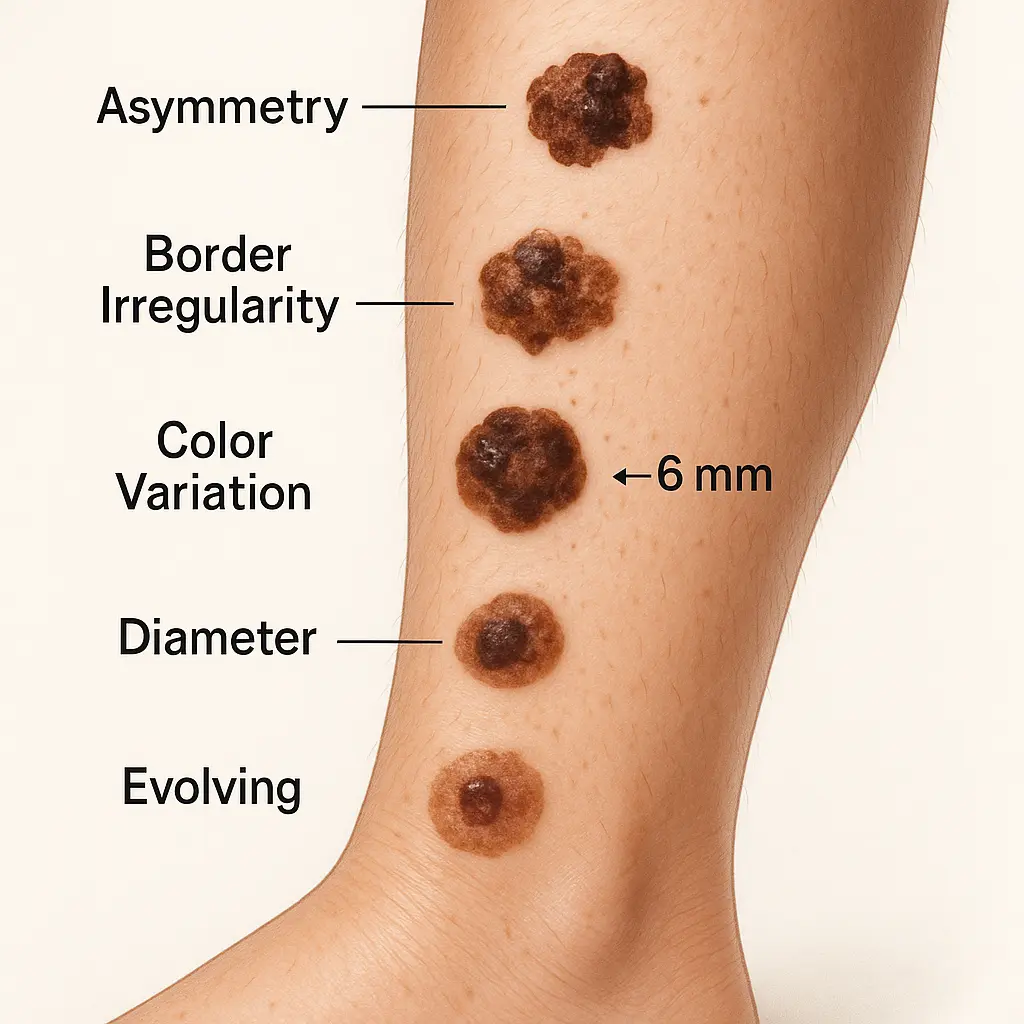
What to Look For: The ABCDEs of Melanoma on Legs
The most widely recognized tool for identifying suspicious moles or spots is the ABCDE rule. When looking at "melanoma pictures on legs," these are the key features you'll be trying to identify.
A: Asymmetry
What it means: If you were to draw a line through the middle of a mole, the two halves wouldn't match. A benign (harmless) mole is usually symmetrical, meaning both halves look the same.
On legs: Imagine a spot on your shin. If one side is round and the other is jagged, or one side is much larger than the other, it's asymmetrical. This is a significant red flag.
B: Border Irregularity
What it means: The edges of the mole are uneven, ragged, notched, or blurred. Benign moles usually have smooth, well-defined borders.
On legs: A mole on your calf that has fuzzy or scalloped edges, rather than a neat, clear outline, would be considered to have an irregular border. Many "melanoma pictures on legs" will clearly show this jagged, non-uniform edge.
C: Color Variation
What it means: The mole has different shades of color within it, such as browns, tans, black, red, white, or blue. Harmless moles are typically a single shade of brown or tan.
On legs: A spot on your thigh that has a mix of dark brown, almost black areas, alongside lighter tan or even reddish patches, is highly suspicious. This multi-colored appearance is a strong indicator seen in many "melanoma pictures on legs."
D: Diameter
What it means: The mole is larger than 6 millimeters (about the size of a pencil eraser). While some melanomas can be smaller, most are larger by the time they are detected.
On legs: If you notice a new or changing spot on your ankle that is bigger than 6mm, it warrants immediate attention. Even if it's smaller but exhibits other ABCDE signs, it should still be checked.
E: Evolving
What it means: The mole is changing in size, shape, color, or elevation. It might also start to itch, bleed, or become tender. This is perhaps the most crucial sign.
On legs: You might have a mole on your calf that has been there for years, but suddenly it starts to grow, gets darker, or becomes itchy. Any change over time, no matter how subtle, is a reason to see a doctor. This "evolving" aspect is often harder to capture in static "melanoma pictures on legs" but is vital for self-assessment.
ABCDE FeatureWhat to Look For (Suspicious)What it Looks Like (Benign)AsymmetryOne half does not match the otherBoth halves matchBorderUneven, ragged, notched, or blurred edgesSmooth, well-defined edgesColorDifferent shades of brown, tan, black, red, white, blueSingle shade of brown or tanDiameterLarger than 6mm (pencil eraser)Usually smaller than 6mmEvolvingChanges in size, shape, color, elevation, or new symptoms (itching, bleeding)Remains stable over time
Beyond ABCDEs: Other Signs and Symptoms on Legs
While the ABCDEs are excellent guidelines, not all melanomas fit this classic description. It's important to be aware of other potential signs, especially when looking at "melanoma pictures on legs" that might not be typical.
Itching, Bleeding, or Tenderness
A mole that suddenly becomes itchy, tender to the touch, or starts to bleed (even without being scratched) is a significant warning sign. These symptoms suggest that the lesion is active and potentially growing. If you have a spot on your leg that develops any of these new sensations, it needs professional evaluation.
Non-Pigmented (Amelanotic) Melanoma
Not all melanomas are dark. Amelanotic melanoma is a type of melanoma that lacks pigment, meaning it can appear as a red, pink, or skin-colored bump or patch. Because it doesn't have the typical dark color, it can be easily mistaken for a benign lesion like a wart, scar, or even a pimple.
"Amelanotic melanoma on the legs can be particularly tricky to identify because it lacks the classic dark color. It often appears as a persistent red or pink lesion that won't heal."
If you see a persistent, non-healing red or pink lesion on your leg, even if it doesn't look like a mole, it's important to have it checked. These lesions can be very aggressive because their atypical appearance often leads to delayed diagnosis. "Melanoma pictures on legs" can sometimes include examples of these less common presentations.
Nodular Melanoma
Nodular melanoma is another type that doesn't always follow the classic ABCDE rule, especially the "Evolving" part, as it can grow very quickly. It often appears as a firm, raised, often dark (though sometimes red or skin-colored) lump on the skin. It can be uniform in color and symmetrical, making it tricky. The key characteristic is its rapid growth and often a "blueberry-like" appearance. If you notice a new, fast-growing lump on your leg, regardless of its color, seek immediate medical attention.
Melanoma Under the Toenails (Acral Lentiginous Melanoma)
While technically not "on" the leg, acral lentiginous melanoma (ALM) often affects the toes and feet, including under the toenails. It appears as a dark streak or discoloration under the nail, often mistaken for a bruise or fungal infection. It can also appear on the soles of the feet or palms of the hands. This type of melanoma is more common in people with darker skin tones but can affect anyone. If you see a new or changing dark streak under a toenail that doesn't grow out with the nail, or a suspicious spot on the sole of your foot, get it checked.
Melanoma vs. Benign Moles/Spots on Legs
It's common to have many moles and spots on your legs. Most of them are harmless (benign). Differentiating between a benign mole and a potential melanoma is often the biggest challenge for individuals. This is where looking at "melanoma pictures on legs" and comparing them to common benign lesions can be helpful for building awareness.
Common Benign Lesions on Legs
Common Moles (Nevi): These are usually small, symmetrical, uniform in color (tan, brown, or black), and have smooth borders. They can be flat or slightly raised.
Seborrheic Keratoses: These are common, benign skin growths that often appear as waxy, "stuck-on" lesions. They can be tan, brown, or black and often have a rough, greasy texture. They are usually harmless but can sometimes be mistaken for melanoma due to their color.
Cherry Angiomas: These are small, bright red or purple bumps caused by clusters of tiny blood vessels. They are harmless and very common, especially with age.
Dermatofibromas: These are firm, harmless bumps that often appear on the lower legs. They can be skin-colored, pink, or reddish-brown and often dimple inwards when squeezed.
Venous Lakes: These are soft, compressible, dark blue or purple bumps that often appear on the lips, ears, or face, but can sometimes be found on the legs. They are harmless dilated blood vessels.
The "Ugly Duckling" Sign
One of the most helpful concepts in differentiating between benign and malignant lesions is the "ugly duckling" sign. This means that a melanoma often looks different from all the other moles on your body. If you have many moles on your legs, and one of them stands out as being odd or different from the rest, it's your "ugly duckling" and should be examined by a doctor. This visual contrast is often clearer than trying to apply the ABCDEs to every single mole. When reviewing "melanoma pictures on legs," try to notice how the suspicious lesion often doesn't "fit in" with typical moles.
The Importance of Self-Exams for Legs
Regular self-skin exams are a vital part of early detection. You are often the first person to notice changes in your skin. Given that legs are a common site for melanoma, dedicating specific attention to them during your self-exam is crucial.
How to Perform a Self-Exam on Your Legs
Find a well-lit room: Good lighting is essential to see subtle changes.
Use a full-length mirror: This helps you see your entire body.
Use a hand mirror: This is critical for hard-to-see areas like the back of your calves, thighs, and behind your knees.
Start systematically:
Begin with your front thighs, shins, and knees.
Turn to examine the outer sides of your thighs and calves.
Use the hand mirror to check the backs of your thighs and calves.
Sit down to examine your ankles, feet, and toes. Don't forget between your toes and the soles of your feet. Also, check under your toenails for any new or changing dark streaks.
Look for the ABCDEs and other signs: Pay attention to any new moles, existing moles that are changing, or any spots that itch, bleed, or are tender.
Take pictures: If you find a suspicious spot, take a picture of it with your phone, noting the date. This can help you track changes over time and show your doctor. When comparing your own spots to "melanoma pictures on legs," remember that your own pictures are for tracking, not self-diagnosis.
How Often Should You Check?
Most dermatologists recommend performing a thorough self-skin exam once a month. This regular habit allows you to become familiar with your skin's normal appearance and quickly spot any new or changing lesions.
What to Do If You Find Something Suspicious
Do not panic! Most suspicious-looking spots turn out to be harmless. However, it's essential to:
Schedule an appointment with a dermatologist: Explain your concerns and mention that you've noticed a suspicious spot on your leg.
Avoid self-diagnosis: While "melanoma pictures on legs" are great for awareness, they cannot replace a professional diagnosis. Only a trained dermatologist can accurately assess a lesion.
Professional Skin Checks: What to Expect
While self-exams are important, they are not a substitute for professional skin checks by a dermatologist. Dermatologists are experts in skin conditions and have specialized tools to examine moles in detail.
When to See a Dermatologist
If you find a suspicious spot: Any mole or lesion on your leg (or anywhere else) that fits the ABCDE criteria, looks like an "ugly duckling," or is new, changing, itching, bleeding, or tender.
Annual full-body skin exams: Everyone should have a professional skin check at least once a year, especially if you have risk factors for melanoma. Your dermatologist might recommend more frequent checks if you are at higher risk.
What a Dermatologist Looks For
During a professional skin check, the dermatologist will:
Examine your entire skin surface: From head to toe, including areas you might miss like your scalp, between your toes, and the soles of your feet. They will specifically pay close attention to your legs, knowing they are a common site for melanoma.
Use a dermatoscope: This is a handheld magnifying tool with a light source that allows the dermatologist to see structures and patterns within the mole that are invisible to the naked eye. This tool is incredibly useful for distinguishing between benign moles and melanoma, often providing a clearer view than any "melanoma pictures on legs" you might find online.
Document findings: They may take digital photos of your moles to track changes over time.
The Biopsy Process
If a dermatologist finds a suspicious lesion on your leg, they will likely recommend a biopsy. This is a simple procedure where a small sample of the mole or the entire mole is removed and sent to a lab for microscopic examination by a pathologist.
Types of biopsies:
Shave biopsy: The top layers of the lesion are shaved off.
Punch biopsy: A small circular piece of the lesion, including deeper layers, is removed.
Excisional biopsy: The entire lesion and a small margin of surrounding healthy skin are removed. This is often preferred for suspected melanoma to ensure complete removal if it is cancerous.
The biopsy results will determine whether the lesion is benign, precancerous, or melanoma, and guide the next steps for treatment.
Types of Melanoma and Their Appearance on Legs
Understanding the different types of melanoma can further help you interpret "melanoma pictures on legs" and recognize the varied ways this cancer can present.
1. Superficial Spreading Melanoma (SSM)
Most Common Type: This accounts for about 70% of all melanomas.
Appearance on Legs: Often appears as a flat or slightly raised patch with irregular borders and varied colors (browns, blacks, tans, reds, blues, whites). It tends to grow outwards on the skin surface for a period before growing downwards.
Why it's common on legs: Its association with intermittent sun exposure makes it a frequent finding on the legs. Many "melanoma pictures on legs" will feature examples of SSM.
2. Nodular Melanoma (NM)
Aggressive and Fast-Growing: This type grows vertically (downwards) into the skin very quickly.
Appearance on Legs: Often appears as a firm, raised, dome-shaped lump. It can be dark (black or blue-black), but can also be red, pink, or skin-colored (amelanotic nodular melanoma). It often feels like a firm, rubbery bump.
Key difference: Unlike SSM, it doesn't spread out on the surface much before growing deep. If you see a new, rapidly growing bump on your leg, even if it's symmetrical and uniform in color, it needs urgent attention.
3. Lentigo Maligna Melanoma (LMM)
On Sun-Damaged Skin: This type typically develops on chronically sun-exposed areas, especially in older adults.
Appearance on Legs: While more common on the face and neck, it can appear on the legs, particularly on areas that have seen a lot of sun over the years. It looks like a flat or slightly raised patch with irregular borders and uneven color, often shades of brown or tan. It grows slowly over many years before becoming invasive.
4. Acral Lentiginous Melanoma (ALM)
On Palms, Soles, and Under Nails: This type is not linked to sun exposure as strongly as others.
Appearance on Legs/Feet: Appears on the soles of the feet, palms of the hands, or under the fingernails or toenails. On the feet, it might look like a dark, irregular patch on the sole or a dark streak under a toenail. It's often mistaken for a bruise, wart, or fungal infection.
Importance: This type is more common in people with darker skin tones, but it can affect anyone. It's often diagnosed at a later stage because it's in less obvious locations or mistaken for other conditions. When you look at "melanoma pictures on legs," make sure you also consider images of the feet and nails.
5. Amelanotic Melanoma
Lack of Pigment: As mentioned earlier, this type lacks melanin, making it appear pink, red, skin-colored, or even clear.
Appearance on Legs: Can mimic benign lesions like scars, warts, or even insect bites. It might be a persistent non-healing sore or a firm, red lump.
Challenge: Its lack of color makes it difficult to identify using the ABCDE rule, making awareness of its varied appearance crucial.
Visualizing Melanoma on Legs: What Pictures Show
When you search for "melanoma pictures on legs," you'll encounter a wide range of visual examples. It's important to understand what these images are trying to convey and their limitations.
Common Visuals in Melanoma Pictures on Legs
Classic Irregular Moles: Many images will show moles on the shins or calves that are clearly asymmetrical, with jagged borders and multiple shades of brown, black, and tan. These are often superficial spreading melanomas. You might see a mole that looks like a splattered inkblot rather than a neat dot.
Raised, Dark Lumps: You'll see pictures of elevated, often uniform dark brown or black bumps. These are frequently examples of nodular melanoma. They might look like a small, firm berry stuck on the skin.
Red or Pink Patches/Bumps: Some "melanoma pictures on legs" will highlight amelanotic melanoma, showing lesions that are surprisingly red, pink, or skin-colored, challenging the idea that all melanomas are dark. These might look like persistent insect bites or unhealing sores.
Lesions on the Soles of Feet or Under Toenails: Images of acral lentiginous melanoma will show dark, irregular patches on the sole of the foot or dark streaks under toenails, sometimes with discoloration spreading to the surrounding skin or cuticle.
Changing Lesions (Sequential Images): While less common in static search results, educational resources might show "melanoma pictures on legs" over time, demonstrating how a spot has grown, changed shape, or altered in color from one month to the next. This highlights the "Evolving" characteristic.
Ulcerated or Bleeding Lesions: Some advanced melanomas might appear as open sores, or have crusting or bleeding. Pictures of these lesions will often show a central break in the skin or signs of dried blood.
The Power and Limitations of Pictures
Power for Awareness: "Melanoma pictures on legs" are incredibly powerful for raising awareness. They help you visualize what to look for, making the ABCDEs more concrete. They can be a wake-up call and encourage people to perform self-exams.
Limitations for Diagnosis:Crucially, looking at pictures online can never diagnose melanoma.
Lighting and Angle: Pictures can be misleading due to lighting, camera quality, and angle. What looks suspicious in a photo might be benign in person, and vice-versa.
Context Missing: Pictures don't show if the mole is new, changing, itchy, or bleeding – vital information for diagnosis.
Professional Expertise: Dermatologists have years of training and specialized tools (like dermatoscopes) to evaluate lesions at a microscopic level that pictures simply cannot replicate.
Emotional Impact: Self-diagnosing from pictures can cause unnecessary anxiety or, worse, false reassurance.
"While 'melanoma pictures on legs' are invaluable for educating yourself about potential signs, they are a guide for suspicion, not a diagnostic tool. Always consult a dermatologist for any concerning skin lesion."
Use "melanoma pictures on legs" as a learning tool to train your eye, but let a medical professional make the final judgment.
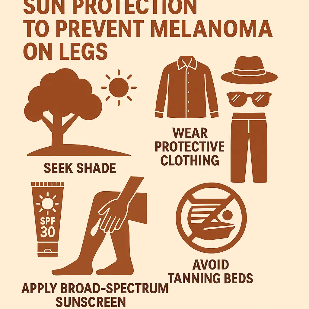
Prevention Strategies for Melanoma on Legs
Preventing melanoma, especially on the legs, largely comes down to smart sun habits and regular vigilance.
Sun Protection: Your First Line of Defense
Seek Shade: The sun's UV rays are strongest between 10 AM and 4 PM. Try to limit your time in direct sunlight during these hours. Seek shade under trees, umbrellas, or awnings.
Wear Protective Clothing:
Long pants or skirts: When outdoors for extended periods, especially during peak sun hours, covering your legs with clothing is highly effective. Look for fabrics with a high UPF (Ultraviolet Protection Factor).
Wide-brimmed hats: While not directly protecting legs, a hat protects your face and neck, contributing to overall sun safety.
UV-blocking sunglasses: Protect your eyes from UV damage.
Apply Sunscreen Generously and Regularly:
SPF 30 or higher: Use a broad-spectrum sunscreen that protects against both UVA and UVB rays.
Water-resistant: If you're swimming or sweating, choose a water-resistant formula.
Apply 15-30 minutes before sun exposure: This allows the sunscreen to form a protective barrier.
Reapply every two hours (or more often): Especially after swimming, sweating, or towel drying. Don't forget your legs, ankles, and feet! It's easy to miss these areas.
Avoid Tanning Beds: Tanning beds emit concentrated UV radiation, significantly increasing your risk of melanoma and other skin cancers. There is no such thing as a "safe" tan from a tanning bed.
Regular Skin Checks
As discussed, perform monthly self-exams and schedule annual professional skin checks with a dermatologist. This two-pronged approach ensures that any new or changing lesions on your legs (or elsewhere) are identified promptly.
Know Your Risk
Understand your personal risk factors. If you have fair skin, a history of sunburns, many moles, or a family history of melanoma, you need to be extra diligent with sun protection and skin checks.
Living with Melanoma: Diagnosis and Treatment
While the focus of this article is detection, it's helpful to have a brief understanding of what happens after a melanoma diagnosis.
Diagnosis Confirmation
If a biopsy confirms melanoma on your leg, further tests may be done to determine the stage of the cancer (how deep it has grown and if it has spread). This might include:
Sentinel lymph node biopsy: To check if cancer cells have spread to nearby lymph nodes.
Imaging tests: Such as CT scans, MRI, or PET scans, to look for spread to distant organs.
Treatment Options
Treatment for melanoma depends on its stage and location. The primary treatment is usually surgery to remove the melanoma and a margin of healthy tissue around it. For more advanced melanomas, other treatments may be necessary:
Chemotherapy: Uses drugs to kill cancer cells throughout the body.
Radiation Therapy: Uses high-energy rays to kill cancer cells.
Targeted Therapy: Uses drugs that target specific genes or proteins involved in cancer growth.
Immunotherapy: Boosts the body's own immune system to fight cancer cells.
Importance of Follow-Up
After treatment, regular follow-up appointments with your dermatologist and oncologist are crucial. This includes frequent skin checks to monitor for recurrence or new melanomas.
Common Misconceptions About Melanoma on Legs
Dispelling myths is important for accurate awareness, especially when people are trying to interpret "melanoma pictures on legs."
"It's just a mole, I've had it forever." While many moles are benign and stable, the "E" in ABCDE (Evolving) is critical. Any change in an existing mole, no matter how old, warrants investigation. Melanoma can also appear as a brand-new spot.
"Only affects fair-skinned people." While fair skin is a major risk factor, melanoma can affect people of all skin tones, including those with darker skin. In individuals with darker skin, melanoma often appears in less sun-exposed areas like the soles of the feet, palms of the hands, or under nails (acral lentiginous melanoma). This highlights the importance of checking all areas of the legs and feet.
"Only where the sun hits directly." While sun exposure is a primary cause, melanoma can develop on skin that rarely sees the sun, such as the inner thigh, between the toes, or under the toenails. This is why a full-body skin exam is essential.
"It doesn't hurt, so it's fine." Early melanomas often cause no pain, itching, or other symptoms. Waiting for symptoms to appear can lead to a delayed diagnosis. The visual changes (ABCDEs) are usually the first signs.
"Tanning beds are safer than the sun." This is absolutely false. Tanning beds emit concentrated UV radiation and significantly increase the risk of melanoma.
The Emotional Impact of a Melanoma Diagnosis
A diagnosis of melanoma, even early-stage, can be a frightening and emotionally challenging experience. It's normal to feel:
Fear and anxiety: About the diagnosis, treatment, and future health.
Stress: About medical appointments, finances, and disruption to daily life.
Sadness or depression: Due to the severity of the diagnosis.
Anger or frustration: About the diagnosis or perceived lack of control.
It's important to remember that you are not alone. Seeking support from family, friends, support groups, or mental health professionals can be incredibly helpful. Open communication with your medical team about your emotional well-being is also vital.
Future of Melanoma Detection and Treatment
The field of melanoma research is constantly evolving, bringing new hope for earlier detection and more effective treatments.
AI in Dermatology: Artificial intelligence (AI) is being developed to assist dermatologists in identifying suspicious lesions. AI algorithms can analyze images of moles, potentially identifying subtle patterns that might be missed by the human eye. While not replacing doctors, AI could become a valuable screening tool. This means that in the future, "melanoma pictures on legs" might be analyzed by advanced AI systems for initial screening.
Advanced Imaging Techniques: New imaging technologies are being explored that can provide even more detailed views of skin lesions without the need for a biopsy.
Personalized Medicine: Genetic research is leading to more personalized treatment approaches, tailoring therapies to the specific genetic makeup of a patient's melanoma.
New Therapies: Ongoing research continues to discover and refine targeted therapies and immunotherapies, offering improved outcomes for advanced melanoma.
These advancements highlight a future where detection is even more precise and treatments more effective, but the fundamental importance of self-awareness and professional checks remains.
Conclusion
Understanding "melanoma pictures on legs" is a powerful step towards protecting your skin health. By learning to identify the ABCDEs and other subtle signs, you empower yourself to be an active participant in early detection. Remember that while visual aids are excellent for awareness, only a trained dermatologist can provide an accurate diagnosis. Prioritize regular self-skin exams, especially focusing on your legs, and don't hesitate to seek professional medical advice for any suspicious spots. Sun protection and avoiding tanning beds are your best defenses against this serious skin cancer. Be vigilant, be informed, and take proactive steps to keep your skin healthy. Your skin is your body's largest organ – protect it! 💖
October 2, 2025
🇨🇦
Our clinic currently provides care to patients within
Canada only.
We apologize for any inconvenience this may cause.