Have you ever noticed a small, soft flap of skin hanging from your body and wondered, "What exactly is that?" 🤔 For many, these are common skin tags, usually harmless little growths that pop up in places where skin rubs together. But what if that growth looks a bit different? What if you're searching online for "cancerous skin tags pictures" because you're worried about a new spot? It's natural to be concerned, especially when the internet is filled with confusing images.
This comprehensive guide is here to help you understand the crucial difference between a benign (harmless) skin tag and a potentially cancerous skin growth. While pictures can offer some clues, they are never a substitute for a professional medical examination. We'll explore what real skin tags look like, what different types of skin cancer can resemble, and most importantly, when it's time to see a doctor. Our goal is to empower you with knowledge, reduce anxiety, and guide you toward proper care.
Before we dive into what might be concerning, let's get clear on what a typical skin tag is. Skin tags, medically known as acrochordons, are incredibly common. They are small, soft, benign (non-cancerous) growths that usually hang off the skin by a tiny stalk. They are often the same color as your skin or slightly darker.
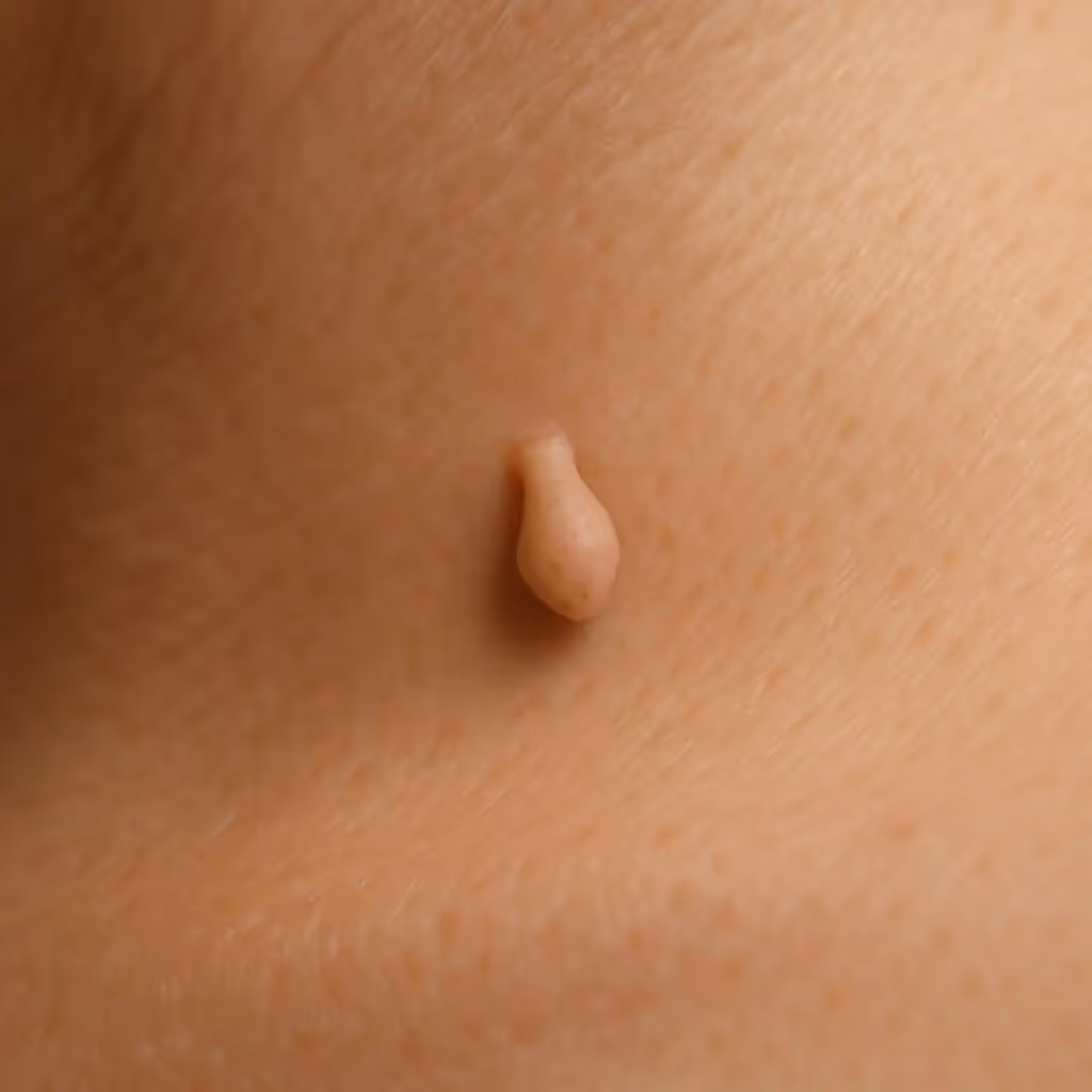
Imagine a tiny, deflated balloon or a small, soft piece of rice hanging from your skin. That's often what a skin tag resembles. Here are their typical characteristics:
While the exact cause isn't always clear, several factors increase your likelihood of developing skin tags:
In almost all cases, no. Skin tags are benign. They don't turn into cancer, and they don't spread. The main issues people have with them are:
"The vast majority of skin tags are completely harmless. Your primary concern should be if a 'skin tag' changes or doesn't fit the typical description."
If a skin tag is bothering you, a doctor can easily remove it in a quick, in-office procedure. This might involve freezing (cryotherapy), cutting (excision), or burning (cauterization). For more information on various skin conditions and their treatments, you might find it helpful to visit resources like The Minor Surgery Center's conditions page.
This is where the term "cancerous skin tags pictures" becomes tricky. True skin tags are not cancerous. However, other skin growths can look similar to skin tags, and some of these can be cancerous. This similarity is why self-diagnosis from pictures is so risky.
Many different types of skin growths exist, and telling them apart can be challenging even for trained eyes without proper tools. Here are a few common benign growths that might be confused with skin tags:
The key takeaway here is that while many growths are benign, some look deceptive. This is why professional help is always recommended if there's any doubt.
When people search for "cancerous skin tags pictures," they are often trying to find images of skin cancers that might resemble a skin tag or other common benign growth. It's vital to understand that skin cancer comes in many forms, and its appearance can vary significantly.
There are three main types of skin cancer:
Let's look at how these might present and why they could be mistaken for something less serious.
Melanoma is dangerous because it can spread quickly to other parts of the body if not caught early. It often develops in existing moles but can also appear as a new spot.
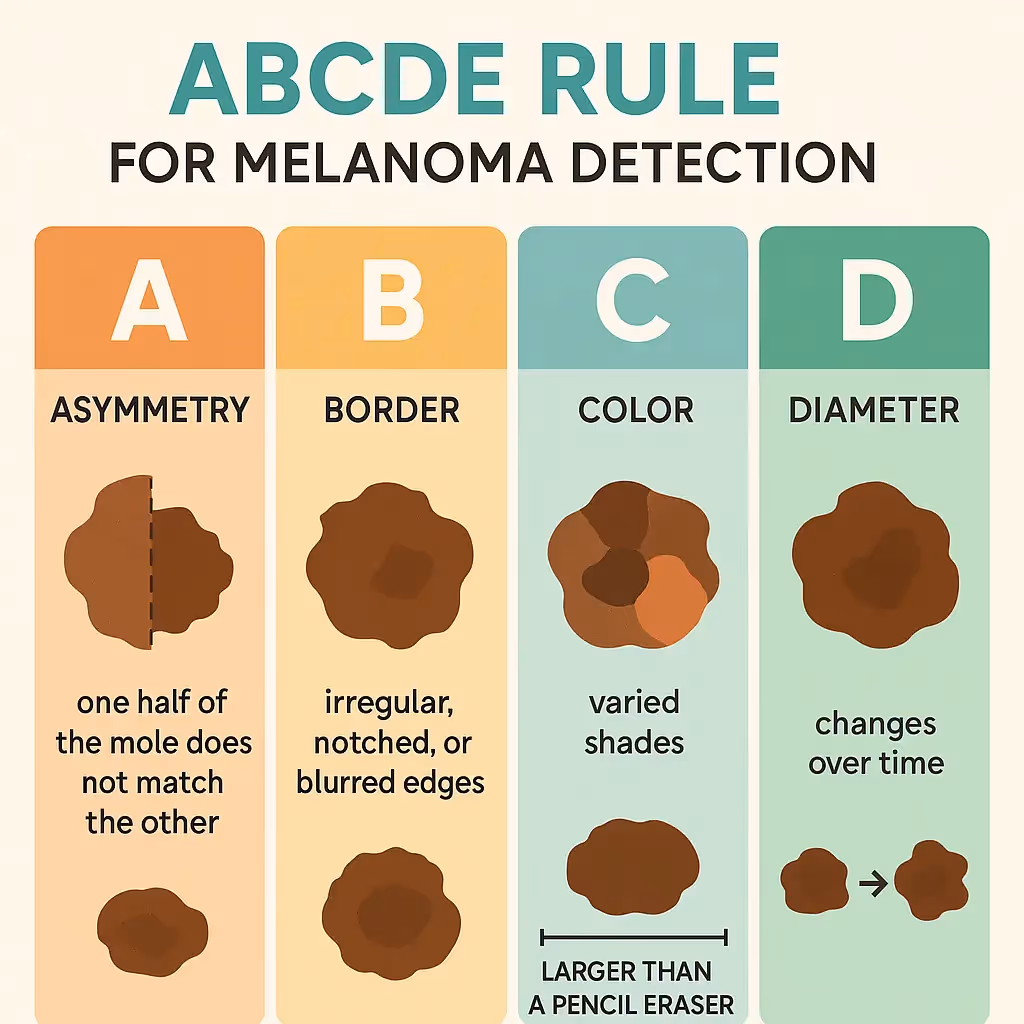
What to look for (The ABCDE Rule): The ABCDE rule is a widely used guide to help people identify potential melanoma. If you notice any of these signs, especially in a new or changing spot, it's time to see a doctor.
How Melanoma might be mistaken for a "skin tag": While less common, some melanomas can be raised (nodular melanoma) and might have a somewhat irregular shape or color that could, to an untrained eye, be dismissed as an "unusual mole" or even a very dark, firm skin tag. However, a true skin tag is soft, uniform in color (even if dark), and typically has a distinct stalk. Melanoma will usually lack the classic stalk and will show at least one of the ABCDE signs.
Example description (not actual picture): Imagine a dark brown or black bump that is slightly raised, feels firm, and has an uneven border. It might have a small crust on top or bleed easily if bumped. It doesn't have a distinct stalk, and if you look closely, its color might not be consistent throughout. This would be very different from a soft, floppy skin tag.
BCC is the most common form of skin cancer. It grows slowly and rarely spreads to other parts of the body, but it can cause significant local damage if left untreated. It often appears on sun-exposed areas like the face, neck, and hands.
What to look for: BCCs can appear in several ways:
How BCC might be mistaken for a "skin tag": A BCC, especially a nodular BCC, can sometimes present as a raised, flesh-colored or slightly pink bump. If it's small and located in a skin fold, it might be dismissed as an irritated skin tag. However, BCCs are typically firmer, often have a "pearly" appearance, and lack the distinct stalk of a skin tag. They might also bleed easily or have visible blood vessels.
Example description (not actual picture): Picture a small, raised bump on your neck or face that looks almost like a clear or pinkish pearl. It might be shiny and have a tiny, visible blood vessel running across it. It doesn't hang off the skin. Or, imagine a flat, reddish patch that looks like a persistent rash but doesn't respond to typical creams. These are concerning and are not skin tags.
SCC is the second most common type of skin cancer. It can grow more quickly than BCC and has a greater chance of spreading if not treated. Like BCC, it often appears on sun-exposed skin.
What to look for:
How SCC might be mistaken for a "skin tag": An SCC, particularly one that is raised and somewhat irregular, could potentially be mistaken for a very inflamed or unusual skin tag, especially if it's crusty or bleeding. However, SCCs are typically firmer, often have a rough or scaly surface, and lack the characteristic soft, dangling nature and stalk of a true skin tag. They are also more likely to be painful, tender, or bleed spontaneously.
Example description (not actual picture): Think of a reddish, scaly patch on your ear or hand that feels rough and might be tender to the touch. It might have a central crust or bleed if you pick at it. It's not soft and doesn't dangle. Alternatively, a firm, red bump that looks like a persistent wart but keeps growing or bleeding.
Searching for "cancerous skin tags pictures" online is a common first step for many people, but it comes with significant limitations and risks.
1. Variability in Appearance: Skin cancers, like many medical conditions, don't always look the same from person to person. A melanoma in one individual might appear slightly different in another. Online pictures often show textbook examples, but real-life presentations can be much more subtle or unusual.
2. Lack of Context: A picture can't tell you:
*   How long the growth has been there.
*   If it has changed over time (the "E" in ABCDE is crucial).
*   If it's painful, itchy, or bleeding.
*   How it feels to the touch (soft vs. firm).
*   The patient's full medical history or risk factors.
3. Quality of Images: Online images can be low resolution, poorly lit, or misleadingly cropped. They might not capture the true color, texture, or subtle features necessary for an accurate assessment.
4. Emotional Impact: Seeing graphic images of severe skin cancers can cause unnecessary anxiety and panic, even if your growth is benign. Conversely, dismissing a concerning growth because it doesn't look exactly like a picture you found online can delay critical diagnosis.
5. No Substitute for Professional Eyes: Dermatologists and other healthcare professionals are trained to identify subtle signs of skin cancer. They use specialized tools like a dermatoscope (a magnified light source) to examine skin growths in detail, seeing features invisible to the naked eye. They also consider your personal risk factors and medical history.
"Relying solely on 'cancerous skin tags pictures' for self-diagnosis is like trying to fix a complex engine just by looking at photos online. You need the right tools, the right training, and a hands-on examination."
This is the most important section. If you have any doubt or concern about a skin growth, it's always best to get it checked by a healthcare professional. Don't wait! Early detection of skin cancer significantly improves treatment outcomes.
You should see a doctor if you notice any of the following:
Where to go: The best specialist to see for skin concerns is a dermatologist. They are experts in skin, hair, and nail conditions. Your primary care physician can also perform an initial assessment and refer you to a dermatologist if needed.
For scheduling an appointment or learning more about local clinics, you can often find helpful information on clinic websites, such as The Minor Surgery Center's contact page or their clinic information page.
When you visit a healthcare professional for a suspicious skin growth, they will follow a systematic process to make an accurate diagnosis.
The doctor will first perform a thorough visual examination of the growth in question. They will also likely examine other parts of your skin, as skin cancers can appear anywhere. They'll note its size, shape, color, texture, and location.
Many dermatologists use a tool called a dermatoscope. This is a handheld device that magnifies the skin and uses special lighting (often polarized) to allow the doctor to see structures and patterns beneath the skin's surface that are invisible to the naked eye. This can help distinguish between benign and malignant lesions without needing an immediate biopsy.
If, after the visual and dermoscopic examination, the doctor is still concerned, they will recommend a biopsy. This is the only definitive way to diagnose skin cancer. A biopsy involves removing a small sample of the suspicious tissue and sending it to a pathology lab for microscopic examination by a pathologist.
There are several types of biopsies:
The biopsy procedure is usually quick and performed under local anesthesia, meaning the area will be numbed so you won't feel pain.
Once the tissue sample is sent to the lab, a pathologist (a doctor who specializes in diagnosing diseases by examining tissues) will analyze it under a microscope. They will determine if cancer cells are present, and if so, what type of cancer it is and its characteristics (e.g., depth of invasion for melanoma).
The results usually take several days to a couple of weeks. Your doctor will contact you to discuss the findings and recommend the next steps if cancer is found. The expertise of the medical team is crucial in this process, and you can often find information about specialists on a clinic's website, such as The Minor Surgery Center's team page.
If a skin cancer diagnosis is confirmed, your doctor will discuss the most appropriate treatment plan. The choice of treatment depends on the type of skin cancer, its size, location, depth, and your overall health.

This is the most common treatment for most skin cancers, especially melanoma, BCC, and SCC. The surgeon removes the cancerous tissue along with a margin of healthy skin around it to ensure all cancer cells are gone. This is often done in an outpatient setting under local anesthesia.
Mohs surgery is a highly specialized technique often used for BCC and SCC, especially on the face, neck, or other cosmetically sensitive areas, or for recurrent cancers. The surgeon removes layers of skin one at a time and examines each layer under a microscope immediately. This process continues until no cancer cells are seen, allowing for precise removal of the cancer while preserving as much healthy tissue as possible.
Depending on the type and stage of cancer, other treatments may be considered:
The goal of treatment is always to remove or destroy the cancer completely while minimizing scarring and preserving function. For general information on services offered, you can explore the main site of The Minor Surgery Center.
While you can't prevent all skin cancers, you can significantly reduce your risk and improve your chances of early detection, which is key to successful treatment.
Exposure to ultraviolet (UV) radiation from the sun and tanning beds is the leading cause of most skin cancers.
Become familiar with your skin and all the moles, freckles, and blemishes on your body. Performing regular self-skin exams (once a month) can help you spot new or changing growths early.
How to do a self-exam:
Pay close attention to any of the ABCDE signs for moles, or any new, changing, or non-healing spots. Take pictures of moles you want to track over time to help you remember their appearance.
Schedule an annual full-body skin exam with a dermatologist, especially if you have:
Your dermatologist has the training and tools to detect suspicious lesions that you might miss. For common questions about skin health and care, you can refer to resources like The Minor Surgery Center's FAQs page.
Understanding the difference between harmless skin tags and potentially cancerous lesions is empowering. Most people will develop skin tags or other benign growths during their lifetime, and knowing what they are can alleviate unnecessary worry. However, the vigilance for changes or new, suspicious growths must remain.
If you have a skin growth that is confirmed to be a benign skin tag, you generally don't need to do anything unless it's causing irritation or you want it removed for cosmetic reasons. If you have concerns about other types of skin conditions or need further information, resources like The Minor Surgery Center's conditions page can be helpful.
For those who receive a skin cancer diagnosis, remember that early detection and treatment offer the best prognosis. Modern medical advancements provide effective treatments for most skin cancers. Regular follow-ups with your dermatologist will be crucial to monitor for recurrence or new lesions.
Ultimately, your skin is your body's largest organ, and it's essential to take care of it. Regular self-checks, sun protection, and professional examinations are your best allies in maintaining skin health and catching any issues early. For more articles and insights on skin health, you can explore The Minor Surgery Center's blog.
The quest for "cancerous skin tags pictures" often stems from a natural human desire to understand and control one's health. While it's excellent to be proactive and informed, it's crucial to recognize the limitations of self-diagnosis, especially when it comes to something as varied and complex as skin lesions.
True skin tags are benign, harmless growths. However, several types of skin cancer can mimic benign lesions, making a visual distinction by an untrained eye nearly impossible. The ABCDE rule for melanoma and knowing the common appearances of BCC and SCC are valuable tools for awareness, but they are not diagnostic.
If you have any doubt, any change in a mole or growth, or any new, suspicious spot on your skin, please do not hesitate. Consult a healthcare professional, ideally a dermatologist. They have the expertise, the tools, and the training to provide an accurate diagnosis and ensure you receive the appropriate care. Your health is too important to leave to chance or to rely solely on online images.


