When individuals notice unusual white spots appearing on their skin, concern about potential cancer often follows. While not all white spots indicate malignancy, understanding the connection between cancer white spots on skin and various skin conditions remains crucial for early detection and proper medical care. These distinctive markings can signal different types of skin cancer or precancerous conditions that require immediate attention from healthcare professionals.
The appearance of white spots on skin can range from harmless cosmetic concerns to serious medical conditions requiring urgent intervention. Distinguishing between benign white spots and those potentially linked to cancer becomes essential for maintaining optimal skin health. Medical professionals emphasize that early recognition and prompt evaluation of suspicious skin changes significantly improve treatment outcomes and prognosis.
This comprehensive guide explores the various aspects of cancer white spots on skin, including identification techniques, diagnostic procedures, treatment options, and prevention strategies. Healthcare experts recommend that individuals familiarize themselves with the warning signs and seek professional evaluation when concerning changes occur.
• Early detection is critical: White spots on skin that change in size, shape, or color may indicate skin cancer and require immediate medical evaluation
• Multiple types exist: Various forms of skin cancer can present as white spots, including basal cell carcinoma, squamous cell carcinoma, and melanoma
• Professional diagnosis is essential: Only qualified dermatologists can accurately distinguish between benign white spots and cancerous lesions through proper examination and testing
• Treatment success depends on timing: Early-stage skin cancers have significantly higher cure rates compared to advanced cases
• Prevention strategies work: Regular skin monitoring, sun protection, and routine dermatological checkups can prevent many cases of skin cancer
Cancer white spots on skin represent a complex dermatological phenomenon that requires careful examination and professional assessment. These spots can manifest in various forms, sizes, and textures, making accurate identification challenging for untrained individuals. Medical research indicates that certain types of skin cancer can present as white or light-colored lesions, particularly in their early stages.
The development of white spots on skin occurs through different mechanisms depending on the underlying cause. Cancerous white spots typically result from abnormal cell growth that disrupts normal pigmentation processes. These cellular changes can create areas of hypopigmentation or complete loss of melanin production, leading to the characteristic white appearance.
Healthcare professionals categorize cancer white spots on skin based on their cellular origin and growth patterns. Understanding these classifications helps medical teams develop appropriate treatment strategies and provide accurate prognoses for patients. The most common types include basal cell carcinoma, squamous cell carcinoma, and certain forms of melanoma that present with white or light-colored characteristics.
The formation of white spots in cancerous conditions involves complex cellular processes that affect melanocyte function and pigment production. Cancer cells can interfere with normal melanin synthesis, leading to areas of reduced pigmentation or complete depigmentation. This process often occurs gradually, making early detection challenging without regular skin monitoring.
Malignant cells may also destroy surrounding healthy tissue, including melanocytes responsible for skin pigmentation. This destruction creates white or light-colored patches that can expand over time as the cancer progresses. Understanding these mechanisms helps healthcare providers identify suspicious lesions and recommend appropriate diagnostic procedures.
Basal cell carcinoma represents the most common form of skin cancer and can frequently present as cancer white spots on skin. These malignancies typically develop in sun-exposed areas and often appear as pearly white or translucent bumps with visible blood vessels. The white coloration results from the tumor's effect on surrounding tissue and altered blood flow patterns.
Early-stage basal cell carcinomas may appear as small, white, scaly patches that patients often mistake for dry skin or minor irritation. As these cancers progress, they can develop central ulceration while maintaining their characteristic white or pink borders. Healthcare professionals emphasize that any persistent white spot that fails to heal within several weeks warrants professional evaluation.
The growth pattern of basal cell carcinoma typically remains localized, rarely spreading to distant body parts. However, these cancers can cause significant local tissue destruction if left untreated. Regular skin examinations help identify suspicious white spots early when treatment options remain most effective and minimally invasive.
Squamous cell carcinoma can also present as cancer white spots on skin, particularly in its early stages or specific subtypes. These cancers often begin as rough, scaly white patches known as actinic keratoses before progressing to invasive carcinoma. The white appearance typically results from abnormal keratin production and cellular changes within the epidermis.
Unlike basal cell carcinoma, squamous cell carcinoma carries a higher risk of metastasis, making early detection and treatment crucial. White spots associated with this cancer type may feel rough or gritty to the touch and often occur in sun-damaged areas of the body. Patients frequently report that these spots bleed easily when scratched or irritated.
Healthcare providers often observe squamous cell carcinomas developing within existing white spots or patches of damaged skin. This progression pattern emphasizes the importance of monitoring any persistent white skin changes and seeking professional evaluation when suspicious features develop.
While melanoma typically presents as dark or multicolored lesions, certain subtypes can appear as cancer white spots on skin. Amelanotic melanoma lacks the characteristic dark pigmentation, appearing instead as white, pink, or flesh-colored growths. These variants pose particular diagnostic challenges due to their atypical appearance.
Hypopigmented melanoma represents another form that can present with white or light-colored areas within darker lesions. These cancers often develop irregular borders and may show variations in color intensity across different regions of the tumor. The white areas typically indicate regions of decreased melanin production or tumor-induced tissue changes.
Medical professionals emphasize that any changing white spot, regardless of size or initial appearance, requires evaluation to rule out amelanotic melanoma. These cancers can grow rapidly and metastasize early, making prompt diagnosis and treatment essential for optimal outcomes.
Cancer white spots on skin typically exhibit specific physical characteristics that distinguish them from benign conditions. These features include irregular borders, asymmetrical shapes, and variable textures that change over time. Healthcare professionals train to recognize these subtle differences during routine skin examinations.

Cancerous white spots often feel different from surrounding healthy skin, with textures ranging from smooth and waxy to rough and scaly. The consistency may vary across different areas of the same lesion, indicating heterogeneous cellular composition. Patients frequently report that these spots feel firmer or more raised than normal skin tissue.
Size variations represent another important characteristic of cancer white spots on skin. While benign white spots typically remain stable in size, cancerous lesions tend to grow progressively over weeks or months. This growth pattern may occur uniformly or show irregular expansion in specific directions.
Medical professionals recommend applying a modified version of the ABCDE rule when evaluating suspicious white spots:
🔍 A - Asymmetry: Cancerous white spots often show asymmetrical shapes where one half doesn't match the other half when divided by an imaginary line.
🔍 B - Border Irregularity: Malignant lesions typically display irregular, scalloped, or poorly defined borders rather than smooth, even edges.
🔍 C - Color Variation: Even within white spots, cancerous lesions may show subtle color variations, including areas of pink, red, or translucent regions.
🔍 D - Diameter: Lesions larger than 6mm (about the size of a pencil eraser) require professional evaluation, though smaller cancerous spots can occur.
🔍 E - Evolution: Any white spot that changes in size, shape, color, or symptoms over time needs immediate medical attention.
Cancer white spots on skin may present with various associated symptoms that help distinguish them from benign conditions. These symptoms can include persistent itching, burning sensations, or tenderness that doesn't resolve with typical skincare measures. Patients often report that these sensations worsen over time rather than improving.
Bleeding represents a significant warning sign associated with cancerous white spots. Malignant lesions frequently bleed spontaneously or with minimal trauma, such as during routine washing or clothing contact. This bleeding pattern differs from normal wound healing and typically recurs despite attempts at care.
Some patients experience pain or discomfort associated with cancer white spots on skin, particularly as the lesions progress. This pain may manifest as a constant ache, sharp shooting sensations, or increased sensitivity to touch. Healthcare providers consider pain an important diagnostic clue, especially when combined with other suspicious features.
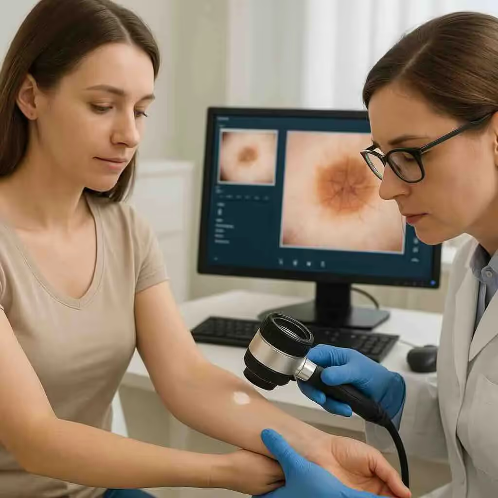
Professional evaluation of cancer white spots on skin begins with a comprehensive clinical assessment performed by qualified dermatologists or healthcare providers. This evaluation includes detailed medical history taking, focusing on risk factors such as sun exposure history, family cancer history, and previous skin cancer diagnoses. Healthcare professionals document the timeline of spot development and any associated symptoms.
Physical examination involves careful inspection of the suspicious white spot using specialized lighting and magnification tools. Dermatologists assess the lesion's size, shape, color variations, texture, and relationship to surrounding tissue. This examination often extends beyond the primary concern to include full-body skin screening for additional suspicious lesions.
Digital photography frequently accompanies initial assessments to provide baseline documentation for future comparison. These images help track changes over time and assist in treatment planning when malignancy is confirmed. Many dermatology practices utilize advanced imaging systems that enhance visualization of subtle features invisible to the naked eye.
For comprehensive evaluation of skin conditions, patients can access specialized care at The Minor Surgery Center, where experienced professionals provide thorough diagnostic services for various dermatological concerns.
Dermoscopy represents a crucial diagnostic tool for evaluating cancer white spots on skin, providing magnified visualization of surface and subsurface structures. This non-invasive technique reveals patterns and features that help differentiate between benign and malignant lesions. Trained dermatologists can identify specific dermoscopic patterns associated with different types of skin cancer.
Advanced imaging techniques, including reflectance confocal microscopy and optical coherence tomography, offer additional diagnostic capabilities for challenging cases. These technologies provide cellular-level detail without requiring tissue removal, helping guide biopsy decisions and treatment planning. Such advanced diagnostic tools are particularly valuable for evaluating subtle white spots with unclear clinical features.
Artificial intelligence-assisted diagnostic tools increasingly support dermatologists in analyzing cancer white spots on skin. These systems analyze thousands of image features simultaneously, providing probability assessments for malignancy risk. While these tools supplement clinical judgment, they don't replace the expertise of qualified medical professionals.
When clinical evaluation suggests possible malignancy, biopsy procedures provide definitive diagnosis for cancer white spots on skin. Several biopsy techniques are available, with selection depending on the lesion's size, location, and suspected diagnosis. Punch biopsies, shave biopsies, and excisional biopsies each offer specific advantages for different clinical scenarios.
Histopathological analysis of biopsy specimens involves microscopic examination by specialized pathologists who identify cellular characteristics indicative of malignancy. This analysis determines the specific cancer type, grade, and other prognostic factors that guide treatment decisions. Advanced techniques, including immunohistochemistry and molecular testing, may provide additional diagnostic information.
Results from biopsy procedures typically become available within one to two weeks, depending on the complexity of analysis required. Patients receive detailed explanations of findings, including cancer staging information when applicable. This information forms the foundation for developing comprehensive treatment plans tailored to individual patient needs.
Patients seeking expert evaluation of concerning skin changes can find detailed information about available services at The Minor Surgery Center's conditions page, which outlines various dermatological conditions and treatment approaches.
Surgical intervention remains the primary treatment for most cancer white spots on skin, offering the highest cure rates when performed appropriately. The specific surgical approach depends on factors including cancer type, size, location, and patient health status. Early-stage cancers often require only minor surgical procedures with excellent cosmetic outcomes.
Excisional Surgery represents the most common treatment for cancerous white spots, involving complete removal of the tumor with surrounding healthy tissue margins. This approach ensures complete cancer removal while minimizing recurrence risk. Surgeons typically remove additional normal-appearing tissue around the visible tumor to eliminate microscopic cancer extensions.
Mohs Micrographic Surgery offers the most precise treatment option for certain types of cancer white spots on skin, particularly those in cosmetically sensitive areas or with high recurrence risk. This technique involves sequential removal and microscopic examination of tissue layers until clear margins are achieved. Mohs surgery combines the highest cure rates with maximal tissue preservation.
Curettage and Electrodesiccation provides an effective treatment option for specific types of superficial skin cancers presenting as white spots. This procedure involves scraping away cancerous tissue followed by electrical burning to destroy remaining cancer cells. Multiple treatment cycles may be necessary to achieve complete cancer elimination.
Topical Chemotherapy offers a non-surgical option for treating certain superficial cancer white spots on skin, particularly those covering large areas or multiple locations. Medications such as 5-fluorouracil and imiquimod work by stimulating immune responses against cancer cells or directly interfering with cancer cell division. Treatment typically requires several weeks of application with careful monitoring.
Photodynamic Therapy (PDT) combines light-sensitive medications with specific wavelength light to selectively destroy cancer cells. This treatment proves particularly effective for superficial cancers and offers excellent cosmetic outcomes. PDT requires multiple treatment sessions but preserves normal tissue architecture better than conventional surgery.
Radiation Therapy serves as an alternative treatment for cancer white spots on skin in patients who cannot undergo surgery due to medical conditions or lesion location. External beam radiation delivers targeted energy to cancer cells while minimizing damage to surrounding healthy tissue. Treatment typically involves multiple sessions over several weeks.
Cryotherapy uses extremely cold temperatures to freeze and destroy cancerous tissue, offering a quick outpatient treatment option for selected cases. Liquid nitrogen application causes cellular destruction through ice crystal formation within cancer cells. This technique works best for superficial lesions with well-defined borders.
Immunotherapy represents an exciting frontier in treating advanced cancer white spots on skin, particularly melanoma cases that have spread beyond the initial site. Checkpoint inhibitor medications help the immune system recognize and attack cancer cells more effectively. These treatments have dramatically improved outcomes for patients with advanced skin cancers.
Targeted Therapy focuses on specific molecular pathways involved in cancer development and progression. These medications work by blocking proteins that promote cancer cell growth and survival. Targeted therapies prove particularly effective for cancers with specific genetic mutations identified through molecular testing.
Combination Therapies increasingly combine multiple treatment modalities to maximize effectiveness against cancer white spots on skin. These approaches might include surgery followed by topical therapy, or immunotherapy combined with targeted medications. Research continues to identify optimal treatment combinations for different cancer types and stages.
For patients considering various treatment options, The Minor Surgery Center's team provides expert consultation and personalized treatment recommendations based on individual patient needs and circumstances.
Preventing cancer white spots on skin begins with comprehensive sun protection strategies that reduce cumulative UV radiation exposure throughout life. Healthcare professionals emphasize that consistent sun protection from early childhood significantly reduces skin cancer risk in later decades. Effective sun protection involves multiple complementary approaches rather than relying on single methods.
Sunscreen Application requires proper selection and consistent use of broad-spectrum products with SPF 30 or higher. Dermatologists recommend applying sunscreen 15-30 minutes before sun exposure and reapplying every two hours or after swimming and sweating. Many people apply insufficient amounts, reducing protection effectiveness significantly.
Protective Clothing offers reliable protection against UV radiation, with tightly woven fabrics providing better coverage than loose weaves. Specialized UV-protective clothing includes built-in sun protection factors, making them particularly valuable for individuals at high risk. Wide-brimmed hats and UV-blocking sunglasses complete comprehensive protective ensembles.
Behavioral Modifications include seeking shade during peak UV hours (10 AM to 4 PM), planning outdoor activities for early morning or late afternoon, and avoiding intentional tanning. These lifestyle changes significantly reduce cumulative sun exposure while maintaining active outdoor lifestyles.
Monthly self-examinations enable early detection of cancer white spots on skin and other suspicious changes before they progress to advanced stages. Healthcare providers recommend systematic approaches that ensure complete skin coverage, including areas typically hidden from view. Proper technique involves good lighting, mirrors, and consistent examination patterns.
Documentation Methods help track skin changes over time through photography, written descriptions, or specialized smartphone applications. These records prove invaluable during medical consultations and help identify subtle changes that might otherwise go unnoticed. Many patients find that systematic documentation increases their awareness of normal skin variations.
Family Involvement can improve self-examination effectiveness, particularly for areas difficult to visualize independently. Partners or family members can assist with examining the back, scalp, and other challenging locations. This collaborative approach often leads to earlier detection of suspicious changes requiring professional evaluation.
Annual Dermatological Examinations provide professional assessment of skin health and early detection of cancer white spots on skin that patients might miss during self-examinations. Dermatologists possess specialized training and equipment that enable identification of subtle changes invisible to untrained observers. High-risk patients may require more frequent screening intervals.
Risk-Stratified Screening tailors examination frequency based on individual risk factors including family history, personal cancer history, skin type, and UV exposure patterns. Patients with multiple risk factors benefit from more intensive monitoring, while those at lower risk may follow standard screening guidelines.
Baseline Documentation during initial professional examinations provides reference points for detecting future changes. Many dermatology practices utilize total body photography systems that create comprehensive visual records for comparison during subsequent visits. This technology significantly improves detection of new or changing lesions.
Patients interested in establishing regular skin screening schedules can contact The Minor Surgery Center to arrange consultations with experienced dermatological professionals.
The prognosis for cancer white spots on skin varies significantly depending on cancer type, stage at diagnosis, and patient factors. Early-stage skin cancers generally have excellent cure rates, with five-year survival rates exceeding 95% for most basal and squamous cell carcinomas when treated appropriately. These favorable outcomes emphasize the importance of early detection and prompt treatment.
Basal Cell Carcinoma outcomes remain consistently positive, with cure rates approaching 99% for early-stage lesions treated with appropriate surgical methods. Even advanced cases typically respond well to treatment, though more extensive procedures may be necessary. Recurrence rates remain low when adequate surgical margins are achieved during initial treatment.
Squamous Cell Carcinoma prognosis depends heavily on tumor characteristics and staging at diagnosis. Early-stage lesions have cure rates exceeding 95%, while advanced cases with lymph node involvement require more aggressive treatment approaches. High-risk features such as large size, poor differentiation, or perineural invasion may affect long-term outcomes.
Melanoma prognosis correlates strongly with tumor thickness and staging at diagnosis. Thin melanomas (less than 1mm thick) have excellent survival rates, while thicker lesions carry increased risks of metastasis and poorer outcomes. Recent advances in immunotherapy and targeted therapy have significantly improved outcomes for advanced melanoma cases.
Long-term follow-up care for patients treated for cancer white spots on skin involves regular monitoring for recurrence and development of new skin cancers. Follow-up schedules vary based on cancer type, stage, and individual risk factors. Most patients require examinations every 3-6 months initially, with intervals potentially extending over time.
Surveillance Imaging may be recommended for certain high-risk cases, particularly advanced melanomas or aggressive squamous cell carcinomas. These studies help detect early signs of recurrence or metastasis before they become clinically apparent. Imaging protocols are tailored to individual patient needs and cancer characteristics.
Patient Education forms a crucial component of long-term care, ensuring patients understand signs and symptoms that warrant immediate medical attention. Healthcare providers emphasize the importance of continued sun protection and regular self-examination throughout life. Patients with personal skin cancer history face increased risks for developing additional skin cancers.
Treatment for cancer white spots on skin generally preserves excellent quality of life, particularly when cancers are detected and treated early. Modern surgical techniques prioritize cosmetic outcomes while maintaining cure rates, resulting in minimal functional or aesthetic impairment for most patients. Advanced reconstruction techniques can address concerns in challenging anatomical locations.
Psychological Support may benefit some patients dealing with cancer diagnosis and treatment, particularly those with visible lesions or extensive procedures. Support groups and counseling services help patients cope with anxiety and adjustment challenges. Many patients find reassurance in connecting with others who have faced similar experiences.
Long-term Lifestyle Modifications typically involve enhanced sun protection practices and increased awareness of skin changes. These adjustments become routine for most patients and contribute to overall health improvement. Many patients report that their cancer experience motivates them to adopt healthier lifestyle practices overall.
For ongoing care and monitoring, patients can access comprehensive follow-up services and find answers to common questions at The Minor Surgery Center's FAQ section, which addresses frequent concerns about skin cancer treatment and recovery.
Certain characteristics of cancer white spots on skin require immediate medical evaluation rather than waiting for routine appointments. These urgent warning signs indicate potentially aggressive cancers or rapidly changing lesions that need prompt professional assessment. Healthcare providers emphasize that early intervention significantly improves treatment outcomes and reduces complications.
🚨 Rapid Growth of white spots over days or weeks suggests aggressive malignancy requiring urgent evaluation. Normal skin changes typically occur gradually over months or years, making rapid changes particularly concerning. Patients should document growth patterns through photography when possible to assist healthcare providers in assessment.
🚨 Bleeding or Ulceration of white spots indicates tissue breakdown that commonly occurs with advancing skin cancers. Spontaneous bleeding or persistent ulceration that fails to heal within several weeks warrants immediate medical attention. These changes often signal invasion into deeper tissue layers.
🚨 Pain or Tenderness associated with white spots may indicate nerve involvement or extensive tissue damage. While early skin cancers typically remain painless, developing discomfort suggests progression requiring urgent intervention. Patients should report any new or worsening pain associated with skin lesions.
🚨 Lymph Node Enlargement in areas draining suspicious white spots may indicate cancer spread beyond the initial site. Swollen, firm, or tender lymph nodes require immediate evaluation to determine appropriate staging and treatment approaches. Early detection of lymph node involvement improves treatment success rates.
Certain individuals face elevated risks for developing cancer white spots on skin and require more frequent monitoring and lower thresholds for seeking medical evaluation. Understanding these risk factors helps patients and healthcare providers develop appropriate surveillance strategies tailored to individual circumstances.
Personal Cancer History significantly increases risks for developing additional skin cancers throughout life. Patients with previous skin cancer diagnoses require regular professional monitoring and should seek evaluation for any new or changing white spots. The risk of developing subsequent skin cancers remains elevated for decades after initial treatment.
Family Cancer History provides important risk information, particularly for melanoma and certain genetic syndromes associated with increased skin cancer susceptibility. Patients with strong family histories benefit from earlier and more frequent screening, along with enhanced sun protection practices throughout life.
Immunosuppression from medical conditions or medications dramatically increases skin cancer risks, requiring intensive monitoring protocols. Organ transplant recipients, patients with autoimmune diseases, and those receiving immunosuppressive medications need specialized care approaches and frequent skin evaluations.
Occupational or Recreational UV Exposure creates cumulative damage that increases skin cancer risks over time. Outdoor workers, athletes, and individuals with extensive sun exposure history should maintain heightened awareness of skin changes and seek evaluation for suspicious white spots promptly.
Building relationships with qualified healthcare providers ensures access to expert evaluation when concerning cancer white spots on skin develop. Primary care physicians can provide initial assessments and referrals to dermatology specialists when indicated. Establishing care relationships before problems arise facilitates prompt access during urgent situations.
Dermatology Specialists offer the most comprehensive expertise in evaluating suspicious skin changes and providing specialized treatment options. Many dermatology practices maintain urgent consultation availability for concerning lesions requiring rapid assessment. Regular relationships with dermatologists enable continuity of care and familiarity with individual patient risk factors.
Multidisciplinary Teams may be necessary for complex cases involving advanced skin cancers or patients with multiple medical conditions. These teams typically include dermatologists, surgical specialists, oncologists, and other healthcare professionals working collaboratively to optimize patient outcomes.
Patients seeking to establish care relationships or requiring urgent evaluation of concerning skin changes can learn more about available services and schedule consultations through The Minor Surgery Center's clinic information.
Successfully managing the risk of cancer white spots on skin requires thoughtful lifestyle adaptations that balance cancer prevention with maintaining quality of life. These modifications become particularly important for individuals with elevated risk factors or personal histories of skin cancer. Healthcare providers emphasize that sustainable lifestyle changes prove more effective than extreme measures that are difficult to maintain long-term.
Daily Sun Protection Routines must become automatic habits rather than occasional practices. This includes applying sunscreen as part of morning routines, keeping protective clothing readily available, and planning outdoor activities around UV exposure considerations. Many patients find that establishing consistent routines makes sun protection feel natural rather than burdensome.
Wardrobe Modifications can provide effective protection while maintaining personal style preferences. UV-protective clothing, wide-brimmed hats, and quality sunglasses offer reliable protection that doesn't require frequent reapplication like sunscreens. Many manufacturers now produce stylish protective clothing suitable for various activities and occasions.
Activity Planning involves considering UV exposure levels when scheduling outdoor activities and events. This doesn't require avoiding outdoor pursuits but rather timing them appropriately and ensuring adequate protection measures. Many patients discover that early morning or late afternoon activities provide enjoyable outdoor experiences with reduced cancer risks.
Dealing with cancer white spots on skin and ongoing cancer risk can create significant emotional challenges requiring attention and support. Many patients experience anxiety about recurrence, guilt about past sun exposure behaviors, or fear about future health outcomes. Acknowledging these emotional responses as normal parts of the cancer experience helps patients seek appropriate support.
Anxiety Management strategies help patients cope with ongoing concerns about skin changes and cancer risk. Techniques such as mindfulness, relaxation exercises, and cognitive behavioral approaches can reduce anxiety while maintaining appropriate vigilance about skin health. Professional counseling may benefit patients experiencing significant distress.
Support Networks provide valuable emotional resources for patients dealing with skin cancer experiences. Support groups, whether in-person or online, connect patients with others facing similar challenges. Family and friends also play crucial roles in providing practical and emotional support throughout the cancer journey.
Positive Coping Strategies focus on aspects of health and life that patients can control rather than dwelling on uncontrollable factors. This includes maintaining overall health through good nutrition and exercise, staying current with medical recommendations, and finding meaning in helping others learn about skin cancer prevention.
Modern technology offers valuable tools for monitoring cancer white spots on skin and supporting early detection efforts. Smartphone applications can help patients track skin changes, set reminders for self-examinations, and document concerning findings for healthcare providers. However, these tools supplement rather than replace professional medical evaluation.
Photography Applications designed specifically for skin monitoring help patients maintain consistent documentation of skin changes over time. These tools often include features for organizing images by body location and tracking changes in lesion characteristics. Some applications provide risk assessment features, though professional evaluation remains essential for definitive diagnosis.
Wearable Technology increasingly includes UV exposure monitoring capabilities that help patients make informed decisions about sun protection needs. These devices can provide real-time feedback about UV levels and cumulative exposure, supporting better sun protection behaviors.
Telemedicine Platforms expand access to dermatological expertise, particularly for patients in areas with limited specialist availability. These platforms enable remote consultations for concerning skin changes, though they cannot completely replace in-person examinations for comprehensive skin cancer screening.
For additional information about managing skin health and accessing ongoing support resources, patients can explore The Minor Surgery Center's blog, which provides current information about skin cancer prevention, treatment advances, and patient care topics.
The recognition and proper management of cancer white spots on skin represents a critical component of comprehensive healthcare that can literally save lives. Throughout this guide, the evidence consistently demonstrates that early detection and prompt treatment of suspicious white spots lead to excellent outcomes for the vast majority of patients. The key lies in understanding warning signs, maintaining vigilant monitoring practices, and seeking professional evaluation when concerning changes occur.
Healthcare professionals emphasize that cancer white spots on skin should never be ignored or dismissed as minor cosmetic concerns. The various types of skin cancer that can present as white spots—including basal cell carcinoma, squamous cell carcinoma, and amelanotic melanoma—all require different treatment approaches but share the common characteristic of responding best to early intervention. Patients who understand these principles and act accordingly significantly improve their chances of successful treatment outcomes.
The diagnostic and treatment landscape for skin cancer continues to evolve, offering patients more options than ever before. From minimally invasive surgical techniques to advanced immunotherapy approaches, medical professionals can now tailor treatment plans to individual patient needs while maximizing cure rates and preserving quality of life. However, these advances prove most beneficial when cancers are detected in their earliest stages.
Prevention strategies remain the most effective approach to reducing the burden of cancer white spots on skin in the population. Comprehensive sun protection, regular skin monitoring, and routine professional screening create multiple layers of protection against skin cancer development. These preventive measures require commitment and consistency but offer substantial returns in terms of reduced cancer risk and improved long-term health outcomes.
The emotional and psychological aspects of dealing with skin cancer risk deserve equal attention alongside the medical components. Patients benefit from understanding that anxiety about skin changes represents a normal response that can be managed effectively with appropriate support and coping strategies. Building strong relationships with healthcare providers and support networks creates the foundation for successfully navigating skin cancer concerns throughout life.
Looking forward, continued research into skin cancer prevention, diagnosis, and treatment promises even better outcomes for future patients. Advances in artificial intelligence, molecular diagnostics, and targeted therapies will likely transform how cancer white spots on skin are detected and treated. However, the fundamental principles of vigilant monitoring, early detection, and prompt treatment will remain cornerstones of effective skin cancer management.
Take action today by scheduling a comprehensive skin examination with a qualified healthcare provider, especially if you have noticed any suspicious white spots or other concerning skin changes. Your proactive approach to skin health could make the difference between a minor medical procedure and a major health challenge.
For expert evaluation and treatment of skin concerns, contact the experienced professionals who can provide the specialized care you deserve. Don't wait—your skin health is too important to delay.
This article is for informational purposes only and does not constitute medical advice. Always consult with qualified healthcare professionals for proper diagnosis and treatment of skin concerns. Individual results may vary, and treatment recommendations should be based on professional medical evaluation.


